Lignocelluloytic activities and composition of bacterial community in the camel rumen
←
→
Page content transcription
If your browser does not render page correctly, please read the page content below
AIMS Microbiology, 7(3): 354–367.
                                                                   DOI: 10.3934/microbiol.2021022
                                                                   Received: 12 July 2021
                                                                   Accepted: 15 September 2021
                                                                   Published: 24 September 2021
http://www.aimspress.com/journal/microbiology

Research article

Lignocelluloytic activities and composition of bacterial community in
the camel rumen

Alaa Emara Rabee1,*, Robert Forster2 and Ebrahim A Sabra3

1
    Animal and Poultry Nutrition Department, Desert Research Center, Cairo, Egypt
2
    Lethbridge Research and Development Centre, Agriculture and Agrifood Canada, Lethbridge, AB,
    Canada
3
    Animal Biotechnology Department, Genetic Engineering and Biotechnology Research Institute,
    University of Sadat City, Sadat City, Egypt

* Correspondence: Email: rabee_a_m@yahoo.com; Tel: +201096884139; Fax: +20226357858.

Abstract: The camel is well-adapted to utilize the poor-quality forages in the harsh desert conditions
as the camel rumen sustains fibrolytic microorganisms, mainly bacteria that are capable of breaking
down the lignocellulosic biomass efficiently. Exploring the composition of the bacterial community
in the rumen of the camel and quantifying their cellulolytic and xylanolytic activities could lead to
understanding and improving fiber fermentation and discovering novel sources of cellulases and
xylanases. In this study, Illumina MiSeq sequencing of the V4 region on 16S rRNA was applied to
identify the bacterial and archaeal communities in the rumen of three camels fed wheat straw and
broom corn. Furthermore, rumen samples were inoculated into bacterial media enriched with xylan
and different cellulose sources, including filter paper (FP), wheat straw (WS), and alfalfa hay (AH)
to assess the ability of rumen bacteria to produce endo-cellulase and endo-xylanase at different
fermentation intervals. The results revealed that the phylum Bacteroidetes dominated the bacterial
community and Candidatus Methanomethylophilus dominated the archaeal community. Also, most
of the bacterial community has fibrolytic potential and the dominant bacterial genera were Prevotella,
RC9_gut_group, Butyrivibrio, Ruminococcus, Fibrobacteres, and Treponema. The highest xylanase
production (884.8 mU/mL) was observed at 7 days. The highest cellulase production (1049.5
mU/mL) was observed when rumen samples were incubated with Alfalfa hay for 7 days.

Keywords: Camel rumen; bacteria; archaea; enzymes; cellulase; xylanase
355
1. Introduction
The camel (Camelus dromedaries) produces milk and meat under desert conditions more than
other ruminants [1]. This unique animal is well adapted to hot desert conditions by its unique feeding
behavior and the functional structure of its digestive tract [2]. The digestion in the rumen depends on the
microbial fermentation that takes place in the rumen, the first compartment in the camel stomach [3]. The
retention time of feed particles in the camel rumen is longer than other ruminants, which prolongs
the exposure of lignocellulosic biomass to the fibrolytic microorganisms that helps the efficient
digestion [4–6]. Moreover, the high-digestion efficiency of the camel rumen could be attributed to
the structure of the microbial community in the rumen, where the lignocellulolytic bacteria dominate
the microbial community in the rumen of the camel [7]. This finding is supported by a metagenomics
analysis in the camel rumen microbiome, which revealed that the camel rumen microbiome contains
higher proportions of glycoside hydrolases compared with other gastrointestinal metagenomes from
other herbivores [8,9].
Therefore, the camel rumen microbiota could be a rich source of cellulase and xylanase
enzymes that could be used in a wide range of biotechnological and industrial applications [10].
Bacteria dominate the microbial community in the rumen and make the greatest contribution to
rumen fermentation [11]. Also, archaea remove the hydrogen (H2) in the rumen by using it to reduce
carbon dioxide (CO2) to methane (CH4) through methanogenesis [7]. The production of methane
increases greenhouse gases emissions [12] and represents a loss in dietary gross energy intake [13].
Therefore, investigation of these microbial communities is the key to understanding their roles and
maximize ruminal fermentation and fiber digestion [14]. The chemical composition of the animal
diet is the main determiner of the structure and abundance of rumen microbiota [1,3]. For instance,
poor-quality feeds that are rich in lignocellulose, including wheat straw stimulate the fibrolytic
bacteria and starchy feeds stimulate amylolytic bacteria [7].
Many rumen bacterial isolates are involved in the production of cellulolytic enzymes
commercially such as Rumminococcus [15], Bacillus [16,17], Clostridium [18]. Therefore, the camel
rumen has received great interest for mining for enzymes with biotechnological and industrial
applications [9,10,19,20]. Cellulases and xylanases have a key role in the bioconversion of
lignocellulosic biomass to animal feed or fermentable sugars for bioethanol production [21].
Lignocellulolytic enzymes are widely used in feed additives to improve the animal digestibility and
gut health [22]. Furthermore, these enzymes have many industrial and biotechnological applications
such as in textiles and detergent industry, and food and pharmaceutical applications [17,23].
Therefore, the demand for cheap, high-active, and stable enzymes are growing rapidly [10,21].
There is a need to understand the ability of camels to utilize the poor-quality forages with a high
content of lignocellulose [5,6] and to discover novel sources of lignocellulolytic enzymes [24].
Therefore, this study aims to explore the composition of the bacterial community in the rumen of
camels fed wheat straw and broom corn and to assess the ability of the camel rumen anaerobic
bacteria to produce cellulase and xylanase enzymes using different cellulose sources.
AIMS Microbiology Volume 7, Issue 3, 354–367.356
2. Materials and methods
2.1. Animals and sampling
Camels in this study (n = 3) were reared in a commercial private farm in Giza, Egypt. They
were housed in shaded pens and fed wheat straw and broom corn and offered free drinking water.
Then the camels were slaughtered in the Giza slaughtering house, Giza, Egypt. The chemical
compositions of wheat straw and broom corn are presented in supplementary table S1. The rumen
samples were obtained after slaughtering and were strained via cheesecloth. Apart of liquid samples
were cryopreserved using glycerol according to the protocol of Phillips and Gordon. (1988) [25] for
cultivation purposes and 5 mL from every liquid sample were frozen using liquid nitrogen and stored
at −80 ℃ for further processing.
2.2. RNA isolation, PCR amplification, and amplicon sequencing
Total RNA was extracted and reverse-transcribed into cDNA using the protocol of Wang et al.
(2017) [26]. The 16S rRNA gene was amplified using primer set 338F and 806R for V4 region [27].
The PCR amplifications were performed by PTC-220 DNA Engine Dyad Peltier Thermal Cycler,
Roche Molecular system. The PCR reaction contained mix of 4 µL template cDNA, 12.5 µL
KAPA2G Robust Hot Start ready mix PCR kit (KAPA BIO), 1.25 µL of forward primer, 1.25 µL of
reverse primer, and 6 µL molecular biology water. The cycling conditions were, 1 cycle at 95 ℃ for 3
min and 30 cycles at 94 ℃ for 20 s, 65 ℃ for 20 s and 72 ℃ for 50 s followed by 72 ℃ for 3 min.
The PCR products were gel-purified using QIAquick Gel Purification Kit (Qiagen) and DNA
concentration was measured using Quant-iTPico Green dsDNA Assay Kit (Invitrogen). Then, the
libraries were finally quantified by 7900HT Fast Real-Time PCR System (Life Technologies
Corporation) using NEBNext Library Quant Kit protocol. The libraries’ amplicons were then
sequenced in the Illumina MiSeq system using MiSeq Reagent Kit v2.
2.3. Data analysis
The analysis of libraries was performed using QIIME Version 1.9.0 [28]. The quality of
generated 2 × 250 paired-end sequence reads was checked using Fast QC version 0.11.4 [29]. The
adaptors, barcodes, and low quality reads were removed using Trimmomatic program version 0.35 [30].
Pear version 0.9.6 [31] was used to merge read1 and read2 in a single dataset. A de novo picking of
Operational Taxonomic Units (OTUs) was performed using SILVA databases as references. Alpha
diversity indices, Chao1, Shannon, inverse Simpson’s, and the number of OTUs were calculated
using QIIME. All Sequences have been deposited in SRA under study code SRP105269 with the
accession numbers SRX2765886, SRX2765885, and SRX2765884.
2.4. Cultivation condition
The growth medium that was used in this experiment was the modification of Medium 10 [32]. The
composition of the growth medium was as follow (per 1000 ml distilled water): 2 g trypticase, 0.5 g yeast
extract, 37 mL solution of K2HPO4•3H2O (0.6 g in 100 mL distilled H2O), 37 mL salt solution [0.16 g
AIMS Microbiology Volume 7, Issue 3, 354–367.357
CaCl2•2H2O, 0.6 g KH2PO4, 1.2 g NaCl, 0.6 g (NH4)2SO4, 0.25 g MgSO4•7H2O in 100 mL distilled
H2O], 1 mL Hemin solution (1 g L-1), 1mL Resazurin solution (1 g L-1), 50 mL solution of
Na2CO3 (8 g in 100 distilled H2O), 1 g L-cysteine HCl, 200 mL clarified rumen fluid, 1 mL vitamin
mix and 1mL trace mineral solution that were described by McSweeney et al. (2005) [33]. Also,
clarified rumen fluid and anaerobic medium were prepared according to the protocol of McSweeney
et al. (2005) [33]. To measure the xylanolytic activities of rumen bacteria, the growth media were
enriched with birchwood xylan (100 mg/bottle) (X). To determine the cellulolytic activities, the
growth media were enriched with one of three fiber sources, filter paper (FP) (2 discs/bottle), wheat
straw (WS) (100 mg/bottle), and alfalfa hay (AH) (100 mg/bottle). The pH was adjusted at 6.8 and
the media were prepared under anaerobic condition. Anaerobic medium (50 mL) was tubed into 120
mL-Serum bottles under steam of CO2; then the bottles were sealed and autoclaved at 121 ℃ for 15
min. Eight bottles were prepared for every sample for four media (X, FP, WS, and AH) (3 animals, 2
replicates and 4 media; 8 bottles per animal). Preserved rumen samples were thawed by warm water,
and then 0.3 mL was inoculated to the growth media. The inoculated bottles were incubated at 39 ℃
and the bacterial growth was checked by the microscopic examination and the degradation of filter
paper. Aliquots for enzyme measurement at three time intervals, 24 h, 48 h, and 7days were
collected.
2.5. Cellulase and xylanase enzyme assay
Samples of growing cultures were collected at different time intervals as shown previously. The
collected samples were centrifuged at 13000 xg, 10 min, 4 ℃ and the supernatant served as the
enzyme source. Cellulase and xylanase activities (mU/mL) were measured using EnzChek Cellulase
substrate kit (Invitrogen, UK) that determines endo-1,4-β-glucanase and EnzChek Ultra Xylanase
Assay Kit (Invitrogen, UK) that determines endo-1,4-β-xylanase according to the manufacturer
recommendations and a blank of media without inoculation was used.
2.6. Statistical analysis
The statistical analyses were performed using the IBM SPSS20 version 20 [34], and the Tukey
test was carried out to determine the significant differences at p < 0.05. The difference in xylanase
production at different incubation times was performed using Repeated Measures ANOVA and the
differences in cellulase production using different cellulose sources at different incubation times
were performed using Mixed ANOVA.
3. Results
3.1. Sequencing information and diversity indices
The sequencing of variable region 4 (V4) of 16S rRNA in three rumen samples resulted in 35310
high-quality sequence reads. The total number of sequence reads was 13450 in animal A, 11770 in
animal B and 10090 in animal C. A total of 8329 OTUs were observed in the three samples with a
total of 3258 OTUs were detected in animal A, 2455 in animal B, and 2616 in animal C. Alpha
diversity analysis of the microbial community was performed using different indices, including
Chao1, Shannon, Inverse Simpson and Phylogenetic Diversity (PD) Whole tree (Table 1). The
AIMS Microbiology Volume 7, Issue 3, 354–367.358
sequence reads in the current study were identified as bacteria (94.58%), archaea (1.07%), and 4.35 %
of sequence reads were not assigned to any specific microbial group.
Table 1. Alpha-diversity indices of microbial community in the rumen of camels.
Animal A Animal B Animal C Overall mean
PD_whole_tree 166.858 151.181 151.966 156.6
Chao1 11885.3 8959.611 10111.83 10318.9
Observed OTUs 3258.4 2455.1 2616.2 2776.5
Shannon 8.826 7.959 8.843 8.54
Simpson 0.986 0.976 0.986 0.98
In the current study, seventeen bacterial and one archaeal phyla were observed in the rumen of
camels under investigation. Phylum Bacteriodetes and Firmicutes represented about 75% of bacteria
community. Other bacterial phyla that represented more than 0.8% of bacterial community were
Fibrobacteres, Spirochaetae, and Elusimicrobia, Proteobacteria , Synergistes and Verrucomicrobia.
Additionally, other phyla that were detected to be less than 0.8% were Actinobacteria, Candidate
division SR1, Candidate division TM7, Cyanobacteria, Chloroflexi, Lentisphaerae, Planctomycetes,
SHA-109, and Tenericutes (Figure 1; Supplementary Table S2). The bacterial community in the
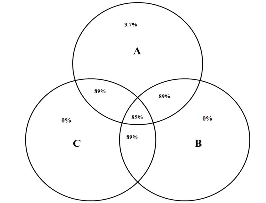
rumen of camels under investigation was assigned into 54 bacterial genera (Supplementary table S3).
Venn diagram showed that 48 bacteria genera (85%) were shared between the three animals (Figure 2).
100%
Cyanobacteria
Chloroflexi
90%
Candidate division TM7
80% Candidate division SR1
Verrucomicrobia
70% Tenericutes
Synergistetes
60% Elusimicrobia
SHA-109
50%
Proteobacteria
Planctomycetes
40%
Lentisphaerae
30% Actinobacteria
Euryarchaeota
20% Unclassified
Spirochaetae
10% Firmicutes
Fibrobacteres
0%
Bacteroidetes
Animal A Animal B Animal C
Figure 1. Stacked bar chart shows the relative abundance of bacterial phyla in animal A, B, and C.
AIMS Microbiology Volume 7, Issue 3, 354–367.359
Figure 2. Venn diagram shows the number of bacterial genera shared between rumen
samples of camel A, B and C. Each circle represents an animal and the overlapping areas
represent the common bacterial genera.
When the microbial community was inspected for family and genus level, the results revealed
that phylum Bacteriodetes was dominated by family Prevotellaceae (27%), and genus Prevotella.
Also, uncultured Bacteriodetes such as RC9 gut group, S24-7, BS11 gut group represented a high
proportion (8.46%) of phylum Bacteriodetes. Phylum Firmicutes was dominated by family
Lachnospiraceae (7.9%), which was dominated by genus Butyrivibrio and family Ruminococcaceae
(10.32%), which was dominated by genus Ruminococcus (Supplementary table S3). Phylum
Actinobacteria was dominated by the genus Atopobium and phylum Proteobacteria was dominated
by Desulfovibrio.
The archaeal community in the rumen of camels under investigation was represented in four genera
Candidatus Methanomethylophilus (0.81%), Methanobrevibacter (0.2%), Methanosphaera (0.04%),
Methanomicrobium (0.01%) (Supplementary Table S3).
3.2. Lignocellulolytic enzymes production
This study investigated the ability of the bacterial community in the rumen of camels to produce
cellulase and xylanase enzymes using rumen samples of camels fed wheat straw.
AIMS Microbiology Volume 7, Issue 3, 354–367.360
3.3. Xylanase production
The bacterial xylanase (endo-1,4-β- xylanase ) production was measured at different incubation
times by inoculating camel rumen samples into anaerobic bacterial medium containing birchwood
xylan for 24 h, 48 h, and 7 days at 38 ℃ and pH = 6.8. The results revealed that xylanase production
was increased from 24 h to 7 days. The overall mean production was 184.8 ± 101.3 mU/mL, (mean ±
SD) at 24 h, 243.5 ± 68 at 48 h, and 884.8 ± 111.3 at 7 days. The difference in xylanase production at
different fermentation times was significant (p < 0.01) (Figure 3).
1100
1049.5
1000
Enzyme production mU/ ml
941.8
900 884.8
839.5
800
700
600 X
500 FP
400 WS
300 266.7
243.5 AH
200 184.8 193.25 164
100 82.4 96.3
0 20.6
24h 48h 168h
Incubation Time (h)
Figure 3. Effect of incubation time on xylanase and cellulase production by rumen
bacteria of dromedary camel using birchwood xylan (x) and different cellulose sources,
filter paper (FP), wheat straw (WS), and alfalfa hay (AH). Data are shown as means of
three samples and asterisk shows the significant different differences at p < 0.05.
3.4. Cellulase production at different incubation time and using different cellulose sources
Bacterial cellulase (endo-1,4-β-glucanase) production was quantified by inoculating the camel
rumen samples into media containing one of three different cellulose sources, FP, WS, and AH at
different incubation times 24 h, 48 h, and 7 days at 38 ℃ and Ph = 6.8. The results showed that
cellulose sources impacted the cellulase production; also prolonging the incubation time increased
the cellulase production and the highest activity was obtained at 7 days except for the WS media,
where the production declined after 48 h (Figure 3; Supplementary Table S4). Regarding the
substrate type, the maximum production was obtained with AH media at 7 days and the lowest
production was observed with FP media at 24 h. The difference between incubation times and
substrate type was significant (p < 0.01) and the interaction between substrate and incubation time
was not significant.
AIMS Microbiology Volume 7, Issue 3, 354–367.361
4. Discussion
Lignocellulosic biomass could be hydrolyzed into fermentable sugars in the animal rumen by
different types of cellulases and xylanases, which work synergistically to break down the cellulose
and xylan in the plant cell wall [16]. The microbial communities in the rumen of dromedary camels
are predominated by fibrolytic bacteria that make the greatest contribution to the fermentation of
poor-quality plant biomass [3,7]. Therefore, understanding the composition of rumen bacteria in the
camels and their ability to produce cellulolytic and xylanolytic enzymes could open the door to
maximizing animal production by improving lignocellulose degradation as well as discovering novel
sources of enzymes with a wide range of applications [21]. Previous studies that were conducted on
microbial community in the rumen of dromedary camels focused on the composition of the bacterial
community using 16S rRNA/rDNA sequencing [1,3,7]. However, there is a need to determine the
ability of the bacterial community to produce lignocellulolytic enzymes. Therefore, the current study
explained the composition of bacteria and archaea in the rumen of three camels fed wheat straw and
broom corn using cDNA-amplicon sequencing by Illumine MiSeq platform. Furthermore, the ability
of rumen bacteria to produce xylanase and cellulase enzymes was determined. Alpha diversity
indices were similar to values were observed in cattle [11,35] and higher than values of Surti Buffalo [36].
Most of the bacterial reads (65.29%) were assigned to the Firmicutes and Bacteroidetes phyla (Figure 1),
which is in agreement with previous studies on the camel rumen [1,7], cattle [35], and Surti Buffalo [36].
Also, phylum Bacteroidetes dominated the bacterial community in this study, which is in agreement
with the results of the camel rumen [3].
Bacteroidetes degrade the protein and polysaccharides such as cellulose, pectin, and xylan [37].
Uncultured members of Bacteroidetes are specialized in lignocellulose degradation [38]. Members of
Bacteroidetes were dominated by Prevotella and RC9_gut_group; these results are similar to
previous findings on bovines and camels [3,39]. The Prevotella degrade hemicelluloses, pectin,
starch, and protein and produce propionate in the rumen [40], which is used as an energy source by
the host animal and declines the methanogenesis in the rumen [41,42]. Thus, Bacteroidetes might
play a key role in the utilization of poor-quality feeds in the rumen. Phylum Firmicutes was
dominated by Ruminococcaceae and Lachnospiraceae families that were found to be active in fiber
digestion in the rumen [37,41]. Also, this phylum was dominated by cellulolytic bacterial genera,
Butyrivibrio and Ruminococcus [3,43].
The Fibrobacteres were observed in a higher proportion in the camel rumen. The percentage of this
phylum in the current study was 18.1%, while it was 4.5–29% in Mehshana buffalo [37], 4.2–14.1% in
wild ruminant [44], 3.09% in the camel in Iran [3]. Fibrobacteres have been shown in previous
studies to be the principal cellulolytic active bacteria in the rumen [41,45]. Genus Treponema, the
dominant genus in phylum Spirochaetes, has the ability of cellulose degradation [46,47]. Genus
Elusimicrobium dominated the phylum Elusimicrobia; this genus was observed in the gut of
cellulose-degrading termite [48]. Therefore, this phylum has a potential role in fiber degradation in
the rumen [7]. Actinobacteria phylum has acetogenic activities and was found also in the rumen of
moose [49]. Lentisphaerae phylum was dominated by Victivallis that ferment cellobiose [50].
The dominant bacterial families in this study were family Prevotellaceae (27%), uncultured
Bacteriodetes (RC9 gut group, S24-7, BS11 gut group), and family Lachnospiraceae. All these
groups have fibrolytic or potential fibrolytic activities [3,7,51,52], which, indicates that most of the
bacterial community (about 80%) in the rumen of the camels under investigation has a role in fiber
AIMS Microbiology Volume 7, Issue 3, 354–367.362
degradation. This could explain the ability of camels to survive in desert harsh conditions with
low-quality forages. Also, this result highlights the camel rumen as a good source of fibrolytic
enzymes and productive bacteria [9,10,53].
The composition of the microbial community in the rumen is mainly influenced by the type of
animal diet [54], and lignocellulolytic diets stimulate the fibrolytic microbes [7,55]. The camels in
this study were fed wheat straw, which is considered poor-quality forage as it has low nutritive value
regarding crude protein, and soluble carbohydrate and it has high lignocellulose content [56], which
might support the high proportion of fibrolytic bacteria.
This study explained the possibility of using the anaerobic bacterial community of the camel
rumen in producing cellulase and xylanase by inoculating the camel rumen contents into anaerobic
bacterial media enriched with xylan and different fiber sources, including filter paper, wheat straw,
and alfalfa hay.
The maximum xylanase production (884.8 mU/mL) was observed at 7 days (Figure 3), this
finding had a similar trend to results on different xylanolytic gut bacteria [57]. On the other hand, the
anaerobic bacterial community in this study produced more xylanase than the aerobic fungi [58] and
anaerobic rumen fungi of the camel gut [21]. Cellulase production by anaerobic bacteria in this study
varied greatly between incubation periods and cellulose sources, which is in agreement with previous
studies [15,59]. In this study, we used three fiber sources, filter paper (FP), wheat straw (WS), and
alfalfa hay (AH). The highest cellulase production (1049.5 mU/mL) was observed by anaerobic
bacteria incubated in AH media at 7 days, similar results were obtained by the cellulolytic bacteria
isolated from goat and swine [16,57,60], and cow manure [17]. Cellulase production by anaerobic
bacterial community in the current study was higher than the production of Bacillus isolated from
cow dung [17], cellulolytic bacteria isolated from goat and swine [57], and aerobic and anaerobic
fungi [24,58]. The decrease in production in WS after 48 hrs could be attributed to the depletion of
nutritional ingredients in the medium [60].
In addition, this study identified the archaeal community in the camel rumen. Understanding the
archaeal community in camel rumen is important as the methanogenic archaea are the main producers
of methane in the rumen by converting the H2 and CO2 produced in the rumen to methane [61]. Also, the
archaeal community is highly impacted by diet [7]. The archaeal community in the rumen of camels
under investigation was dominated by Methanomethylophilus, Methanobrevibacter, Methanosphaera,
and Methanomicrobium, this result is consistent with other studies on camel [3,7].
Methanobrevibacter was found dominant in the rumen of a wide range of ruminant animals and
correlated with high methane production [54,62,63]. Additionally, Methanomicrobium correlated
positively with high lignocellulose in the animal diet in buffalo [64], and camels [7].
5. Conclusions
This study applied Illumina-amplicon sequencing and batch incubation technique to get insight
into the composition of bacterial and archaeal communities in the rumen of camels fed low-quality
forages and to quantifying the cellulolytic and xylanolytic activities of rumen bacteria. Most of the
rumen bacteria in the camel rumen have fibrolytic activities. The production of cellulase and
xylanase was impacted by incubation time and cellulose source where the alfalfa hay was associated
with high-cellulase production. This study highlights the camel rumen as a promising source for
fibrolytic enzymes.
AIMS Microbiology Volume 7, Issue 3, 354–367.363
Acknowledgments
We would like to thank the administration of Desert Research Center and the staff of Lethbridge
Research Center. This study received no specific funding.
Conflict of interest
The authors declare no conflict of interest.
Author Contributions
Alaa Emara Rabee conceived and designed the experiments, performed the experiments,
analyzed the data, prepared figures and/or tables, authored or reviewed drafts of the
paper, and approved the final draft.
Ebrahim Sabra conceived and designed the experiments, analyzed the data, authored or
reviewed drafts of the paper, and approved the final draft.
Robert Forster conceived and designed the experiments, analyzed the data, authored or
reviewed drafts of the paper, and approved the final draft.
References
1. Samsudin AA, Evans PN, Wright AD, et al. (2011) Molecular diversity of the foregut bacteria
community in the dromedary camel (Camelus dromedarius). Environ Microbiol 13: 3024–3035.
2. Kay RNB, Maloiy GMO (1989) Digestive secretions in camels. Options Méditerranéennes–Série
Séminaires-n.°2, 83–87.
3. Gharechahi J, Zahiri HS, Noghabi KA, et al. (2015) In-depth diversity analysis of the bacterial
community resident in the camel rumen. Syst Appl Microbiol 38: 67–76.
4. Lechner-Doll M, Engelhardt WV (1989) Particle size and passage from the forestomach in camels
compared to cattle and sheep fed a similar diet. J Anim Physiol Anim Nutr 61: 120–128.
5. Iqbal A, Khan BB (2001) Feeding behaviour of camel. Pak J Agric Sci 38: 58–63.
6. Samsudin AA, Wright ADG, Al Jassim R (2012) Cellulolytic bacteria in the foregut of the
dromedary camel (Camelus dromedarius). Appl Environ Microbiol 78: 8836–8839.
7. Rabee AE, Forster RJ, Elekwachi CO, et al. (2020) Comparative analysis of the metabolically
active microbial communities in the rumen of dromedary camels under different feeding
systems using total rRNA sequencing. Peer J 8: e10184.
8. Bhatt VD, Dande SS, Patil NV, et al. (2013) Molecular analysis of the bacterial microbiome in
the forestomach fluid from the dromedary camel (Camelus dromedarius). Mol Biol Rep 40:
3363–3371.
9. Gharechahi J, Salekdeh GH (2018) A metagenomic analysis of the camel rumen’s microbiome
identifies the major microbes responsible for lignocellulose degradation and fermentation.
Biotechnol Biofuels 11: 216.
10. Ameri R, Laville E, Potocki-VeÂronèse G, et al. (2018) Two new gene clusters involved in the
degradation of plant cell wall from the fecal microbiota of Tunisian dromedary. PLoS One 13:
e0194621.
AIMS Microbiology Volume 7, Issue 3, 354–367.364
11. Jami E, White BA, Mizrahi I (2014) Potential role of the bovine rumen microbiome in modulating
milk composition and feed ffficiency. PLoS One 9: e85423.
12. Moss AR, Jouany JP, Newbold J (2000) Methane production by ruminants: its contribution to
global warming. Ann Zootech 49: 231–253.
13. Van Nevel CJ, Demeyer DI (1996) Control of rumen methanogenesis. Environ Monit Assess 42:
73–97.
14. Lee K, Webb RI, Janssen PH, et al. (2009) Phylum Verrucomicrobia representatives share a
compartmentalized cell plan with members of bacterial phylum Planctomycetes. BMC Microbiol
9: 5.
15. Ekinci MS, Ӧzcan N, ӦzkӦse E, et al. (2001) A Study on cellulolytic and hemicellulolytic
enzymes of anaerobic rumen bacterium Ruminococcus flavefaciens Strain 17. Turk J Vet Anim
Sci 25: 703–709.
16. Seo JK, Park TS, Kwon IH, et al. (2013) Characterization of cellulolytic and xylanolytic
enzymes of Bacillus licheniformis JK7 isolated from the rumen of a native Korean goat.
Asian-Aust J Anim Sci 26: 50–58.
17. Sadhu S, Ghosh PK, Aditya G, et al. (2014) Optimization and strain improvement by mutation
for enhanced cellulase production by Bacillus sp. (MTCC10046) isolated from cow dung. J
King Saud UnivSci 26: 323–332.
18. Khatab MSA, Abd El Tawab AM, Fouad MT (2017) Isolation and characterization of anaerobic
bacteria from frozen rumen liquid and its potential characterization. Int J Dairy Sci 12: 47–51.
19. Selinger LB, Fosberg CW, Cheng KJ (1996) The rumen: A unique source of enzymes for
enhancing livestock production. Anaerobe 2: 263–284.
20. Hess M, Sczyrba A, Egan R, et al. (2011) Metagenomic discovery of biomass degrading genes
and genomes from cow rumen. Science 331: 463–467.
21. Rabee AE, Al Ahl AAS, Sabra EA, et al. (2019a) Assessment of xylanolytic and cellulolytic
activities of anaerobic bacterial community in the rumen of camel using different substrates.
Menoufia J Animal Poultry Fish Prod 3: 69–82.
22. Molina-Guerrero CE, de la Rosa G, Gonzalez Castañeda J, et al. (2018) Optimization of culture
conditions for production of cellulase by Stenotrophomonas maltophilia. Bio Res 13:
8358–8372.
23. Sethi S, Datta A, Gupta BL, et al. (2013) Optimization of Cellulase Production from Bacteria
Isolated from Soil. Inter Scholarly Res Not 2013.
24. Rabee AE, Forster RJ, Elekwachi CO, et al. (2019b) Community structure and fibrolytic activities
of anaerobic rumen fungi in dromedary camels. J Basic Microbiol 49: 1–10.
25. Phillips MW, Gordon GLR (1988) Sugar and polysaccharide fermentation by rumen anaerobic
fungi from Australia, Britain and New Zealand. BioSystems 21: 377–383.
26. Wang Z, Elekwachi C, Jiao J, et al. (2017) Changes in Metabolically Active Bacterial
Community during Rumen Development, and Their Alteration by Rhubarb Root Powder
Revealed by 16S rRNA Amplicon Sequencing. Front Microbiol 8: 159.
27. Liu K, Xu Q, Wang L, et al. (2016) Comparative studies of the composition of bacterial
microbiota associated with the ruminal content, ruminal epithelium and in the faeces of lactating
dairy cows. Microb Biotechnol 9: 257–268.
28. Caporaso JG, Kuczynski J, Stombaugh J, et al. (2010) QIIMEE allows analysis of
high-throughput community sequencing data. Nat Methods 7: 335–336.
AIMS Microbiology Volume 7, Issue 3, 354–367.365
29. Andrews S (2010) Fast QC: a quality control tool for high throughput sequence data. Available
from: http://www.bioinformatics.babraham.ac.uk/projects/fastqc.
30. Bolger AM, Lohse M, Usadel B (2014) Trimmomatic: A flexible trimmer for Illumina Sequence
Data. Bioinformatics 30: 2114–2120.
31. Zhang J, Kobert K, Flouri T, et al. (2014) PEAR: a fast and accurate Illumina Paired-End reAd
mergeR. Bioinformatics 30: 614–620.
32. Caldwell DR, Bryant MP (1966) Medium without rumen fluid for nonselective enumeration and
isolation of rumen bacteria. Appl Microbiol 14: 794–801.
33. McSweeney CS, Denman SE, Mackie RI (2005) Rumen bacteria. In: Makkar H.P., McSweeney
C.S. (eds) Methods in Gut Microbial Ecology for Ruminants. Springer, Dordrecht.
34. IBM Corp. Released (2011) IBM SPSS Statistics for Windows, Version 20.0. Armonk, NY: IBM
Corp.
35. Petri RM, Schwaiger T, Penner GB, et al. (2013) Characterization of the core rumen
microbiome in cattle during transition from forage to concentrate as well as during and after an
acidotic challenge. PLoS One 8: e83424.
36. Pandya PR, Singh KM, Parnerkar S, et al. (2010) Bacterial diversity in the rumen of Indian Surti
buffalo (Bubalus bubalis), assessed by 16S rDNA analysis. J Appl Genet 51: 395–402.
37. Pitta DW, Kumar S, Veiccharelli B, et al. (2014) Bacterial diversity associated with feeding dry
forage at different dietary concentrations in the rumen contents of Mehshana buffalo (Bubalus
bubalis) using 16S pyrotags. Anaerobe 25: 31–41.
38. Naas AE, Mackenzie AK, Mravec J, et al. (2014) Do rumen Bacteroidetes utilize an alternative
mechanism for cellulose degradation? mBio 5: e01401–e01414.
39. Fouts DE, Szpakowski S, Purushe J, et al. (2012) Next generation sequencing to define
prokaryotic and fungal diversity in the bovine rumen. PLoS One 7: e48289.
40. Russell JB, Rychlik JL (2001) Factors that alter rumen microbial ecology. Science 292:
1119–1122.
41. Nathani NM, Patel AK, Mootapally CS, et al. (2015) Effect of roughage on rumen microbiota
composition in the efficient feed converter and sturdy Indian Jaffrabadi buffalo (Bubalus bubalis).
BMC Genomics 16: 1116.
42. Koike S, Yoshitani S, Kobayashi Y, et al. (2003) Phylogenetic analysis of fiber-associated
rumen bacterial community and PCR detection of uncultured bacteria. FEMS Microbiol Lett 229:
23–30.
43. Liu K, Xu Q, Wang L, et al. (2017) The impact of diet on the composition and relative
abundance of rumen microbes in goat. Asian-Australas J Anim Sci 30: 531–537.
44. Gruninger RJ, McAllister TA, Forster RJ (2016) Bacterial and archaeal diversity in the
gastrointestinal tract of the orth American beaver (Castor canadensis). PLoS One 11: e0156457.
45. Ransom-Jones E, Jones DL, McCarthy AJ, et al. (2012) The Fibrobacteres: an important phylum
of cellulose-degrading bacteria. Microb Ecol 63: 267–281.
46. Ishaq SL, Wright AG (2012) Insight into the bacterial gut microbiome of the North American
moose (Alces alces). BMC Microbiol 12: 212.
47. Leahy S, Kelly W, Ronimus R, et al. (2013) Genome sequencing of rumen bacteria and archaea
and its application to methane mitigation strategies. Animal 7: 235–243.
AIMS Microbiology Volume 7, Issue 3, 354–367.366
48. Herlemann DPR, Geissinger O, Ikeda-Ohtsubo W, et al. (2009) Genomic analysis of
“Elusimicrobium minutum,” the first cultivated representative of the phylum “Elusimicrobia”
(formerly termite group 1). Appl Environ Microbiol 70: 2841–2849.
49. Ishaq S, Sundset M, Crouse J, et al. (2015) High-throughput DNA sequencing of the moose rumen
from different geographical locations reveals a core ruminal methanogenic archaeal diversity and
a differential ciliate protozoal diversity. Microb Genom 1: e000034.
50. Zoetendal E, Plugge CM, Akkermans ADL, et al. (2003) Victivallisvadensis gen. nov., sp. nov.,
a sugar-fermenting anaerobe from human faeces. Int J Syst Evol Microbiol 53: 211–215.
51. Jewell KA, McComirck C, Odt CL, et al. (2015) Ruminal bacterial community composition in
dairy cows is dynamic over the course of two lactations and correlates with feed efficiency. Appl
Environ Microbiol 18: 4697–4710.
52. Noel SJ, Højberg O, Urich T, et al. (2016) Draft genome sequence of “Candidatus
Methanomethylophilus” sp. 1R26, enriched from bovine rumen, a methanogenic archaeon
belonging to the Methanomassiliicoccales order. Genome Announc 4: e01734– e01715.
53. Zorec M, Vodovnik M, MarinŠek-Logar R, et al. (2014) Potential of selected rumen bacteria for
cellulose and hemicellulose degradation. Food Technol Biotechnol 52: 210–221.
54. Henderson G, Cox F, Ganesh S, et al. (2015) Rumen microbial community composition varies
with diet and host, but a core microbiome is found across a wide geographical range. Sci Rep 5:
14567.
55. Carberry CA, Kenny DA, Han S, et al. (2012) Effect of phenotypic residual feed intake and
dietary forage content on the rumen microbial community of beef cattle. Appl Environ
Microbiol 78: 4949–4958.
56. Shrivastava B, Jain KK, Kalra A, et al. (2014) Bioprocessing of wheat straw into nutritionally
rich and digested cattle feed. Sci Rep 4: 6360.
57. Asem D, Leo VV, Passari AK, et al. (2017) Evaluation of gastrointestinal bacterial population for
the production of holocellulose enzymes for biomass deconstruction. PLoS One 12: e0186355.
58. Salmon DNX, Spier MR, Soccol CR, et al. (2014) Analysis of inducers of xylanase and
cellulase activities production by Ganoderma applanatum LPB MR-56. Fungal Biol 118:
655–662.
59. Williams AG, Withers SE (1982) The production of plant cell wall polysaccharide-degrading
enzymes by hemicellulolytic rumen bacterial isolates grown on a range of carbohydrate
substrates. J Appl Bacteriol 52: 377–387.
60. Yang W, Meng F, Peng J, et al. (2014) Isolation and identification of a cellulolytic bacterium
from the Tibetan pig’s intestine and investigation of its cellulase production. Electron J
Biotechnol 17: 262–267.
61. Hook SE, Wright ADG, McBride BW (2010) Methanogens: methane producers of the rumen and
mitigation strategies. Archaea 2010: 945785.
62. St-Pierre B, Wright AG (2012) Molecular analysis of methanogenic archaea in the forestomach
of the alpaca (Vicugna pacos). BMC Microbiol 12: 1.
63. Salgado-Flores A, Bockwoldt M, Hagen L, et al. (2016) First insight into the faecal microbiota
of the high Arctic muskoxen (Ovibos moschatus). Microb Genom 2.
AIMS Microbiology Volume 7, Issue 3, 354–367.367
64. Franzolin R, Wright AG (2016) Microorganisms in the rumen and reticulum of buffalo (Bubalus
bubalis) fed two different feeding systems. BMC Research Notes 9: 243.
© 2021 the Author(s), licensee AIMS Press. This is an open access
article distributed under the terms of the Creative Commons
Attribution License (http://creativecommons.org/licenses/by/4.0)
AIMS Microbiology Volume 7, Issue 3, 354–367.You can also read