Lupus miliaris disseminatus faciei with complete response to isotretinoin
←
→
Page content transcription
If your browser does not render page correctly, please read the page content below
Volume 27 Number 1| January 2021
Dermatology Online Journal || Case Presentation                                                                              27(1):9

Lupus miliaris disseminatus faciei with complete response to
isotretinoin
María Rogel-Vence1 MD, Marcos Carmona-Rodríguez1 MD, Violeta Herrera-Montoro2 MD, Lucía González-
Ruiz1 MD, Maria Pilar Cortina-de la Calle1 MD, Maria Prado Sánchez-Caminero1 MD
Affiliations:1Department of Dermatology, Hospital General Universitario de Ciudad Real, Spain, 2Department of Pathology,
Hospital Universitario de Ciudad Real, Spain
Corresponding Author: María Rogel-Vence, Hospital General Universitario de Ciudad Real, Obispo Rafael Torija, Ciudad Real, 13005,
Spain, Tel: 34-600863475, Email: mrogelvence@gmail.com

                                                                       characterized by the presence of reddish-brown,
  Abstract                                                             dome-shaped papules involving the central face. It is
  Lupus miliaris disseminatus faciei is an uncommon                    a chronic inflammatory disorder which affects young
  type of granulomatous rosacea characterized by a                     adults of both sexes. Histopathological features
  papular eruption in the central regions of the face. A
                                                                       include dermal epithelioid cell granulomas with
  43-year-old woman presented with an asymptomatic
  papular eruption on the face that had developed                      central necrosis. Treatment is frequently
  over a period of five months. Physical examination                   unsatisfactory. Several treatments have been
  revealed multiple, small, reddish-brown papules,                     reported, however no treatment has consistently
  distributed symmetrically on the central area of the                 been able to prevent the desfiguring scars. We report
  face. A biopsy was taken, showing dermal epithelioid                 a new case of LMDF successfully treated with
  cell granulomas with central necrosis and                            isotretinoin without residual scaring.
  surrounding        lymphocytic        infiltrate    with
  multinucleate giant cells. No foreign bodies were
  found in granulomas and no mycobacterial or fungal
  components were detected. On the basis of these                      Case Synopsis
  findings, the diagnosis of lupus miliaris disseminatus               A 43-year-old woman presented to the dermatology
  faciei was made. The patient was given oral                          clinic with an asymptomatic papular eruption on the
  isotretinoin 20mg/day with initial slow response.
                                                                       face that had developed over a period of five
  After 6 months’ treatment the lesions completely
  disappeared. Many authors consider this entity to be                 months. Physical examination revealed multiple
  a variant of granulomatous rosacea. It is a chronic                  small      reddish-brown,     papules      distributed
  condition that primarily affects young adults.                       symmetrically on the central face, namely the eyelids
  Treatment is usually unsatisfactory. Therapies with                  and perioral area (Figure 1). There was
  corticosterois, tetracyclines, retinoids, clofazimine or             accompanying scaling. A complete blood count with
  topical tacrolimus have been described but there is a                renal and hepatic panel were normal. A biopsy was
  lack of controlled studies and convincing results. Our
  success with a 6-month course of low dose                            taken, showing dermal epithelioid cell granulomas
  isotretinoin suggests consideration of a longer trial                with central necrosis and surrounding lymphocytic
  prior to abandoning this as treatment.                               infiltrate with multinucleate giant cells. No foreign
                                                                       bodies were found in the granulomas. No
                                                                       mycobacterial or fungal components were detected
Keywords: lupus miliaris disseminates faciei,                          in dermal tissues by Ziehl-Neelsen or periodic acid-
granulomatous rosacea, isotretinoin, granuloma                         Schiff staining (Figure 2). On the basis of these
                                                                       findings, the diagnosis of LMDF was made. The
                                                                       patient was given oral isotretinoin 20 mg/day with
Introduction                                                           initial slow response. By three months, a moderate
Lupus miliaris disseminatus faciei (LMDF) is an                        improvement had been achieved with flattening of
uncommon dermatosis of unknown etiology,                               the papules. After 6 months of treatment with

                                                                 -1-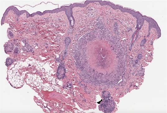
Volume 27 Number 1| January 2021
Dermatology Online Journal || Case Presentation 27(1):9
on histopathologic studies, but the use of
polymerase chain reaction and culture have shown
no evidence of this bacterium [2]. Similarly, LMDF
A
A
B
Figure 1. A) First visit. The patient presented with multiple small
reddish-brown, papules distributed symmetrically on the central
face. B) On closer view, multiple papules around the eyelids can
B
be appreciated.
isotretinoin the lesions had resolved, leaving no
disfiguring scars on the face (Figure 3). The patient
maintained isotretinoin treatment 6 more months
with a lower dosis (5mg/day) and treatment was
then discontinued. At present the patient has no
active treatment and has maintained a complete
response after 12 months of medical monitoring.
C
Case Discussion
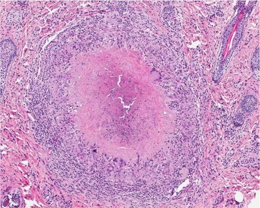
Lupus miliaris disseminatus faciei is a rare Figure 2. A) Panoramic image of dermal granuloma with central
necrosis. H&E, 10×. B) Epithelioid cell granuloma with central
inflammatory dermatosis first described by Fox in
necrosis and surrounding lymphocytic infiltrate on higher
1878 [1]. The etiology is unknown. At first, it was magnification. H&E, 100×. C) No mycobacterial or fungal
associated with Mycobacterium tuberculosis infection components were detected in dermal tissues by Ziehl-Neelsen
owing to the presence of central caseating necrosis technique, 100×.
-2-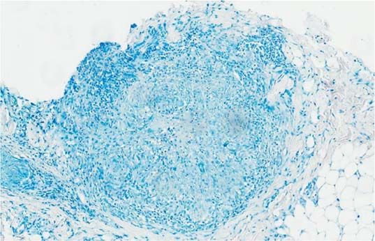
Volume 27 Number 1| January 2021
Dermatology Online Journal || Case Presentation 27(1):9
[2,10] often leaving numerous pitted scars and
pigmentation. Despite the usual spontaneous
resolution, no treatment has been able to prevent
the disfiguring scars.
Histopathological studies usually show dermal
epithelioid cell granulomas with central necrosis and
surrounding lymphocytic infiltrate with
multinucleate giant cells [10]. Foreign bodies,
mycobacteria, and fungi should be eliminated by
Ziehl-Neelsen and periodic acid-Schiff staining.
Histopathological findings in granulomatous
rosacea and LMDF frequently show overlap, but t he
A
LMDF subset particularly exhibits granulomas with
central necrosis.
Many therapies have been reported in the literature
but there is no established treatment for this entity.
Unfortunately, despite treatment or spontaneous
involution, residual scars are a frequent outcome.
Reported treatments include oral glucocorticoids
[11], dapsone [12], tetracyclines (doxycycline,
minocycline) [13], isotretinoin [14,15], and
clofazimine [16]. Successful treatment of LMDF with
combined oral metronidazole or oral dapsone
B combined with topical tacrolimus also have yielded
good results [17]. More recently, laser therapy also
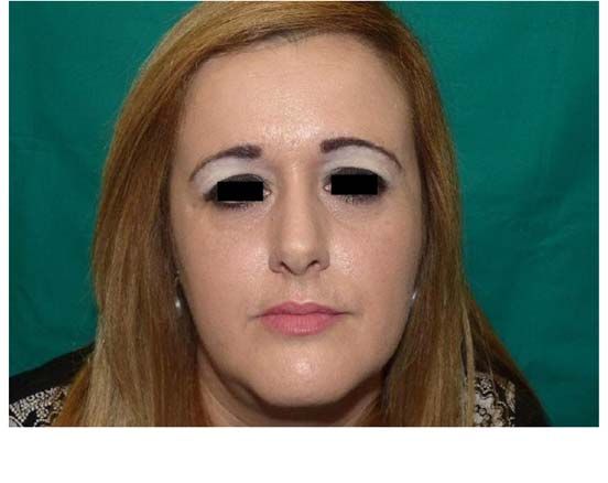
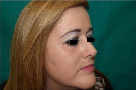
Figure 3. A) Front view. After 6 months of treatment with
isotretinoin, examination revealed a complete resolution of lupus may have a role in the treatment of LMDF with a
miliaris disseminatus faciei. The patient presented neither pitted 1,450nm diode laser [18] and a non-ablative
scars nor residual pigmentation. B) Lateral view. fractionated 1,565nm laser [19].
was also believed to be a form of sarcoidosis.
However, LMDF is currently considered as a type of Conclusion
granulomatous rosacea, or in some cases a Lupus miliaris disseminatus faciei is considered as an
granulomatous periorificial dermatitis. The incidence uncommon variant of granulomatous rosacea. Some
of this disease is unknown but is considered to be cases might also be an example of granulomatous
uncommon [3]. It occurs most commonly in young periorificial dermatitis. It is necessary to rule out
adults, between the second and the fourth decades other granulomatous diseases. Treatment of LMDF is
of life [3]. It affects both genders, although most usually unsatisfactory. We report a new case of LMDF
cases have been reported in men [2]. Rare cases have in an adult woman successfully treated with low
been described in children [4] and the elderly [5,6]. dose oral isotretinoin. Slow improvement was noted
Clinically, it is characterized by an asymptomatic at first, but eventually she achieved and maintained
yellow-brown or brown-redish papular eruption a complete response without recurrences after 12
mainly involving the central face, especially the months of monitoring. Pitted or disfiguring scars did
eyelids [2,7]. Extrafacial m anifestations have been not result. Oral isotretinoin should be considered a
described in other localizations such as axillae [8] or good option for treatment of LMDF and should be
hands [9]. Lupus miliaris disseminatus faciei usually initiated as soon as posible in hopes of preventing
involutes spontaneously within 12 to 24 months scarring.
-3-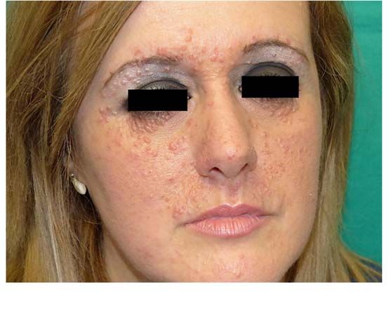
Volume 27 Number 1| January 2021
Dermatology Online Journal || Case Presentation                                                                                            27(1):9

Potential conflicts of interest                                                  The authors declare no conflicts of interests.

References
1.  Fox T. Disseminated follicular lupus (simulating acne) Lancet.               11. Uesugi Y, Aiba S, Usuba M, Tagami H. Oral prednisone in the
    1878;112:75–76. [DOI: 10.1016/S0140-6736(02)42942-X].                            treatment of acne agminata. Br J Dermatol. 1996;134:1098-1100.
2. Cymerman R, Rosenstein R, Shvartsbeyn M, Meehan SA, Kornreich                     [PMID: 8763432].
    C. Lupus miliaris disseminatus faciei. Dermatol Online J.                    12. Kumano K, Tani M, Murata Y. Dapsone in the treatment of miliary
    2015;21:13030/qt6b83q5gp. [PMID: 26990343].                                      lupus of the face. Br J Dermatol. 1983;109:57-62. [PMID: 6860572].
3. Toda-Brito H, Aranha JMP, Tavares ES. Lupus miliaris disseminatus             13. Goh BK, Tan HH. Doxycycline in the treatment of acne agminata.
    faciei. An Bras Dermatol. 2017;92:851-853. [PMID:29364447].                      Clin Exp Dermatol. 2003;28:677-679. [PMID: 14616848].
4. Misago N, Nakafusa J, Narisawa Y. Childhood granulomatous                     14. Berbis P, Privat Y. Lupus miliaris disseminatus faciei: efficacy of
    periorificial dermatitis: lupus miliaris disseminatus faciei in                  isotretinoin. J Am Acad Dermatol. 1987;16:1271-1272. [PMID:
    children?. J Eur Acad Dermatol Venereol. 2005;19:470-473. [PMID:                 3474247].
    15987296].                                                                   15. Daneshpazhooh M, Ehsani A, Toosi S, Robati RM. Isotretinoin in
5. Dekio S, Jidoi J, Imaoka C. Lupus miliaris disseminatus faciei--                  acne agminata. Saudi Med J. 2007;28:1600-1602. [PMID:
    report of a case in an elderly woman. Clin Exp Dermatol.                         17914528].
    1991;16:295-296. [PMID: 1794175].                                            16. Seukeran DC, Stables GI, Cunliffe WJ, Sheehan-Dare RA. The
6. Rocas D, Kanitakis J. Lupus miliaris disseminatus faciei: report of a             treatment of acne agminata with clofazimine. Br J Dermatol.
    new case and brief literature review. Dermatol Online J. 2013;19:4.              1999;141:596-597. [PMID: 10583095].
    [PMID: 23552001].                                                            17. Al-Mutairi N. Nosology and therapeutic options for lupus miliaris
7. Dev T, Thami T, Longchar M, Sethuraman G. Lupus miliaris                          disseminatus faciei. J Dermatol. 2011;38:864-873. [PMID:
    disseminatus faciei: a distinctive facial granulomatous eruption.                21714812].
    BMJ Case Rep. 2017;2017:bcr2017221118. [PMID: 28710244].                     18. Jih MH, Friedman PM, Kimyai-Asadi A, Friedman ES, Hymes SR,
8. Nemer KM, McGirt LY. Extrafacial lupus miliaris disseminatus.                     Goldberg LH. Lupus miliaris disseminatus faciei: treatment with
    JAAD Case Rep. 2016;2:363-365. [PMID: 27699199].                                 the 1450-nm diode laser. Arch Dermatol. 2005;141:143-145. [PMID:
9. Choi JY, Chae SW, Park JH. Lupus Miliaris Disseminatus Faciei with                15724009].
    Extrafacial Involvement. Ann Dermatol. 2016;28:791-794. [PMID:               19. Beleznay K, Friedmann DP, Liolios AM, Perry A, Goldman MP.
    27904291].                                                                       Lupus miliaris disseminatus faciei treated with 1,565 nm
10. Alonso V, Ramón D, Martín JM, Monteagudo C, Molina I, Jordá E.                   nonablative fractionated laser resurfacing: a case report. Lasers
    Lupus miliar diseminado de la cara [Lupus miliaris disseminatus                  Surg Med. 2014;46:663-665. [PMID: 25263633].
    faciei]. Actas Dermosifiliogr. 2005;96:182-185. [PMID: 16476363].

                                                                           -4-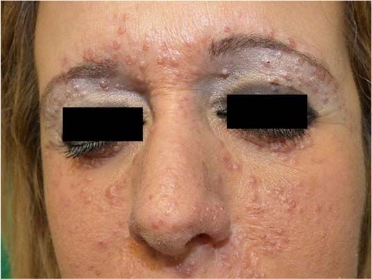
You can also read