Case Report Appendiceal Diverticulitis in a Young Female Diagnosed on Pathology after Laparoscopic Appendectomy for Acute Appendicitis
←
→
Page content transcription
If your browser does not render page correctly, please read the page content below
Hindawi
Case Reports in Medicine
Volume 2021, Article ID 2508956, 3 pages
https://doi.org/10.1155/2021/2508956

Case Report
Appendiceal Diverticulitis in a Young Female Diagnosed on
Pathology after Laparoscopic Appendectomy for
Acute Appendicitis

          Oluwatobi O. Onafowokan ,1,2 Aboubakr Khairat ,1,3 and Hugo J. R. Bonatti                                          1,3

          1
            University of Maryland Community Medical Group, Easton, MD, USA
          2
            Royal Lancaster Infirmary, Lancaster, UK
          3
            Meritus Surgical Specialists, Hagerstown, MD, USA

          Correspondence should be addressed to Hugo J. R. Bonatti; hugo.bonatti@dr.com

          Received 1 May 2020; Revised 30 January 2021; Accepted 1 March 2021; Published 8 March 2021

          Academic Editor: John Kortbeek

          Copyright © 2021 Oluwatobi O. Onafowokan et al. This is an open access article distributed under the Creative Commons
          Attribution License, which permits unrestricted use, distribution, and reproduction in any medium, provided the original work is
          properly cited.
          Background. Appendiceal diverticulitis is a rare cause of inflammation of the appendix, which may mimic acute appendicitis. Its
          diagnosis is often delayed, and its occurrence carries an increased risk of significant complications, such as perforation. Case
          Presentation. A 23-year-old woman presented with sudden onset, severe, right lower quadrant abdominal pain and nausea. Her
          WBC was elevated, and abdominal CT showed findings indicative of acute appendicitis with a 13 mm fluid-filled appendix and
          local stranding. During laparoscopic appendectomy, significant inflammation was found around the appendix with some mucous
          material around the tip. The appendix base was not involved, and an endoloop was used to secure the stump. No other intra-
          abdominal abnormalities were observed. The patient recovered uneventfully. Pathology showed no classic appendicitis but
          appendiceal diverticulitis with signs of perforation. Discussion. Appendiceal diverticulitis is a rare condition which cannot be
          distinguished from acute appendicits clinically and on imaging. Diagnosis may be established based on pathology such as in our
          case. Appendectomy is indicated in appendiceal diverticulitis, and an appendix diverticulum is incidentally found during surgery
          or other investigations. This is due to the increased risk of perforation and the reported development of malignant tumors,
          including the appendix carcinoid.

1. Introduction                                                        occurrence carries an increased risk of significant complica-
                                                                       tions, including perforation [8] and a higher risk of mortality
Appendiceal diverticulosis is an uncommon pathology, first              [2, 4, 9]. Progression of diverticulosis to diverticulitis may occur
described in 1893 [1–3]. Congenital appendiceal diverticulosis         following a partial or complete obstruction of the appendix
is a true diverticulum, with a rare incidence of 0.014% [4].           lumen. This obstruction may be due to inflammation, mucosal
Acquired appendiceal diverticulosis is a false diverticulum on         swelling, fecaliths, torsion, or fibrous strictures [2].
the mesenteric border of the appendix, with a relatively more               Appendectomy is recommended when appendiceal di-
common incidence of 1.9%. Pathogenesis of acquired                     verticulosis is incidentally discovered on imaging or during
appendiceal diverticulosis is not completely understood, but it        surgery as up to 66% of patients will progress to an acute
may be associated with diverticulosis of other colonic seg-            inflammation [2]. Other authors have considered prophy-
ments, and various pathologies of the appendix [3, 5].                 lactic appendectomy less beneficial [10]. The predominant
    Appendiceal diverticulitis occurs due to the inflammation           scenario in the majority of publications involves patients
of an appendiceal diverticulum [2, 6]. It is a rare cause of           diagnosed with acute appendicitis and undergoing appen-
inflammation of the appendix, which may mimic acute ap-                 dectomy, but with final pathology showing appendiceal
pendicitis [5, 7]. Its diagnosis may be delayed, and its               diverticulitis and not appendicitis. It should be noted that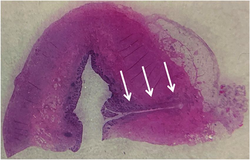
2                                                                                                          Case Reports in Medicine

                                          (a)                                             (b)

Figure 1: CT scan (transverse and coronal cut). Thickened fluid-filled appendix with localized fat stranding; an appendiceal diverticulum
cannot be seen.

both inflammatory processes may occur simultaneously
according to the classification proposed by Lipton et al. [8].
     We report the clinical course of a young female who
presented with an episode of right lower quadrant pain with
a clinical and computed tomography (CT) scan diagnosis of
acute appendicitis. She underwent laparoscopic appendec-
tomy and was found to have appendiceal diverticulitis on
final pathology.

2. Case Presentation
A 23-year-old healthy woman presented to the emergency               Figure 2: Intraoperative findings: periappendicitis and acute in-
room (ER) with acute onset, severe right lower quadrant              flammation of the appendix with mucinous secretions.
(RLQ) pain and nausea. On examination, she was alert and
oriented, her abdomen was soft, but she had significant
tenderness in the RLQ and a positive Murphy’s sign. White            such as cystic fibrosis [7]. Diagnosis is most frequently inci-
blood count was normal, and abdominal computed tomog-                dental but may be made my ultrasound or CT scan in selected
raphy (CT) scan showed a fluid-filled and dilated appendix             cases [9]. Appendiceal diverticulitis has been shown to be more
with thickened walls and localized inflammatory changes in            than four times as likely to perforate compared with acute
the RLQ indicative of acute appendicitis (Figure 1).                 appendicitis, increasing mortality 30-fold compared with
    She was started on antibiotics in the ER (ertapenem 1 g)         simple appendicitis [2, 8]. Therefore, correct diagnosis and
and consented for laparoscopic appendectomy. Laparoscopy             urgent management with antibiotics and appendectomy are
was done with 5 mm left (L) UQ and umbilical trocars and a           currently favoured with laparoscopy being the preferred ap-
suprapubic Teleflex MiniLap Alligator Grasper [11]. The               proach [12, 13]. In contrast to diverticulitis of the colon or small
appendix was dilated and acutely inflamed with mucosal                bowel, no large series demonstrating successful nonoperative
secretions on the tip of the appendix (Figure 2). The vascular       management of appendiceal diverticulitis are available, and this
pedicle and appendix base were secured with endoloops.               is most likely due to the fact that appendicitis and appendiceal
    The patient was discharged the next morning and had an           diverticulitis are difficult to distinguish on imaging [14]. Ra-
uneventful recovery. Final pathology revealed a diverticulum in      diographic diagnosis of appendiceal diverticulosis/diverticulitis
the tip of the appendix, with active inflammation (Figure 3).         is difficult, but CT scan may identify an appendiceal diver-
                                                                     ticulum with the pericaecal fat showing increased density.
3. Discussion                                                            It should be recognized that diverticula of the appendix
                                                                     are also associated with an increased risk of appendiceal
Acute appendicitis due to appendiceal diverticulitis is a rare       neoplasms including carcinoid tumors, adenocarcinoma,
condition, with an incidence of 0.004–2.1% [7]. Risk factors         and mucinous adenomas (pseudomyxoma peritonei) [4];
may include age >30 years old, male gender, and comorbidities        strengthening the argument for appendectomy [13, 15].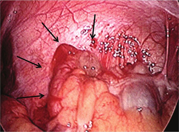
Case Reports in Medicine 3
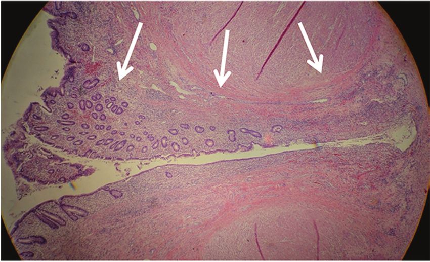
(a) (b)
Figure 3: Pathology findings: section of the appendix tip (a) and close up (b) of the diverticulum (arrows) showing acute inflammation.
To summarise, acute appendicitis due to appendiceal [8] S. Lipton, J. Estrin, and I. Glasser, “Diverticular disease of the
diverticulitis is an uncommon condition, which is often appendix,” Surgery, Gynecology & Obstetrics, vol. 168, no. 1,
diagnosed on pathology. Appendiceal diverticulitis may be pp. 13–16, 1989.
associated with significant complications. Currently, ap- [9] M. L. Altieri, G. N. Piozzi, P. Salvatori, M. Mirra, G. Piccolo,
and N. Olivari, “Appendiceal diverticulitis, a rare relevant
pendectomy is favoured for acute appendiceal diverticulitis
pathology: presentation of a case report and review of the
and incidentally discovered appendiceal diverticulosis, with literature,” International Journal of Surgery Case Reports,
laparoscopy being the preferred approach. vol. 33, pp. 31–34, 2017.
[10] H. Kabiri, L. E. Clarke, and C. D. Tzarnas, “Appendiceal
Disclosure diverticulitis,” The American Surgeon, vol. 72, no. 3,
pp. 221–223, 2006.
Parts of this article were presented as a poster at the 60th [11] H. J. R. Bonatti, “Development of a two port laparoscopic
Annual Meeting of the Austrian Society of Surgery in June appendectomy technique at a rural hospital,” Minimally In-
vasive Surgery, vol. 2019, no. 8, 7 pages, Article ID 9761968,
2019 in Innsbruck, Austria (https://link.springer.com/
2019.
article/10.1007/s10353-019-0600-2). [12] M. Z. Albeeshi, A. A. Alwanyan, A. A. Salim, and
I. T. Albabtain, “Appendiceal diverticulitis presenting as acute
Conflicts of Interest appendicitis diagnosed postoperatively,” Journal of Surgical
Case Reports, vol. 2019, no. 12, p. rjz332, 2019.
The authors declare that they have no conflicts of interest. [13] J. L. Ng, S. L. Wong, and R. Mathew, “Appendiceal diver-
ticulosis: a harbinger of underlying primary appendiceal
adenocarcinoma?” Journal of Gastrointestinal Oncology, vol. 9,
References no. 2, pp. E1–E5, 2018.
[14] A. H. Yardimci, C. T. Bektas, E. Pasaoglu et al., “Retrospective
[1] T. N. Kelynack, “A contribution to the pathology of the study of 24 cases of acute appendiceal diverticulitis: CT
vermiform appendix,” Glasgow Medical Journal, vol. 41, no. 1, findings and pathological correlations,” Japanese Journal of
pp. 67-68, 1894. Radiology, vol. 35, no. 5, pp. 225–232, 2017.
[2] B. J. Phillips and C. W. Perry, “Appendiceal diverticulitis,” [15] S. A. Käser, N. Willi, and C. A. Maurer, “Prevalence and
Mayo Clinic Proceedings, vol. 74, no. 9, pp. 890–892, 1999. clinical implications of diverticulosis of the vermiform ap-
[3] D. C. Collins, “A study of 50,000 specimens of the human pendix,” Journal of International Medical Research, vol. 41,
vermiform appendix,” Surgery, Gynecology & Obstetrics, no. 4, pp. 1350–1356, 2013.
vol. 101, no. 4, pp. 437–445, 1955.
[4] M. P. Dupre, I. Jadavji, E. Matshes, and S. J. Urbanski,
“Diverticular disease of the vermiform appendix: a diagnostic
clue to underlying appendiceal neoplasm,” Human Pathology,
vol. 39, no. 12, pp. 1823–1826, 2008.
[5] I. Yamana, S. Kawamoto, K. Inada, S. Nagao, T. Yoshida, and
Y. Yamashita, “Clinical characteristics of 12 cases of appen-
diceal diverticulitis: a comparison with 378 cases of acute
appendicitis,” Surgery Today, vol. 42, no. 4, pp. 363–367, 2012.
[6] D. S. Heffernan, N. Saqib, and M. Terry, “A case of appen-
diceal diverticulitis, and a review of the literature,” Irish
Journal of Medical Science, vol. 178, no. 4, pp. 519–521, 2009.
[7] B. Abdullgaffar, “Diverticulosis and diverticulitis of the ap-
pendix,” International Journal of Surgical Pathology, vol. 17,
no. 3, pp. 231–237, 2009.You can also read