Management of Adenomyosis - Endovascular Today
←
→
Page content transcription
If your browser does not render page correctly, please read the page content below
WOMEN’S
                                                                                                                  H E A LT H

Management of
Adenomyosis
A review of characteristic imaging findings and treatment options, with an emphasis on the
use of uterine artery embolization.

BY THERESA M. CARIDI, MD, AND JAMES B. SPIES, MD, MPH

A
         denomyosis is defined as ectopic endometrial tis-      myosis on MRI include thickening of the junctional
         sue within the musculature of the uterus.1 It is a     zone exceeding 12 mm and high-signal-intensity foci
         challenging condition in that it often overlaps in     on T2/T1-weighted images similar to the case shown in
         symptoms and is found in conjunction with other        Figure 1.4 Many studies have evaluated the diagnostic
gynecologic disorders, including endometriosis and uterine      accuracy of TVUS and MRI techniques for adenomyosis.
leiomyoma (fibroids).2 The typical clinical manifestations      TVUS has a sensitivity of approximately 72% and speci-
of adenomyosis occur in women who are 40 to 50 years of         ficity of 81% versus 77% and 89% for MRI, respectively.8
age and include abnormal uterine bleeding and dysmenor-         In addition, MRI can provide greater detail about the
rhea (65% of patients).1 The exact pathogenesis is not fully    extent of disease and additional uterine lesions, as well
defined but is thought to be the result of direct invagina-     as information about the less common presentation of
tion of the endometrium into the myometrium.3 The               adenomyosis, where focal disease in the form of an ade-
interventionalist’s role is to offer uterine artery emboliza-   nomyoma is present rather than the more typical diffuse
tion (UAE), which provides some benefits over traditional       adenomyosis findings.
medical and surgical therapies for adenomyosis.

IMAGING FOR ADENOMYOSIS
   Transvaginal ultrasound (TVUS) and MRI are often uti-
lized for the noninvasive diagnosis of adenomyosis.4 Just
as the clinical symptomatology can overlap with other
pelvic conditions, imaging also has limitations because
there are no standard diagnostic criteria for adenomyosis
and many uterine lesions can coexist on imaging.1,5
   Classic findings of adenomyosis on ultrasound include
heterogeneous myometrial echotexture, asymmetric
thickening of the wall of the myometrium, myometrial
cysts, subendometrial echogenic linear striations, and
poor definition of the endometrial–myometrial junc-
tion.6 With TVUS, the findings of adenomyosis are often
observer dependent and can be difficult for the less
experienced clinician.7 It has been suggested that use of
sonohysterography allows for an important distinction
between myometrial and endometrial lesions because
of the added benefit of a distended endometrial cavity,         Figure 1. Preembolization T2-weighted MRI showing a dif-
leading to improved diagnostic accuracy.6                       fusely thickened junctional zone with several high-signal foci
   The question then becomes what, if any, benefit is           consistent with diffuse adenomyosis. Small scattered fibroids
there to performing an MRI? Typical findings of adeno-          are also seen.

                                                                          VOL. 17, NO. 1 JANUARY 2018 ENDOVASCULAR TODAY 57
WOMEN’S
  H E A LT H

   TREATMENT OPTIONS                                               consultation, which allows for review of the symptom-
      Management of symptomatic adenomyosis can include            atology, a physical examination, and imaging. The first
   medication, hysterectomy, conservative surgery, or UAE.9        question that is answered in this setting is, “Does this
   Conventional treatment with hysterectomy allows for defin-      imaging finding of adenomyosis explain the patient’s
   itive diagnosis and treatment; however, medications, con-       symptoms?” If the patient has dysfunctional uterine
   servative surgery, and UAE are less invasive techniques that    bleeding such as bleeding between cycles, endometrial
   permit preservation of the uterus and possible fertility.10     biopsy is sometimes recommended to be performed by
      Medical therapies using suppressive hormonal treat-          a gynecologist before considering embolization, based on
   ments, such as continuous use of oral contraceptive pills,      the American College of Obstetricians and Gynecologists
   high-dose progestins, selective estrogen/progesterone           guidelines.
   receptor modulators, the levonorgestrel intrauterine               Each patient will be asked about their desire for future
   device, aromatase inhibitors, danazol, and gonadotropin-        fertility, and a brief overview of the aforementioned
   releasing hormone receptor agonists, can improve symp-          alternative options will be presented when appropriate.
   toms by temporarily inducing regression of adenomyosis.11       Simplified pictures of the pelvis and catheter place-
   Unfortunately, many of these medical options do not             ment are used during description of the procedure. The
   provide sustained improvement and are limited by their          outcomes of the procedure and possible risks are also
   menopausal-like side effects as well as temporarily blocking    covered. Active untreated infection, pregnancy, and
   the ability to conceive.1                                       suspected gynecologic malignancy are considered abso-
      For reproductive-age women and those with focal              lute contraindications to UAE, whereas contrast allergy,
   adenomyoma, conservative surgical approaches may be             coagulopathy, desire for future fertility, and renal impair-
   a better option. A recent systematic review of excisional       ment are relative contraindications.13 Additional risks
   and nonexcisional surgical approaches, including adeno-         discussed with the patient include onset of menopause,
   myomectomy with or without myometrial reduction,                arterial injury at the puncture site, clot formation, and
   endomyometrial ablation or resection, electrocoagulation        in the case of concomitant fibroids, missed malignant
   of adenomyoma, and myometrial excision, found that              tumor (leiomyosarcoma), and fibroid passage.
   more than 75% of women experienced symptom relief                  Results of laboratory testing, including complete blood
   with these conservative surgical treatments.10 However,         count for platelet count, basic metabolic panel for potas-
   recurrence rates varied from 9% to 32%, depending on the        sium and creatinine, and international normalized ratio,
   technique.                                                      are reviewed or tests are ordered if not available. Patients
      Although hysterectomy is the gold standard for diagno-       are then evaluated for their ability to undergo the proce-
   sis and treatment of adenomyosis, it is reserved for women      dure using moderate sedation.
   who have completed childbearing.1 Vaginal, laparoscopic,
   and abdominal hysterectomies vary in recovery time and          Procedure
   postoperative morbidity. Although hysterectomy is known            A pregnancy test is performed on the day of the pro-
   as the definitive treatment for adenomyosis, interestingly,     cedure for all women of childbearing age. Patients refrain
   pelvic pain is not necessarily eliminated in patients who       from eating and drinking 6 hours prior to UAE, and the
   undergo hysterectomy. Stovall et al found that despite a        procedure is performed under moderate sedation with
   histologic confirmation of uterine pathology in patients        fentanyl and midazolam. A Foley catheter is inserted into
   with chronic pelvic pain, nearly one-quarter of patients had    the urinary bladder prior to the start of the procedure.
   continued pelvic pain after hysterectomy.12                        Depending on the preference of the interventionalist,
      With the limitations of the aforementioned techniques,       unilateral common femoral artery, bilateral common
   several studies have investigated the use of UAE for adeno-     femoral artery (Figure 2), or left transradial access is
   myosis and have shown promise. Overall, there is a lack of      obtained. The patient is provided prophylactic treat-
   sufficient randomized trial data to support one treatment       ment with ketorolac and an antiemetic. The internal iliac
   over another, and several factors, including age, severity of   artery is catheterized with a 4- to 5-F catheter. Either a
   symptoms, desire for future fertility, and associated comor-    4- to 5-F catheter or a high-flow microcatheter is used to
   bidities, should be considered in the evaluation.1              catheterize and embolize the uterine arteries. Attention
                                                                   is given to begin embolization distal to the origin of the
   UTERINE ARTERY EMBOLIZATION                                     cervicovaginal branches of the uterine artery, if visualized.
   Preprocedural Evaluation                                           The embolic agent we use for embolization is
     When considering UAE for the treatment of adenomy-            Embosphere microspheres (Merit Medical Systems, Inc.)
   osis, all patients at our institution have a preprocedural      measuring 300–500 µm in diameter, although other

58 ENDOVASCULAR TODAY JANUARY 2018 VOL. 17, NO. 1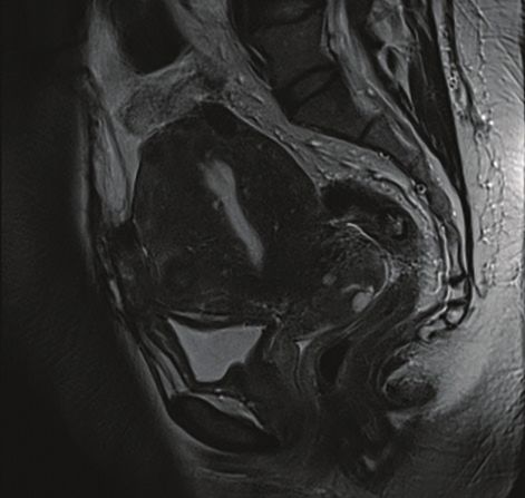
WOMEN’S
  H E A LT H

                                                                                        undergo embolization for fibroids alone,
                                                                                        and it is important to have a pain man-
                                                                                        agement protocol in place. Patients are
                                                                                        immediately provided with a patient-
                                                                                        controlled analgesia pump following
                                                                                        the procedure. Intravenous ketorolac is
                                                                                        given at scheduled intervals to address
                                                                                        postembolization inflammation, and
                                                                                        patients are admitted for 23-hour
                                                                                        observation. More recently, there are
                                                                                        proponents for scheduling same-day
                                                                                        discharge, particularly for those who
                                                                                        undergo the procedure using a radial
                                                                                        artery approach or arterial closure
                                                                                        device. It is the opinion of the authors
                                                                                        that significant pain and nausea control
                                                                                        is commonly needed for the first night
                                                                                        after UAE for adenomyosis, and this is
                                                                                        done most safely within the hospital set-
                                                                                        ting. Further investigation is needed to
                                                                                        evaluate newer pain control methods in
   Figure 2. Digital subtraction angiogram with micro-      Figure 3. Digital sub-      conjunction with maintaining successful
   catheters in the bilateral uterine arteries showing      traction angiogram of       UAE outcomes.
   asymmetric filling of the uterus.                        the right ovarian artery       The morning after the procedure, the
                                                            showing supply to the       patient-controlled analgesia is discontin-
                                                            right fundal region.        ued and a trial of oral analgesics is pre-
                                                                                        sented before the patient is discharged
   embolics such as particulate polyvinyl alcohol and                 home on scheduled ibuprofen with an oral narcotic
   Embozene microspheres (Boston Scientific Corporation)              for breakthrough pain, if needed. Patients then receive
   can also be used. Once two vials of Embosphere micro-              a phone call approximately 48 hours after discharge,
   spheres have been administered, the size of the micro-             and a 3-month clinical follow-up is scheduled. MRI at
   spheres may be upsized in diameter at the discretion of            3 months is reviewed during the clinic visit for degree of
   the operator. The recommended endpoint for embo-                   infarction, decrease in uterine size, and any complicating
   lization is near-stasis, as defined by the visualization of        factors (Figure 4).
   contrast within the transverse segment of the uterine
   artery for duration of time equivalent to five heartbeats.         Outcomes
   Preservative-free lidocaine (50 mg) is then administered              Previous case studies have shown favorable short-term
   into the uterine artery. A recent prospective randomized outcomes for symptom relief after UAE for adenomyo-
   study showed improvement in postprocedural pain at                 sis; however, recurrence rates are higher than for the
   4 hours with the use of intra-arterial lidocaine.14                treatment of fibroids.15 Popovic et al revealed 83.8% of
      Aortography may be performed in patients who                    pure adenomyosis patients experienced symptom relief
   have previously undergone pelvic surgery, those with a             with a median follow-up of 9.4 months, but only 64.9%
   suspected ovarian artery or other collateral (Figure 3),           of patients experienced sustained improvement after
   or those with an outside MRI that was performed with-              a median follow-up of 40.6 months.15 Overall uterine
   out the MRA component. At the completion of the                    volumes decreased by 23% to 32% (including those with
   procedure, all catheters are removed and hemostasis is             combined adenomyosis and fibroids). Complications
   achieved with manual compression or with use of an                 included amenorrhea (20.9%) and eventual hysterecto-
   arterial closure device at the discretion of the operator.         my (12.8%). Of note, all amenorrheic patients were older
                                                                      than 45 years.
   Postprocedural Management                                             A recent review and meta-analysis showed over-
      Embolization of adenomyosis is often associated with            all symptom improvement in 83.1% of patients who
   greater pain than typically experienced by patients who            underwent UAE for adenomyosis.16 Symptom improve-

60 ENDOVASCULAR TODAY JANUARY 2018 VOL. 17, NO. 1
WOMEN’S
H E A LT H
ditions. Pain control in the postprocedure period is an
important aspect to consider, and adjunctive techniques
such as the use of intra-arterial lidocaine may be useful.
UAE offers favorable short-term outcomes for patients
with adenomyosis, but further randomized controlled
trials are needed to determine if symptom resolution is
sustainable and explore the impact on fertility. Although
recurrence rates are higher than for fibroids, UAE for
adenomyosis offers symptom relief in two-thirds of
patients and is uterine sparing. n
1. Struble J, Reid S, Bedaiwy MA. Adenomyosis: a clinical review of a challenging gynecologic condition. J Minim
Invasive Gynecol. 2016;23:164-185.
2. Ascher SM, Jha RC, Reinhold C. Benign myometrial conditions: leiomyomas and adenomyosis. Top Magn Reson
Imaging. 2003;14:281-304.
3. Bergeron C, Amant F, Ferenczy A. Pathology and physiopathology of adenomyosis. Best Pract Res Clin Obstet
Gynaecol. 2006;20:511-521.
4. Agostinho L, Cruz R, Osório F, et al. MRI for adenomyosis: a pictorial review [published online October 4, 2017].
Insights Imaging.
5. Hanafi M. Ultrasound diagnosis of adenomyosis, leiomyoma, or combined with histopathological correlation.
J Hum Reprod Sci. 2013;6:189-193.
6. Verma SK, Lev0Toaff AS, Baltarowich OH, et al. Adenomyosis: sonohysterography with MRI correlation. AJR Am
J Roentgenol. 2009;192:1112-1116.
Figure 4. Postembolization T2-weighted MRI showing 7. Dueholm M, Lundorf E. Transvaginal ultrasound or MRI for diagnosis of adenomyosis. Curr Opin Obstet Gynecol.
decreased overall size of the uterus and decreased prominence 2007;19:505-512.
8. Champaneria R, Abedin P, Daniels J, et al. Ultrasound scan and magnetic resonance imaging for the diagnosis of
of the diffuse adenomyosis seen in Figure 1.
adenomyosis: systematic review comparing test accuracy. Acta Obstet Gynecol Scand. 2010;89:1374-1384.
9. Radzinsky VE, Khamoshina MB, Nosenko EN, et al. Treatment strategies for pelvic pain associated with adeno-
myosis. Gynecol Endocrinol. 2016;32(suppl 2):19-22.
ment was separated into four groups: short-term pure 10. Younes G, Tulandi T. Conservative surgery for adenomyosis and results: a systematic review [published online
adenomyosis (89.6%), short-term adenomyosis with July 21, 2017]. J Minim Invasive Gynecol.
11. Pontis A, D’Alterio MN, Pirarba S, et al. Adenomyosis: a systematic review of medical treatment. Gynecol
fibroids (94.3%), long-term pure adenomyosis (74.0%), Endocrinol. 2016;32:696-700.
and long-term combined adenomyosis (84.5%), and 12. Stovall TG, Ling FW, Crawford DA. Hysterectomy for chronic pelvic pain of presumed uterine etiology. Obstet
Gynecol. 1990;75:676-679.
good initial outcomes were found but with less of a 13. Keung JJ, Spies JB, Caridi TM. Uterine artery embolization: a review of current concepts [published online
sustained response. Percentage of uterine volume reduc- September 29, 2017]. Best Pract Res Clin Obstet Gynaecol.
14. Noel-Lamy M, Tan KT, Simons ME, et al. Intraarterial lidocaine for pain control in uterine artery embolization:
tion was greater and statistically significant in the pure a prospective, randomized study. J Vasc Interv Radiol. 2017;28:16-22.
adenomyosis group at 3 months, but results were simi- 15. Popovic M, Puchner S, Berzaczy D, et al. Uterine artery embolization for the treatment of adenomyosis:
a review. J Vasc Interv Radiol 2011;22:901-909.
lar between the groups at 6- and 12-month follow-up. 16. de Bruijn AM, Smink M, Lohle, PNM, et al. Uterine artery embolization for the treatment of adenomyosis:
Amenorrhea was reported in 6.3% of all patients, and all a systematic review and meta-analysis [published online October 9, 2017]. J Vasc Interv Radiol.
of these patients were older than 40 years. The need for
hysterectomy varied between groups in the short term,
with 2.6% of pure adenomyosis patients and only 1.4% of Theresa M. Caridi, MD
combined adenomyosis patients undergoing hysterecto- Assistant Professor
my; however, there was no significant difference in these Division of Vascular and Interventional Radiology
patient groups in the long term (7.2% vs 7%). MedStar Georgetown University Hospital
Washington, DC
CONCLUSION theresa.m.caridi@gunet.georgetown.edu
Patients with adenomyosis often experience abnormal Disclosures: Consultant to Vascular Solutions.
uterine bleeding and dysmenorrhea. A range of treat-
ment options is available depending on the patient’s James B. Spies, MD, MPH
age and comorbidities, type of adenomyosis, desire for Professor and Chairman
future fertility, and desire to maintain their uterus. UAE Department of Radiology
presents a useful option for those who wish to avoid Division of Vascular and Interventional Radiology
hysterectomy and prefer a minimally invasive technique MedStar Georgetown University Hospital
with a short hospital stay. Symptomatology should cor- Washington, DC
relate to characteristic imaging findings on TVUS or spiesj@gunet.georgetown.edu
MRI, with MRI preferred because it provides additional Disclosures: None.
information about the frequent concomitant pelvic con-
VOL. 17, NO. 1 JANUARY 2018 ENDOVASCULAR TODAY 61You can also read