Traumatic Brain Injury and its Findings on Computed Tomography
←
→
Page content transcription
If your browser does not render page correctly, please read the page content below
EAS Journal of Radiology and Imaging Technology
Abbreviated Key Title: EAS J Radiol Imaging Technol
ISSN: 2663-1008 (Print) & ISSN: 2663-7340 (Online)
Published By East African Scholars Publisher, Kenya
Volume-3 | Issue-3 | May-Jun-2021 |                                                                      DOI: 10.36349/easjrit.2021.v03i03.001

 Original Research Article

Traumatic Brain Injury and its Findings on Computed Tomography
Talha Zafar1*, Nosheen Arshad2, Rehan Afsar3, Syed Muhammad Yousaf Farooq4, Akash John5, Muhammad Ahmad
Naeem6, Hamna Zafar7
17
  , Medical Imaging Doctor, Department of Radiological Sciences and Medical Imaging, the University of Lahore, Gujrat, Pakistan
2356
  , , , Lecturer, Department of Radiological Sciences and Medical Imaging, the University of Lahore, Gujrat, Pakistan
4
  Lecturer, Research Incharge; University Institute of Radiological Sciences and Medical Imaging Technology, University of Lahore,
Lahore Pakistan

                                            Abstract: Background: Traumatic brain injury is leading cause of death in developing
             Article History                countries. TBI mostly occurs due to RTA. Patients with TBI must assessed thoroughly
            Received: 17.03.2021
            Accepted: 24.04.2021
                                            and must notice the changes if present. Ct scan has become the best implement in
            Published: 05.05.2021           radiological assessment due to a feature that it can properly characterize the
                                            temperament and site of the lesions. Method: It is retro-respective cross sectional study
           Journal homepage:                stated that 200 patients, admit in ED, from October 2019- January 2021 Medcare
     https://www.easpublisher.com           International Hospital Gujranwala, Pakistan. The results were evaluated by computed
                                            tomography for the type and location of the lesions identified. Results: By performing
          Quick Response Code
                                            CT-scan it has been evaluated that Scalp hematoma was seen more. About 4.5% were
                                            seen in extra-Dural hematoma, 4% in non-hemorrhagic contusion, 12.5% hemorrhagic
                                            contusion, 82.5% in Scalp hematoma, 8% in subarachnoid hemorrhage, 17% with age
                                            related atrophy, 13.5% in subdural hematoma, 4% in inflammatory changes, skull
                                            fracture about 30% and plain examination were seen about 44.5% and mostly man
                                            (n=58) are involved in RTA as compare to the female (n=14). Conclusion: In
                                            Conclusion the frequency of scalp hematoma is 82.5% and the incidence of non-
                                            hemorrhage contusions and extradural hematoma has the same frequency. RTA patients
                                            have a higher risk of developing intra cranial hemorrhage and mostly occur in males as
                                            compare to females because mostly males are exposed to outer environment. Tables
                                            were used to describe the results.
                                            Key words: Computed Tomography, RTA, Skull fractures, lesions, Hemorrhage, Intra
                                            cranial hemorrhage.
Copyright © 2021 The Author(s): This is an open-access article distributed under the terms of the Creative Commons Attribution 4.0 International
License (CC BY-NC 4.0) which permits unrestricted use, distribution, and reproduction in any medium for non-commercial use provided the original
author and source are credited.

                                                                             years age. Male to female ratio is 4.41: 1. The death rate
INTRODUCTION                                                                 in 2013-2017 is about 33.3% because of RTA [8]. Male
          Head injuries, poly-trauma, multi-trauma are                       are highly involve because they are mostly outdoor like
mainly due to huge accidents in road (road traffic                           driving, vehicles, working outdoors [5, 9]. Previous
accident) RTA [1]. High illness and death rate in low                        study found intra-cerebral hematoma (46.33%), skull
and middle income countries are present with traumatic                       fracture (62.04%), subdural hematoma (19.37%), brain
head injuries [2]. Traumatic brain Injury can be defined                     swelling and edema (63.35%), midline shift (24.34%),
as changed brain function, confusion, coma and change                        subarachnoid hematoma (28.79%), epidural hematoma
in consciousness or neuromotor deficit [3]. TBI is very                      (30.36%) and pneumocranium (12.04%) [1]. All type of
much related to traumatic head injuries which occur                          injuries but basically death and disability mostly occur
mostly due to RTA in young people [4]. and fall history                      due to brain injuries [10]. Brain injures involves the
in children [5]. Any patient with a head injury and alter                    contusions, intracranial injuries, skull fracture, bruising,
state of consciousness should be evaluated for brain                         hematomas, brain swelling, edema and hemorrhages [1,
trauma. The radiological valuation changed affectedly                        11].
with the aid of computed tomography as the location
and type of lesions were carefully examined [6]. Head                                In the study researchers have find that CT
injuries are mostly seen in males as compare to females                      procedures demonstrates more predictable radiography.
[7, 21]. In the previous studies about 81% in male and                       RTA is about 62% mild injuries includes 76% moderate
19% in females and the age group with 20% include 20-                        and severe head injuries are about 14 % to 10 % [11].
29 years old males and 21% included for the 19-20                            The decrease in the amount and seriousness of injuries
*Corresponding Author: Talha Zafar                                                                                                        126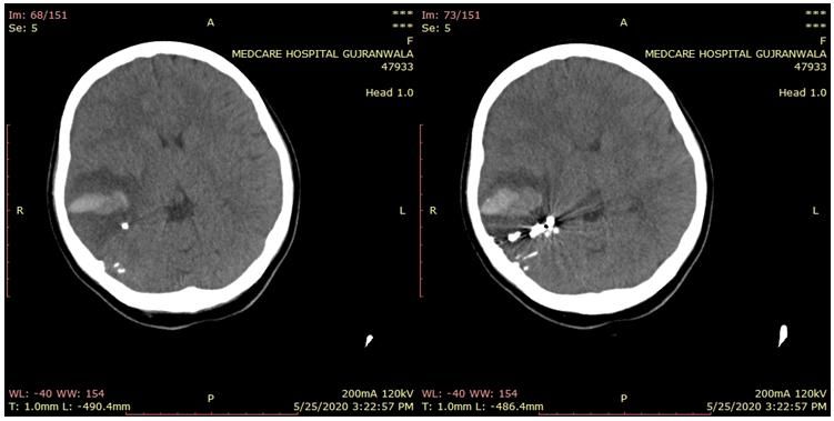
Talha Zafar et al; EAS J Radiol Imaging Technol; Vol-3, Iss-3 (May-Jun, 2021): 126-131

advances the amount of operative to develop the health                Medcare International Hospital Gujranwala, Pakistan.
repute of society [12]. In RTA patients hyper dense                   Patients included in this study were those who met the
subdural hematoma is seen frequently but non-                         inclusion criteria. Patient with traumatic brain injury,
accidental patients showed mixed density subdural                     RTA and history of fall with scale of 13 to 15 GCS with
hematoma [13]. Patients with brain injuries usually                   symptoms of dizziness, nausea, headache, vomiting and
involve the symptoms of loss of consciousness, short                  altered state of consciousness were included. Patients
term memories loss, amnesia, behavior change,                         who died before the stage of Computed tomography
irritability, vomiting and headache        are all after              arriving at ED after 24 hours of injury and alcoholic
traumatic injuries but the post injuries includes traffic             were excluded. Data was collected from emergency
accidents, slipped down, fall down injuries etc [14-16].              departments with consent. Data was analyzed by SPSS
CT scan examination may be important in some cases                    software 21 version. Numerical data was describing in
however in most cases it is challenging to achieve as for             mean and standard deviation. Frequencies and
the troubles with radioactivity contact and bulk                      percentage was used to display the qualitative data. Chi
motions. In addition, if no intracranial abnormality is               square test were applied to evaluate the relationship
detected immediately after injury, irregular findings                 between variables. P value ≤ 0.05 were measured as
might seem several hours later [17]. The study purpose                noteworthy value. All outcomes were calculated at 95%
was to evaluate frequency of traumatic brain injury on                confidence level.
computed tomography including RTA and fall history
patients.
                                                                      RESULTS
                                                                               It has been seen that male as compared to
MATERIAL AND METHODS                                                  females had more risk of Traumatic brain injury and
         It is retro-respective cross sectional study                 mostly seen in 1 to 14 years olds patients as shown in
stated that 200 patients had the head injurynwho were                 table 1 and 2.
admit in the ED from October 2019 to January 2021 in

                                                        Table-1: Gender
                       Gender
                                          Frequency      Percent    Valid Percent    Cumulative Percent
                                male      143            71.5       71.5             71.5
                       Valid    female    57             28.5       28.5             100.0
                                Total     200            100.0      100.0

                                                         Table-2: Age
                   Age of patients
                                                  Frequency        Percent   Valid         Cumulative
                                                                             Percent       Percent
                   Valid         1 to 14          74               37.0      37.0          37.0
                                 15 to 30         51               25.5      25.5          62.5
                                 31 to 50         25               12.5      12.5          75.0
                                 51 to 65         33               16.5      16.5          91.5
                                 Above 65         17               8.5       8.5           100.0
                                 Total            200              100.0     100.0

© East African Scholars Publisher, Kenya                                                                             127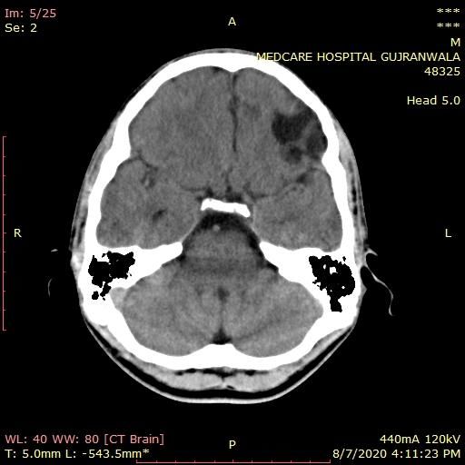
Talha Zafar et al; EAS J Radiol Imaging Technol; Vol-3, Iss-3 (May-Jun, 2021): 126-131

                                           Table-3: Findings seen in CT scan
                                                                Findings       n (%)
                                                   Extra dural hematoma      9(4.5%)
                                             Non hemorrhagic contusion         8(4%)
                                                  Hemorrhagic contusion    25(12.5%)
                                                         Scalp hematoma 165(82.5%)
                                               Subarachnoid hemorrhage        16(8%)
                                             Age related cerebral atrophy   34(17%)
                                                    Subdural Hematoma      27(13.5%)
                                                   Inflammatory changes        8(4%)
                                    Normal plain CT examination of brain   89(44.5%)
                                                            Skull fracture  60(30%)

        It was seen that Extra Dural hematoma is                              The results were concluded that mostly in
9(4.5%), non-hemorrhagic contusion is 8(4%),                         Traumatic head injuries, scalp hematoma were seen
hemorrhagic contusion is 25(12.5%), Scalp hematoma                   about 82.5% and skull fracture were 30% respectively.
were 165(82.5%), subarachnoid hemorrhage were
16(8%), age related cerebral atrophy 34(17%), subdural                         Out of total it has seen that the most cause of
hematoma were 27(13.5%), inflammatory changes were                   injury is due to fall 128(64%) and the RTA were seen
seen about 8(4%), normal plain CT examination                        about 72(36%) in patients.
80(454.5%) and skull fracture were seen about 60(30%)
as shown below table 3.

                                                  Table-4: Cause of injuries
                Cause of injury
                                                  Frequency     Percent    Valid Percent   Cumulative Percent
                Valid    fall                     128           64.0       64.0            64.0
                         Road traffic accident    72            36.0       36.0            100.0
                         Total                    200           100.0      100.0

© East African Scholars Publisher, Kenya                                                                             128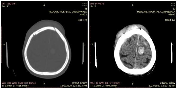
Talha Zafar et al; EAS J Radiol Imaging Technol; Vol-3, Iss-3 (May-Jun, 2021): 126-131
Fig-1: 75 Years, Male, CT BRAIN, H/O RTA
Evidence of multiple fracture involving frontal appreciated involving anterior cortical and subcortical
bone and top parietal bone. Brain parenchymal portion of both frontoparietal lobes anteriorly. Thin
hemorrhage is appreciated in left top centrum semiovale strip of subdural hematoma is also appreciated around
area measuring 2.7 x 2.0 cm with mild surrounding right frontal lobe.
oedema. Areas of hemorrhagic contusion are also
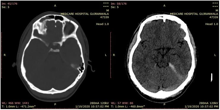
Fig-2: 20 Year, Male, H/O RTA, VOMTING
Evidence of small linear non depressed appreciated in right frontal lobe. Tiny areas of
fracture involving frontal bone including anterior and hemorrhagic contusions are noted along inferior surface
posterior bony boundary of right frontal sinus. It of both frontal lobes specially on left side. Very tiny
extends caudally upto the junction of frontal and strip of subdural hematoma is appreciated along
ethmoid bone including medial edge of roof of orbit on tentorium cerebelli and posterior portion of the falx
right side. Very tiny stirp of extra dural hematoma is cerebri.
Fig-3:15 Year, Male, H/O Fall, Unbalance, Headache
© East African Scholars Publisher, Kenya 129Talha Zafar et al; EAS J Radiol Imaging Technol; Vol-3, Iss-3 (May-Jun, 2021): 126-131
Evidence of low density lesion involving outer porencephalic cyst either because of previous trauma or
aspect of left frontal lobe just anterior to left sylvian infection or ischemic insult.
fissure. Possibility of differential would be with
Fig-4: 16 Years, H/O Fall, FITS
Evidence of brain parenchymal hemorrhage percent. Non-hemorrhage contusions and extradural
involving right temporoparietal lobe with surrounding hematoma have almost an equivalent frequency. Males
oedema. Irregular highly dens calcification is have a higher rate of intracranial hemorrhage than
appreciated in adjacent brain parenchyma which could females. Patients who have been in road traffic accident
be calcified thrombosed vascular malformation. have a higher risk of developing a scalp hematoma than
those who have had other types of traumatic injuries.
GCS reliability is insufficient since computed
DISCUSSION tomography is needed for diagnosis and confirmation of
In our consequences it is reliable throughout the patient's condition.
prior studies that showed head injury is common in
RTA the majority dynamic time of years. A study According to my results, it is suggested that
reported that head injury during RTA were seen 63% patients who have suffered a brain injury must undergo
[18]. Whereas in another study it was reported about an acute non-contrast computed tomography to
that 59-69% of head injuries happen in adolescent [19]. determine the best course of treatment. It is very helpful
During another research it accomplished that behind in unconscious patients and who have allergies to
head injury to affect results age is solitary reason. contrast media.
Result was not as good as through the rising generation.
In order to determine the mode of injury, the
In recent study the result shows that about patient's history should be thoroughly examined. A
71.5% injury occurs in male as compared to female and follow-up scan should be performed within 24-48 hours
they were about 28.5% and mostly the incidence is of the incident to identify the effects of bleeding. The
present from age 1 to 14 about 37%. CT-scan had GCS ranking system isn't enough for accident
become the one to find the changes that occurs in brain classification. For such patients, a CT scan is prescribed
after trauma. Our research reports that the mostly as the first line of examination.
trauma occurs in man as compared to female. It is
concluded that extra dural hematoma were about 4.5%,
non-hemorrhagic contusion 4%, age related cerebral REFERENCES
atrophy 17%, inflammatory changes were seen about 1. Gupta, P. K., Krishna, A., Dwivedi, A. N., Gupta,
4%, fracture in skull were seen in about 30%. K., Madhu, B., Gouri, G., & Shivani, A. (2011).
CT scan findings and outcomes of head injury
It was initially stated that head trauma patients: A cross sectional study. Journal of
sufferers were mostly men as compare to females Pioneering Medical Sciences, 1(3), 78.
because they have more outside exposures outside in 2. Onwuchekwa, C. R., & Alazigha, N. S. (2017).
roads and other activities that are out doors compared to Computed tomography pattern of traumatic head
females seen in Pakistan. In another research who injury in Niger Delta, Nigeria: A multicenter
report that men largely occupied with head injuries evaluation. International journal of critical illness
(86%) [18]. One study found that epidural hematomas and injury science, 7(3), 150.
were associated with skull fractures in approximately 3. Paci, M., Infante-Rivard, C., & Marcoux, J. (2017).
91% of patients [20]. Traumatic brain injury in the workplace. Canadian
journal of neurological sciences, 44(5), 518-524.
CONCLUSION 4. Hans, P., Mehrotra, A., Kumar, P., Agarwal, M.,
According to my research, the prevalence of Kumar, L., Parakh, P., & Tyagi, S. (2017). Role of
scalp hematoma for all types of head trauma is 82.5 Computerized Tomography as Prime Imaging
© East African Scholars Publisher, Kenya 130Talha Zafar et al; EAS J Radiol Imaging Technol; Vol-3, Iss-3 (May-Jun, 2021): 126-131
Modality in the Evaluation of Traumatic Brain accidental and nonaccidental traumatic head injury
Injury. Int J AdvInteg Med Sci, 2(1), 17-23. in children on noncontrast computed
5. Hassan, N., Ali, M., Haq, N. U., Azam, F., Khan, tomography. Pediatrics, 118(2), 626-633.
S., Khan, Z., & Ahmad, S. (2017). Etiology, 14. Du Su Kim, M. H. K., Jang, S. Y., Kim, J. H.,
clinical presentation and outcome of traumatic Kang, D. S., & Song, K. Y. (2013). The usefulness
brain injury patients presenting to a teaching of brain magnetic resonance imaging with mild
hospital of Khyber Pakhtunkhwa. Journal of head injury and the negative findings of brain
Postgraduate Medical Institute (Peshawar- computed tomography. Journal of Korean
Pakistan), 31(4). Neurosurgical Society, 54(2), 100.
6. AHMAD, I., RAZA, M. H., ABDULLAH, A., & 15. Polinder, S., Cnossen, M. C., Real, R. G., Covic,
SAEED, S. (2020). Intracranial CT Scan Findings A., Gorbunova, A., Voormolen, D. C., ... & Von
in the Patients of Head Injury: An Early Experience Steinbuechel, N. (2018). A multidimensional
at Dera Ghazi Khan Teaching Hospital. Pakistan approach to post-concussion symptoms in mild
Journal Of Neurological Surgery, 24(3), 248-252. traumatic brain injury. Frontiers in neurology, 9,
7. Khan, M., Yaqoob, U., Hassan, Z., & Uddin, M. M. 1113.
(2020). Immediate Outcomes of Traumatic Brain 16. Cnossen, M. C., van der Naalt, J., Spikman, J. M.,
Injury at a Tertiary Care Hospital Of Pakistan-A Nieboer, D., Yue, J. K., Winkler, E. A., ... &
Retrospective Study. Lingsma, H. F. (2018). Prediction of persistent
8. Naheed, K., Pal, M. I., Naeem, M., Qasim, A. P., post-concussion symptoms after mild traumatic
Yunis, S., & Misbah, Z. (2019). ANALYSIS OF brain injury. Journal of neurotrauma, 35(22), 2691-
DEATHS DUE TO ROAD TRAFFIC 2698.
ACCIDENTS IN FAISALABAD CITY- 17. Shiomi, N., Echigo, T., Hino, A., Hashimoto, N., &
PAKISTAN. Journal of University Medical & Yamaki, T. (2016). Criteria for CT and initial
Dental College, 10(3), 38-43. management of head injured infants: a
9. Mehta, R. A., & Bambhaniya, A. B. (2018). Profile review. Neurologia medico-chirurgica, ra-2015.
of Fatal Head Injuries in and Around Jamnagar 18. Bharti, P., Nagar, A. M., & Umesh, T. (1993).
Region. Indian Journal of Forensic Medicine and Pattern of trauma in western Uttar
Pathology, 11(3), 187. Pradesh. Neurology India, 41, 49-50.
10. El Hendawy, M. M., Mohammed, M. S., & Saad, 19. Reverdin, A. (1990). Head injury in
A. H. (2020). Surgical Management of Open children. NIMS: Head injury, clinical management
Traumatic Head Injury. The Egyptian Journal of and research. Elizabeth Frost (ed), Geneva,
Hospital Medicine, 78(1), 42-47. Switzerland: Airsen, 193-204.
11. Ibrahim, S. Y. A. (2018). Study of Traumatic Head 20. Phonprasert, C. H. A. R. E., Suwanwela, C. H. A.
Injuries Using Computerized Tomography Among R. A. S., Hongsaprabhas, C. H. A. T. U. R. A. P. O.
sudanese Population (Doctoral dissertation, Sudan R. N., Prichayudh, P. R. A. C. H. A., & O'Charoen,
University of Science and Technology). S. U. P. A. T. (1980). Extradural hematoma:
12. Syed, A. T., Lone, N. A., Wani, M. A., & Bhat, A. analysis of 138 cases. The Journal of trauma, 20(8),
S. (2007). Clinical management of patients with 679-683.
minor head injuries. International journal of health 21. Nazeeha, W. (2020). East African Scholars J Med
sciences, 1(1), 131. Surg; 2(11) (Dec, 2020): 205-211.
13. Tung, G. A., Kumar, M., Richardson, R. C., Jenny,
C., & Brown, W. D. (2006). Comparison of
Cite This Article: Talha Zafar et al (2021). Traumatic Brain Injury and Its Findings on Computed Tomography. EAS J
Radiol Imaging Technol, 3(3), 126-131.
© East African Scholars Publisher, Kenya 131You can also read