Neural bases of focused attention and open monitoring during meditation
←
→
Page content transcription
If your browser does not render page correctly, please read the page content below
Neural bases of focused attention and open monitoring
                            during meditation
          Antonietta Mannaa,b, Antonino Raffonec,e, Mauro G. Perruccia,b, Davide Nardoc,d,
   Antonio Ferrettia,b, Alessandro Londeic,d, Cosimo Del Grattaa,b, Marta Olivetti Belardinellic,d and
                                        Gian Luca Romania,b
    a
        ITAB, Institute for Advanced Biomedical Technologies, “G. D’Annunzio” University Foundation, Chieti, Italy.
                     b
                       Department of Clinical Sciences and Bioimaging, University of Chieti, Chieti, Italy.
                               c
                                 Department of Psychology, “Sapienza” University, Rome, Italy.
          d
            ECONA (Interuniversity Center for Cognitive Processing in Natural and Artificial Systems), Rome, Italy.
             e
               Perceptual Dynamics Laboratory, RIKEN Brain Science Institute, Wako-shi Saitama Japan, Japan.
                Correspondence: Antonietta Manna, University of Chieti “G. d’Annunzio”, Via dei Vestini, 31, 66100, Chieti, Italy.
                                  E-mail: amanna@unich.it, phone +39 08713556952, fax+39 08713556930

Abstract. Meditation refers to a family of complex emotional and attentional regulatory practices, which can be
classified into two main styles - focused attention and open monitoring - involving different attentional, cognitive
monitoring and awareness processes. Despite the increasing number of studies on neural correlates of meditation, the
differential brain activity patterns in focused attention and open monitoring meditation forms have not been
investigated yet in a unitary neuroimaging experiment. We studied brain activity patterns in both focused attention
and open monitoring meditation in Theravada Buddhist monks and lay novices, by functional magnetic resonance
imaging. A massive deactivation of left brain activity during focused attention meditation, involving the activation of
right midfrontal areas, was observed in the monks. By contrast, open monitoring meditation was associated to the
activation of left fronto-temporo-parietal areas. Brain activity in focused attention meditation sharply contrasted with
the rest state. These highly differentiated brain activity patterns were not found in the novices.
Keywords: Meditation, attention, awareness, access consciousness, prefrontal cortex, neural correlates of consciousness, functional magnetic
resonance imaging.
Note: the first two authors (A.M. and A.R.) have contributed equally to this work.

1. Introduction
     Number Meditation can be conceptualized as a family of complex emotional and attentional regulatory practices,
in which mental events are affected by engaging a specific attentional set. Many recent behavioral,
electroencephalographic and neuroimaging studies have revealed the importance of investigating meditation states
and traits to achieve an increased understanding of cognitive and affective neuroplasticity, attention and self-
awareness, as well as for relevant clinical implications [Cahn & Polich, 2006; Lutz, Slagter, Dunne & Davidson, in
press].
     Given that regulation of attention is the central commonality across the many different meditation methods
[Davidson & Goleman, 1977], meditation practices can be usefully classified into two main styles – focused attention
(FA) and open monitoring (OM) – depending on how the attentional processes are directed [Cahn & Polich, 2006;
Lutz, Slagter, Dunne & Davidson, in press]. In the FA (‘concentrative’) style, attention is focused on an intended
object in a sustained fashion. The second style, OM (‘mindfulness-based’) meditation, involves the non-reactive
monitoring of the content of experience from moment to moment, primarily as a means to recognize the nature of
emotional and cognitive patterns. The present functional magnetic resonance imaging (fMRI) experiment examined
the different neural bases of FA and OM meditation, with the participation of Theravada Buddhist monks, who are
expert in practicing both these meditation styles. The evidence of commonalities and differences in the neural
correlates of FA and OM meditation, in the same experimental context and subjects, can shed light on fundamental
processes of attention and awareness. FA meditation entails the capacities of monitoring the focus of attention and
detecting distraction, disengaging attention from the source of distraction, and (re)directing and engaging attention to
the intended object [Lutz, Slagter, Dunne & Davidson, in press]. These attentional and monitoring functions have
been related to dissociable systems in the brain involved in conflict monitoring, selective and sustained attention
(Lutz, Slagter, Dunne & Davidson, in press]. OM meditation involves no explicit attentional focus, and therefore does
not seem associated to brain areas implicated in sustained or focused attention, but to brain regions involved in
vigilance, monitoring and disengagement of attention from sources of distraction from the ongoing stream of
experience [Lutz, Slagter, Dunne & Davidson, in press]. OM practices are based on an attentive set that is
characterized by an open presence and a nonjudgmental awareness of sensory, cognitive and affective fields of
experience in the present moment, and involves a higher-order awareness or observation of the ongoing mental
processes [Cahn & Polich, 2006]. Behavioral studies have shown a more distributed attentional focus, enhanced
conflict monitoring and reduced attentional blink or more efficient resource allocation to serially-presented targets in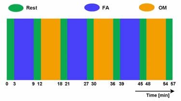
OM meditation practitioners [Lutz, Slagter, Dunne & Davidson, in press]. Despite the increasing number of studies
on neural correlates of meditation states and traits, the differential brain activity patterns in focused attention and open
monitoring meditation forms have not been contrasted yet in a unitary neuroimaging experiment. Therefore, in a
fMRI experiment we studied the brain activity patterns of Buddhist monks who are expert in Samatha (FA) and
Vipassana (OM) meditation forms, and follow the oldest (Theravada) currently active Buddhist tradition. Vipassana
(insight) meditation is central in mindfulness-based clinical interventions and studies [Teasdale et al., 2002].
Although lay practitioners of Vipassana have participated in recent research, to our knowledge this is the first study in
which Theravada Buddhist monks are involved. The brain activation patterns of the monks were compared with the
patterns of lay novice meditators with 10 days of practice of both Samatha (FA) and Vipassana (OM) meditation
styles. A non-meditative Rest condition was also run. Participants alternated performance of FA (Samatha) and OM
(Vipassana) meditation blocks, preceded and followed by a (non-meditative) resting state (‘Rest’) block (see
Methods).
2. Material and Methods
    2.1 Participants.
     Participants included 8 Theravada Buddhist monks (males, mean age 40.9 years, ages 25-58 years, SD 11.6
years), with 17.0 years as mean number of years of Samatha (FA) and Vipassana (OM) meditation practice in
Theravada monasteries (SD 9.7 years). The monks were from the Santacittarama monastery, in central Italy,
following a Thai Forest Tradition (the order was funded by Ajahn Chah, one of the most influential Buddhist teachers
in the 20th century). In this tradition, monks experience regular intensive meditation retreats and typically practice
Samatha-Vipassana meditation two hours per day with the monastery community. Individual meditation practice is
also emphasized. Participants also included a group of 8 novice meditators (males, mean age 31 years, ages 22-34
years, SD 4 years), recruited from the local community. All novice subjects were interested in meditation but had no
prior meditation experience. The novice participants were given oral and written instructions on how to perform
Samatha and Vipassana meditation styles, and during the ten days before the fMRI scan session practiced each of the
two meditation styles 30 minutes per day. All participants were right-handed. Subjects gave their written informed
consent according to the Declaration of Helsinki [World Medical Association Declaration of Helsinki, 1997].
    2.2 Task and Protocol.
    Experimental paradigm consisted of 6min FA (Samatha) and 6min OM (Vipassana) meditation blocks, each
preceded and followed by a 3min non-meditative resting state block (Rest), for three times (see Figure 1). The total
duration of the experiment was 57 minutes. The condition switch was instructed by an auditory word-signal during
the experiment. During all the conditions, the participants kept eyes closed. At the end of the experiment, all
participants reported they could perform the task conditions according to the given instructions.

                          Figure 1. Sequence of the experimental conditions during the experiment..
    2.3 Functional MRI recording
    Functional MRI scans were acquired on a Siemens Magnetom Vision scanner at 1.5 T, equipped with a standard
receiver head coil. BOLD contrast functional imaging was performed using a T2*-weighted echo planar (EPI) free
induction decay (FID) sequence with: TR=4 s, 28 slices, voxel size 4x4x4 mm3, 860 functional volumes for each run.
A high-resolution T1-weighted whole-brain image was also acquired at the end of each session via a 3D-MPRAGE
sequence (sagittal matrix=256x256, FOV= 256 mm, slice thickness =1mm, no gap, in-plane voxel size=1x1 mm2, flip
angle=12°, TR/TE= 9.7/4.0 ms).
    2.4 Physiological measures.
    Respiration rate and ECG were recorded throughout each scanning session in all subjects. EEG was also
recorded, with data to be analyzed for a subsequent report.
    2.5 Data analysis.
    Raw data were analyzed using Brain Voyager QX 1.7 software (Brain Innovation, The Netherlands). The first
three scans of each run were discarded to avoid the T1 saturation effect. Preprocessing consisted in a 3D motion
correction and in a temporal filtering of voxel time series. The data set of one of the monks was discarded from
further analysis due to excessive motion. Preprocessed functional volumes were coregistered with the corresponding
structural data set. Temporal filtering included linear and non-linear (high-pass filter of two cycles per time course)
trends removal. Structural and functional volumes were than transformed into the Talairach space [Talairach and
Tournoux, 1998]. No spatial or temporal smoothing was applied. Statistical analysis was carried out for individual
subjects and condition using the General Linear Model [Friston at al., 1995]. To account for the hemodynamic delay,
the boxcar waveform of each task condition was convolved with the Boynton empirically founded hemodynamic
response function [Boynton et al., 1996]. In order to search for activated areas common to the entire group of
subjects, a voxel-wise random effect group analysis was also performed, distinguishing between monks and novice
meditators. To this purpose, all the subjects’ time series were z-normalized and then concatenated prior the GLM.
Group statistical maps were thresholded at an overall significance level (the probability of a false detection for the
entire functional volume) of pRest (Figure 3) revealed three
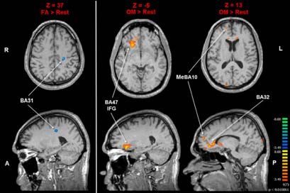
activations in the left hemisphere: medial aPFC (BA10), STG (BA22) and precuneus (BA7).
3.2 Contrasts in the novices group
As regards the novices, the contrast FA>Rest (Figure 4) showed a single activation in the left posterior cingulate
(BA31). The contrast OM>Rest (Figure 4) showed activations in the left dorsal ACC (BA32), the right rostral ACC
(BA32), the right lateral orbitofrontal cortex (IFG, BA47) and the right medial aPFC (BA10).
4. Discussion and Conclusions
For the first time brain activity patterns in FA and OM meditation were contrasted in a neuroimaging (fMRI)
experiment, in expert (Buddhist monks) and lay novices. Overall, we found striking differences between the patterns
of brain activity of monks and novices, in OM and FA meditation styles. The brain activity patterns of the monks in
OM meditation resembled their ordinary brain resting state, whereas their brain activity in focused attention
meditation sharply contrasted with the resting state. It has been recently argued that meditative states are associated to
transient hypofrontality or deactivation in executive networks [Lou, Nowak & Kjaer, 2005; Dietrich, 2003] . In
contrast, other authors have emphasized the activation of executive areas in meditation [Cahn & Polich, 2006; Lutz,
Slagter, Dunne & Davidson, in press]. As expected, the results with our experimental design resolve this controversy.
We conclude that FA meditation is associated to an enhanced (predominantly right) midfrontal and a reduced
(predominantly left) lateral prefrontal activation, and OM meditation to an increased (predominantly left) midfrontal
activation, as compared to rest. We also conclude that OM meditation, as compared to FA meditation, is characterized
by a lateral prefrontal activation in both hemispheres, with a more subtle differentiation in midfrontal brain
activations associated to these fundamental meditation styles. At a macroscopic level of functional organization, a
relative left-lateralization of brain activity patterns resulted in our experiment. Most of the deactivations in FA>Rest
and all the activations in OM>Rest, were in the left hemisphere. We also found a pronounced deactivation of left
anterior and posterior insula in FA>Rest.These patterns may be explained in terms of the emergence of a dynamical global brain state in FA meditation
[Lutz, Slagter, Dunne & Davidson, in press], implying the deactivation of the left insula, probably to prevent a
‘broadcasting’ of conscious access to body states other than breathing sensory states.
GROUP/CONTRAST/AREA x y z k T p
MONKS
FA meditation > Rest
Left SFG, BA10 -10 66 19 135 -10.040 0.0001
Left Dorsal ACC, BA24 -9 26 16 333 10.470 0.0001
Left MFG, BA46 -48 38 16 567 -7.770 0.0001
Left MFG, BA9 -47 32 28 216 -12.300 0.0001
Right MeFG, BA10 12 50 13 270 5.666 0.001
Right Dorsal ACC, BA24 12 32 14 756 10.190 0.0001
Right IFG, BA44 54 15 16 324 -6.034 0.001
Right IFG, BA46 51 32 10 369 -6.480 0.001
Left Precuneus, BA7 0 -70 49 342 -7.913 0.0001
Left TTG, BA41 -41 -25 10 315 -13.960 0.0001
Right STG, BA22 57 -51 13 306 -11.790 0.0001
Left Insula, BA13 -39 8 -1 1647 -6.877 0.0001
Left Anterior Insula, BA13 -42 17 1 540 -7.254 0.0001
Left Posterior Insula, BA13 -44 -10 16 108 -5.712 0.001
OM meditation > Rest
Left MeFG, BA10 -3 53 10 522 7.002 0.0001
Left Precuneus, BA7 -18 -64 43 288 8.911 0.0001
Left STG, BA22 -57 -37 7 243 6.090 0.001
NOVICES
FA meditation > Rest
Left Posterior Cingulate, BA31 -23 -25 37 216 -8.889 0.0001
OM meditation > Rest
Left Dorsal ACC, BA32 -12 20 22 117 4.809 0.002
Right Rostral ACC, BA32 12 39 -4 378 7.892 0.0001
Right MeFG, BA10 15 56 14 243 6.812 0.0001
Right IFG, BA47 21 23 -5 2125 8.035 0.0001
Table 1. Results revealed by the three contrasts.
Figure 2. Activations and deactivations revealed by the FA>Rest contrast, in the monks group. Note the deactivation of insula
(BA13), MFG (BA46), TTG (BA41) and precuneus (BA7) in the left hemisphere, and of IFG (BAs44/46) and STG
(BA22) in the right hemisphere.
Figure 3. Activations revealed by the OM>Rest contrast, in the monks group, including medial aPFC (BA10), precuneus (BA7)
and STG (BA22), in the left hemisphere.Figure 4. Activations and deactivations revealed by the FA>Rest and OM>Rest contrasts, in the novice group.
The process of broadcasting in conscious access is central in the global workspace [Baars, 1998] and dynamic
core [Tononi & Edelman, 1998] models of consciousness. The monks might control the level of cognitive
engagement and broadcasting in conscious access to sensory-related, thought and emotion contents, by massive self-
regulation of fronto-parietal and insular areas in the left hemisphere, in a meditation state-dependent fashion.
Moreover, we found that the (left) medial aPFC exhibited a prominent activation in OM>Rest.. These results suggest
that medial aPFC is involved in monitoring the stream of present experience.
As regards the novices, we only found the deactivation of the left posterior cingulate cortex in FA>Rest.
Considering this result and the evidence about the precuneus in the monks, consistent with the recent proposal that the
precuneus/posterior cingulate cortex plays a pivotal role in the ‘default mode network’ [Fransson & Marrelec, 2008],
it can be hypothesized that the left precuneus/posterior cingulate region is the component of the ‘default mode
network’ which can be more sensitively affected by a goal-independent task, such as FA meditation. Four activated
clusters in the OM>Rest contrast were found in the novice group. Overall, it seems that in novices open monitoring
mostly involved right prefrontal areas. The activation of the left dorsal ACC might be explained by the executive
demand to novices in OM meditation performance.The activations in novices of (right) rostral ACC and (right) lateral
orbitofrontal cortex (IFG), which were not found in the monks group, suggest that in novices open monitoring may
reflect an evaluation-based stance rather than being non-judgmental as in the monks. With reference to the literature,
our study reveals macroscopic qualitative changes in monk brains in terms of differentiation and lateralization of
activity patterns associated to awareness. Finally, our results lead to the suggestion that Samatha (FA) meditation
might be associated with a ‘hyper-default mode’ of brain activity, with extensive deactivation (with reference to the
ordinary resting state) of associative brain areas implied in access consciousness and self-referential processing. This
‘hyper-default mode’ of brain activity can well be associated to the teachings of the Buddha about the importance to
calm and control the flow of mental processes, and about wholesome mental states going beyond the experience of a
separated self [Ajahn Sumedho, 2004].
References
Ajahn Sumedho. Intuitive Awareness. Amaravati Buddhist Monastery, Hemel Hempstead (UK)., 2004
Baars, B.J. In the theatre of consciousness. Journal of Consciousness Studies, 4, 292-309, 1997
Boynton, G.M., Engel, S.A., Glover, G.H., Heeger, D.J., Linear systems analysis of functional magnetic resonance imaging in human V1. Journal
of Neuroscience, 16, 4207–4241, 1996.
Cahn, B.R., Polich, J. Meditation states and traits: EEG, ERP, and neuroimaging studies. Psychological Bullettin, 132, 180–211, 2006.
Davidson, R.J., Goleman, D.J. The role of attention in meditation and hypnosis: A psychobiological perspective on transformations of
consciousness. International Journal of Clinical and Experimental Hypnosis, 25, 291-308, 1977.
Forman, S.D., Cohen, J.D., Fitzgerald, M., Eddy, W.F., Mintun, M.A., Noll, D.C. Improved assessment of significant activation in functional
magnetic resonance imaging (fMRI): use of a cluster-size threshold. Magnetic Resonance in Medicine, 33 (5), 636–647, 1995.
Fransson, P., Marrelec, G. The precuneus/posterior cingulate cortex plays a pivotal role in the default mode network: evidence from a partial
correlation network analysis. NeuroImage, 42, 1178-1184, 2008.
Friston, K.J., Holmes, A.P., Worsley, K.J., Poline, J.P., Frith, C.D., Frackowiak, R.S.J. Statistical parametric maps in functional imaging: a general
linear model approach. Human Brain Mapping, 2, 173–181, 1995.
Lou, H.C., Nowak, M., Kjaer, T.W. The mental self. Progress in Brain Research, 150, 197-204, 2005.
Lutz, A., Slagter H.A., Dunne, J.D., Davidson, R.J. Attention regulation and monitoring in meditation. Trends in Cognitive Neurosciences (in
press), 2008.
Talairach, J., Tournoux, P. Co-planar Stereotaxic Atlas of the Human Brain. New York., Thieme, 1998.
Teasdale, J.D., Williams, J.M.G., Soulsby, J.M., Segal, Z.V., Ridgeway, V.A., Lau, M.A. Prevention of relapse/recurrence in major depression by
mindfulness-based cognitive therapy. Journal of Consulting and Clinical Psychology, 68, 615–623., 2000.
Tononi, G., Edelman, G.M.. Consciousness and Complexity. Science, 282, 1846-1851, 1998.You can also read