Management of venous leg ulcers: Clinical practice guidelines of the Society for Vascular Surgery Ò and the American Venous Forum - Squarespace
←
→
Page content transcription
If your browser does not render page correctly, please read the page content below
Management of venous leg ulcers: Clinical practice
    guidelines of the Society for Vascular SurgeryÒ and
    the American Venous Forum
    Endorsed by the American College of Phlebology and the Union Internationale de Phlébologie

    Thomas F. O’Donnell Jr, MD, Marc A. Passman, MD, William A. Marston, MD, William J. Ennis, DO,
    Michael Dalsing, MD, Robert L. Kistner, MD, Fedor Lurie, MD, PhD, Peter K. Henke, MD,
    Monika L. Gloviczki, MD, PhD, Bo G. Eklöf, MD, PhD, Julianne Stoughton, MD, Sesadri Raju, MD,
    Cynthia K. Shortell, MD, Joseph D. Raffetto, MD, Hugo Partsch, MD, Lori C. Pounds, MD,
    Mary E. Cummings, MD, David L. Gillespie, MD, Robert B. McLafferty, MD,
    Mohammad Hassan Murad, MD, Thomas W. Wakefield, MD, and Peter Gloviczki, MD

SVS/AVF Joint Clinical Practice Guidelines CommitteedVenous Leg Ulcer
    Chair:
       Thomas F. O’Donnell Jr, MD (Cardiovascular Center, Tufts Medical Center, Boston, Mass)
    Vice Chair:
       Marc A. Passman, MD (Division of Vascular Surgery and Endovascular Therapy. University of Alabama at
       Birmingham, Birmingham, Ala; Birmingham Veterans Administration Medical Center, Birmingham, Ala)
    Committee Members:
       Mary E. Cummings, MD (University of Michigan, Ann Arbor, Mich)
       Michael C. Dalsing, MD (Indiana University School of Medicine, IU Health Care System, Indianapolis, Ind)
       Bo G. Eklöf, MD, PhD (Lund University, Sweden)
       William J. Ennis, DO (University of Illinois Hospital and Health Science, Chicago, Ill)
       David L. Gillespie, MD (Department of Vascular Surgery, Cardiovascular Care Center, Southcoast Healthcare
          Systems, Fall River, Mass; Uniformed Services University of the Health Sciences F. Edward Hebert School of
          Medicine, Bethesda, Md)
       Monika L.Gloviczki, MD, PhD (Gonda Vascular Center, Mayo Clinic, Rochester, Minn)
       Peter Gloviczki, MD (Division of Vascular and Endovascular Surgery, Mayo Clinic, Rochester, Minn)
       Peter K. Henke, MD (University of Michigan, Ann Arbor, Mich)
       Robert L. Kistner, MD (Honolulu, Hawaii)
       Fedor Lurie, MD, PhD (Jobst Vascular Institute, Toledo, Ohio)
       William A. Marston, MD (University of North Carolina, Chapel Hill, NC)
       Robert B. McLafferty, MD (Portland Veterans Administration Medical Center, Portland, Ore; Oregon Health
          Sciences University, Portland, Ore)
       M. Hassan Murad, MD (Division of Preventive Medicine, Mayo Clinic, Rochester, Minn)
       Hugo Partsch, MD (Medical University of Vienna, Austria)
       Lori C. Pounds, MD (University of Texas Health Science Center, San Antonio, Tex)
       Joseph D. Raffetto, MD (Harvard Medical School, Boston Mass; Veterans Administration Boston Healthcare
          System, Boston Mass; Brigham and Women’s Hospital, Boston, Mass)
       Sesadri Raju, MD (The Rane Center, Jackson, Miss)
       Cynthia K. Shortell, MD (Division of Vascular Surgery, Duke University Medical Center, Durham, NC)
       Julianne Stoughton, MD (Division of Vascular and Endovascular Surgery, Massachusetts General Hospital,
          Boston, Mass)
       Thomas W. Wakefield, MD (University of Michigan, Ann Arbor, Mich)

Author conflict of interest: none.
J Vasc Surg 2014;60:3S-59S
0741-5214/$36.00
Copyright Ó 2014 by the Society for Vascular Surgery.
http://dx.doi.org/10.1016/j.jvs.2014.04.049

                                                                                                                 3S
JOURNAL OF VASCULAR SURGERY
4S O’Donnell et al August Supplement 2014
Sub-CommitteedClinical Evaluation:
Marc A. Passman, MD (chair); Peter K. Henke, MD; William A. Marston, MD; Robert B. McLafferty, MD; Lori
Pounds, MD
Sub-CommitteedWound Care:
William A. Marston, MD (co-Chair); William J. Ennis, DO (co-Chair); Emily Cummings, MD; Thomas F.
O’Donnell Jr, MD; Lori C. Pounds, MD
Sub-CommitteedCompression:
Fedor Lurie, MD, PhD (co-Chair); Thomas W. Wakefield, MD (co-Chair); MD; Monika L. Gloviczki, MD, PhD;
Hugo Partsch, MD; Cynthia Shortell, MD
Sub-CommitteedSurgery:
Michael C. Dalsing, MD (co-Chair); Robert Kistner, MD (co-Chair); Bo G. Eklöf, MD, PhD; David Gillespie, MD;
Peter Gloviczki, MD; Julianne Stoughton, MD; Sesadri Raju, MD
Sub-CommitteedAncillary:
Monika L. Gloviczki, MD, PhD (Chair); Cynthia K. Shortell, MD; Julianne Stoughton, MD; William J. Ennis, DO
Sub-CommitteedPrimary Prevention:
Peter K. Henke, MD (Chair); Emily Cummings, MD; Michael C. Dalsing, MD; Fedor Lurie, MD, PhD
Section Contributors
Editors: Thomas F. O’Donnell Jr, MD; Marc A. Passman, MD
Summary of Guidelines: SVS/AVF Committee
Need for Intersociety Consensus Guidelines: Thomas F. O’Donnell Jr, MD
Methodology of Guidelines: Thomas F. O’Donnell Jr, MD; Mohammad Hassan Murad, MD
DefinitiondVenous Leg Ulcer: Thomas F. O’Donnell Jr, MD; Robert Kistner, MD
Venous Anatomy and Pathophysiology: Marc A. Passman, MD (Anatomy); Joseph D. Raffetto, MD (Pathophysiology)
Clinical Evaluation: Marc A. Passman, MD; William A. Marston, MD; Peter Henke, MD; Robert B. McLafferty, MD;
Lori C. Pounds, MD; William J. Ennis, MD
Wound Care: William A. Marston, MD; William J. Ennis, DO; Emily Cummings, MD; Lori Pounds, MD; Thomas F.
O’Donnell Jr, MD
Compression: Fedor Lurie, MD, PhD; Thomas W. Wakefield, MD; Monika L. Gloviczki, MD, PhD; Hugo Partsch,
MD; Cynthia Shortell, MD; Andrea Obi, MD (University of Michigan, Ann Arbor, Mich)
Surgery: Michael C. Dalsing, MD; Robert Kistner, MD; Bo G. Eklöf, MD, PhD; Peter Gloviczki, MD; Sesadri Raju,
MD; Julianne Stoughton, MD; David Gillespie, MD
Ancillary: Monika L. Gloviczki, MD, PhD; Cynthia K. Shortell, MD; Julianne Stoughton, MD; William J. Ennis, MD;
William A. Marston, MD
Primary Prevention: Peter Henke, MD; Fedor Lurie, MD, PhD; Emily Cummings, MD; Michael C. Dalsing, MD
SUMMARY OF GUIDELINES FOR MANAGEMENT OF VENOUS ULCER
DEFINITION VENOUS LEG ULCER
Guideline 1.1: Venous Leg Ulcer Definition
We suggest use of a standard definition of venous ulcer as an open skin lesion of the leg or foot that occurs in an
area affected by venous hypertension. [BEST PRACTICE]
VENOUS ANATOMY AND PATHOPHYSIOLOGY
Guideline 2.1: Venous Anatomy Nomenclature
We recommend use of the International Consensus Committee on Venous Anatomical Terminology for stan-
dardized venous anatomy nomenclature. [BEST PRACTICE]
Guideline 2.2: Venous Leg Ulcer Pathophysiology
We recommend a basic practical knowledge of venous physiology and venous leg ulcer pathophysiology for all
practitioners caring for venous leg ulcers. [BEST PRACTICE]JOURNAL OF VASCULAR SURGERY
Volume 60, Number 2S O’Donnell et al 5S
CLINICAL EVALUATION
Guideline 3.1: Clinical Evaluation
We recommend that for all patients with suspected leg ulcers fitting the definition of venous leg ulcer, clinical
evaluation for evidence of chronic venous disease be performed. [BEST PRACTICE]
Guideline 3.2: Nonvenous Causes of Leg Ulcers
We recommend identification of medical conditions that affect ulcer healing and other nonvenous causes of
ulcers. [BEST PRACTICE]
Guideline 3.3: Wound Documentation
We recommend serial venous leg ulcer wound measurement and documentation. [BEST PRACTICE]
Guideline 3.4: Wound Culture
We suggest against routine culture of venous leg ulcers and only to obtain wound culture specimens when clin-
ical evidence of infection is present. [GRADE - 2; LEVEL OF EVIDENCE - C]
Guideline 3.5: Wound Biopsy
We recommend wound biopsy for leg ulcers that do not improve with standard wound and compression
therapy after 4 to 6 weeks of treatment and for all ulcers with atypical features. [GRADE - 1; LEVEL OF
EVIDENCE - C]
Guideline 3.6: Laboratory Evaluation
We suggest laboratory evaluation for thrombophilia for patients with a history of recurrent venous thrombosis
and chronic recurrent venous leg ulcers. [GRADE - 2; LEVEL OF EVIDENCE - C]
Guideline 3.7: Arterial Testing
We recommend arterial pulse examination and measurement of ankle-brachial index on all patients with venous
leg ulcer. [GRADE - 1; LEVEL OF EVIDENCE - B]
Guideline 3.8: Microcirculation Assessment
We suggest against routine microcirculation assessment of venous leg ulcers but suggest selective consideration
as an adjunctive assessment for monitoring of advanced wound therapy. [GRADE - 2; LEVEL OF EVIDENCE - C]
Guideline 3.9: Venous Duplex Ultrasound
We recommend comprehensive venous duplex ultrasound examination of the lower extremity in all patients
with suspected venous leg ulcer. [GRADE - 1; LEVEL OF EVIDENCE - B]
Guideline 3.10: Venous Plethysmography
We suggest selective use of venous plethysmography in the evaluation of patients with suspected venous leg
ulcer if venous duplex ultrasound does not provide definitive diagnostic information. [GRADE - 2; LEVEL OF
EVIDENCE - B]
Guideline 3.11: Venous Imaging
We suggest selective computed tomography venography, magnetic resonance venography, contrast venography,
and/or intravascular ultrasound in patients with suspected venous leg ulceration if additional advanced venous
diagnosis is required for thrombotic or nonthrombotic iliac vein obstruction or for operative planning before
open or endovenous interventions. [GRADE - 2; LEVEL OF EVIDENCE - C]
Guideline 3.12: Venous Disease Classification
We recommend that all patients with venous leg ulcer be classified on the basis of venous disease classification
assessment, including clinical CEAP, revised Venous Clinical Severity Score, and venous diseaseespecific quality of
life assessment. [BEST PRACTICE]
Guideline 3.13: Venous Procedural Outcome Assessment
We recommend venous procedural outcome assessment including reporting of anatomic success, venous hemo-
dynamic success, procedure-related minor and major complications, and impact on venous leg ulcer healing. [BEST
PRACTICE]
WOUND CARE
Guideline 4.1: Wound Cleansers
We suggest that venous leg ulcers be cleansed initially and at each dressing change with a neutral, nonirritating,
nontoxic solution, performed with a minimum of chemical or mechanical trauma. [GRADE - 2; LEVEL OF
EVIDENCE - C]JOURNAL OF VASCULAR SURGERY
6S O’Donnell et al August Supplement 2014
Guideline 4.2: Débridement
We recommend that venous leg ulcers receive thorough débridement at their initial evaluation to remove obvious
necrotic tissue, excessive bacterial burden, and cellular burden of dead and senescent cells. [GRADE - 1; LEVEL OF
EVIDENCE - B] We suggest that additional maintenance débridement be performed to maintain the appearance
and readiness of the wound bed for healing. [GRADE - 2; LEVEL OF EVIDENCE - B] We suggest that the health
care provider choose from a number of débridement methods, including sharp, enzymatic, mechanical, biologic, and
autolytic. More than one débridement method may be appropriate. [GRADE - 2; LEVEL OF EVIDENCE - B]
Guideline 4.3: Anesthesia for Surgical Débridement
We recommend that local anesthesia (topical or local injection) be administered to minimize discomfort asso-
ciated with surgical venous leg ulcer débridement. In selected cases, regional block or general anesthesia may be
required. [GRADE - 1; LEVEL OF EVIDENCE - B]
Guideline 4.4: Surgical Débridement
We recommend that surgical débridement be performed for venous leg ulcers with slough, nonviable tissue, or
eschar. Serial wound assessment is important in determining the need for repeated débridement. [GRADE - 1;
LEVEL OF EVIDENCE - B]
Guideline 4.5 Hydrosurgical Débridement
We suggest hydrosurgical débridement as an alternative to standard surgical débridement of venous leg ulcers.
[GRADE - 2; LEVEL OF EVIDENCE - B]
Guideline 4.6: Ultrasonic Débridement
We suggest against ultrasonic débridement over surgical débridement in the treatment of venous leg ulcers.
[GRADE - 2; LEVEL OF EVIDENCE - C]
Guideline 4.7: Enzymatic Débridement
We suggest enzymatic débridement of venous leg ulcers when no clinician trained in surgical débridement is
available to débride the wound. [GRADE - 2; LEVEL OF EVIDENCE - C] We do not suggest enzymatic débride-
ment over surgical débridement. [GRADE - 2; LEVEL OF EVIDENCE - C]
Guideline 4.8: Biologic Débridement
We suggest that larval therapy for venous leg ulcers can be used as an alternative to surgical débridement.
[GRADE - 2; LEVEL OF EVIDENCE - B]
Guideline 4.9: Management of Limb Cellulitis
We recommend that cellulitis (inflammation and infection of the skin and subcutaneous tissue) surrounding the
venous leg ulcer be treated with systemic gram-positive antibiotics. [GRADE - 1; LEVEL OF EVIDENCE - B]
Guideline 4.10: Wound Colonization and Bacterial Biofilms
We suggest against systemic antimicrobial treatment of venous leg ulcer colonization or biofilm without clinical
evidence of infection. [GRADE - 2; LEVEL OF EVIDENCE - C]
Guideline 4.11: Treatment of Wound Infection
We suggest that venous leg ulcers with >1 3 106 CFU/g of tissue and clinical evidence of infection be treated
with antimicrobial therapy. [GRADE - 2; LEVEL OF EVIDENCE - C] We suggest antimicrobial therapy for
virulent or difficult to eradicate bacteria (such as beta-hemolytic streptococci, pseudomonas, and resistant
staphylococcal species) at lower levels of colony-forming units per gram of tissue. [GRADE - 2; LEVEL OF
EVIDENCE - C] We suggest a combination of mechanical disruption and antibiotic therapy as most likely to be
successful in eradicating venous leg ulcer infection. [GRADE - 2; LEVEL OF EVIDENCE - C]
Guideline 4.12: Systemic Antibiotics
We recommend that venous leg ulcers with clinical evidence of infection be treated with systemic antibiotics
guided by sensitivities performed on wound culture. [GRADE - 1; LEVEL OF EVIDENCE - C] Oral antibiotics
are preferred initially, and the duration of antibiotic therapy should be limited to 2 weeks unless persistent evidence
of wound infection is present. [GRADE - 1; LEVEL OF EVIDENCE - C]
Guideline 4.13: Topical Antibiotics for Infected Wounds
We suggest against use of topical antimicrobial agents for the treatment of infected venous leg ulcers.
[GRADE - 2; LEVEL OF EVIDENCE - C]
Guideline 4.14: Topical Dressing Selection
We suggest applying a topical dressing that will manage venous leg ulcer exudate and maintain a moist, warm
wound bed. [GRADE - 2; LEVEL OF EVIDENCE - C] We suggest selection of a primary wound dressing that will
absorb wound exudate produced by the ulcer (alginates, foams) and protect the periulcer skin. [GRADE - 2;
LEVEL OF EVIDENCE - B]JOURNAL OF VASCULAR SURGERY
Volume 60, Number 2S O’Donnell et al 7S
Guideline 4.15: Topical Dressings Containing Antimicrobials
We recommend against the routine use of topical antimicrobial-containing dressings in the treatment of nonin-
fected venous leg ulcers. [GRADE - 2; LEVEL OF EVIDENCE - A]
Guideline 4.16: Periulcer Skin Management
We suggest application of skin lubricants underneath compression to reduce dermatitis that commonly affects
periulcer skin. [GRADE - 2; LEVEL OF EVIDENCE -C] In severe cases of dermatitis associated with venous leg
ulcers, we suggest topical steroids to reduce the development of secondary ulcerations and to reduce the symptoms
of dermatitis. [GRADE - 2; LEVEL OF EVIDENCE - C]
Guideline 4.17: Anti-inflammatory Therapies
We suggest against use of anti-inflammatory therapies for the treatment of venous leg ulcers. [GRADE - 2;
LEVEL OF EVIDENCE - C]
Guideline 4.18: Indications for Adjuvant Therapies
We recommend adjuvant wound therapy options for venous leg ulcers that fail to demonstrate improvement
after a minimum of 4 to 6 weeks of standard wound therapy. [GRADE - 1; LEVEL OF EVIDENCE - B]
Guideline 4.19: Split-thickness Skin Grafting
We suggest against split-thickness skin grafting as primary therapy in treatment of venous leg ulcers. [GRADE -
2; LEVEL OF EVIDENCE - B] We suggest split-thickness skin grafting with continued compression for selected
large venous leg ulcers that have failed to show signs of healing with standard care for 4 to 6 weeks. [GRADE - 2;
LEVEL OF EVIDENCE - B]
Guideline 4.20: Cellular Therapy
We suggest the use of cultured allogeneic bilayer skin replacements (with both epidermal and dermal layers) to
increase the chances for healing in patients with difficult to heal venous leg ulcers in addition to compression ther-
apy in patients who have failed to show signs of healing after standard therapy for 4 to 6 weeks. [GRADE - 2;
LEVEL OF EVIDENCE - A]
Guideline 4.21: Preparation for Cellular Therapy
We suggest a therapeutic trial of appropriate compression and wound bed moisture control before application
of cellular therapy. [GRADE - 2; LEVEL OF EVIDENCE - C] We recommend that adequate wound bed prepa-
ration, including complete removal of slough, debris, and any necrotic tissue, be completed before the application
of a bilayered cellular graft. [GRADE - 1; LEVEL OF EVIDENCE - C] We recommend additional evaluation and
management of increased bioburden levels before the application of cellular therapy. [GRADE - 1; LEVEL OF
EVIDENCE - C]
Guideline 4.22: Frequency of Cellular Therapy Application
We suggest reapplication of cellular therapy as long as the venous leg ulcer continues to respond on the basis of
wound documentation. [GRADE - 2; LEVEL OF EVIDENCE - C]
Guideline 4.23: Tissue Matrices, Human Tissues, or Other Skin Substitutes
We suggest the use of a porcine small intestinal submucosal tissue construct in addition to compression therapy
for the treatment of venous leg ulcers that have failed to show signs of healing after standard therapy for 4 to 6
weeks. [GRADE - 2; LEVEL OF EVIDENCE - B]
Guideline 4.24: Negative Pressure Therapy
We suggest against routine primary use of negative pressure wound therapy for venous leg ulcers. [GRADE - 2;
LEVEL OF EVIDENCE - C]
Guideline 4.25: Electrical Stimulation
We suggest against electrical stimulation therapy for venous leg ulcers. [GRADE - 2; LEVEL OF EVIDENCE - C]
Guideline 4.26: Ultrasound Therapy
We suggest against routine ultrasound therapy for venous leg ulcers. [GRADE - 2; LEVEL OF EVIDENCE - B]
COMPRESSION
Guideline 5.1: CompressiondUlcer Healing
In a patient with a venous leg ulcer, we recommend compression therapy over no compression therapy to in-
crease venous leg ulcer healing rate. [GRADE - 1; LEVEL OF EVIDENCE - A]
Guideline 5.2: CompressiondUlcer Recurrence
In a patient with a healed venous leg ulcer, we suggest compression therapy to decrease the risk of ulcer recur-
rence. [GRADE - 2; LEVEL OF EVIDENCE - B]JOURNAL OF VASCULAR SURGERY
8S O’Donnell et al August Supplement 2014
Guideline 5.3: Multicomponent Compression Bandage
We suggest the use of multicomponent compression bandage over single-component bandages for the treat-
ment of venous leg ulcers. [GRADE - 2; LEVEL OF EVIDENCE - B]
Guideline 5.4: CompressiondArterial Insufficiency
In a patient with a venous leg ulcer and underlying arterial disease, we do not suggest compression bandages or
stockings if the ankle-brachial index is 0.5 or less or if absolute ankle pressure is less than 60 mm Hg. [GRADE - 2;
LEVEL OF EVIDENCE - C]
Guideline 5.5: Intermittent Pneumatic Compression
We suggest use of intermittent pneumatic compression when other compression options are not available,
cannot be used, or have failed to aid in venous leg ulcer healing after prolonged compression therapy. [GRADE - 2;
LEVEL OF EVIDENCE - C]
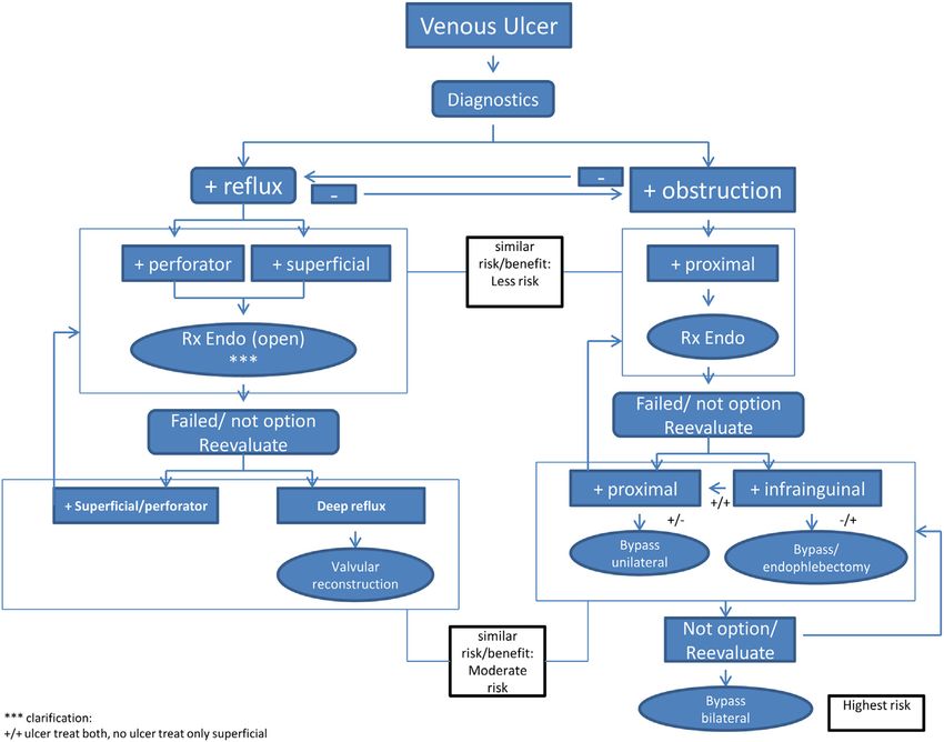
OPERATIVE/ENDOVASCULAR MANAGEMENT
Guideline 6.1: Superficial Venous Reflux and Active Venous Leg UlcerdUlcer Healing
In a patient with a venous leg ulcer (C6) and incompetent superficial veins that have axial reflux directed to the
bed of the ulcer, we suggest ablation of the incompetent veins in addition to standard compressive therapy to
improve ulcer healing. [GRADE - 2; LEVEL OF EVIDENCE - C]
Guideline 6.2: Superficial Venous Reflux and Active Venous Leg UlcerdPrevent Recurrence
In a patient with a venous leg ulcer (C6) and incompetent superficial veins that have axial reflux directed to the
bed of the ulcer, we recommend ablation of the incompetent veins in addition to standard compressive therapy to
prevent recurrence. [GRADE - 1; LEVEL OF EVIDENCE - B]
Guideline 6.3: Superficial Venous Reflux and Healed Venous Leg Ulcer
In a patient with a healed venous leg ulcer (C5) and incompetent superficial veins that have axial reflux directed
to the bed of the ulcer, we recommend ablation of the incompetent veins in addition to standard compressive ther-
apy to prevent recurrence. [GRADE - 1; LEVEL OF EVIDENCE - C]
Guideline 6.4: Superficial Venous Reflux With Skin Changes at Risk for Venous Leg Ulcer (C4b)
In a patient with skin changes at risk for venous leg ulcer (C4b) and incompetent superficial veins that have axial
reflux directed to the bed of the affected skin, we suggest ablation of the incompetent superficial veins in addition to
standard compressive therapy to prevent ulceration. [GRADE - 2; LEVEL OF EVIDENCE - C]
Guideline 6.5: Combined Superficial and Perforator Venous Reflux With or Without Deep Venous Reflux and
Active Venous Leg Ulcer
In a patient with a venous leg ulcer (C6) and incompetent superficial veins that have reflux to the ulcer bed in
addition to pathologic perforating veins (outward flow of >500 ms duration, with a diameter of >3.5 mm) located
beneath or associated with the ulcer bed, we suggest ablation of both the incompetent superficial veins and
perforator veins in addition to standard compressive therapy to aid in ulcer healing and to prevent recurrence.
[GRADE - 2; LEVEL OF EVIDENCE - C]
Guideline 6.6: Combined Superficial and Perforator Venous Reflux With or Without Deep Venous Disease and
Skin Changes at Risk for Venous Leg Ulcer (C4b) or Healed Venous Ulcer (C5)
In a patient with skin changes at risk for venous leg ulcer (C4b) or healed venous ulcer (C5) and incompetent
superficial veins that have reflux to the ulcer bed in addition to pathologic perforating veins (outward flow of >500
ms duration, with a diameter of >3.5 mm) located beneath or associated with the healed ulcer bed, we suggest abla-
tion of the incompetent superficial veins to prevent the development or recurrence of a venous leg ulcer. [GRADE -
2; LEVEL OF EVIDENCE - C] Treatment of the incompetent perforating veins can be performed simultaneously
with correction of axial reflux or can be staged with re-evaluation of perforator veins for persistent incompetence
after correction of axial reflux. [GRADE - 2; LEVEL OF EVIDENCE - C]
Guideline 6.7: Pathologic Perforator Venous Reflux in the Absence of Superficial Venous Disease, With or
Without Deep Venous Reflux, and a Healed or Active Venous Ulcer
In a patient with isolated pathologic perforator veins (outward flow of >500 ms duration, with a diameter of
>3.5 mm) located beneath or associated with the healed (C5) or active ulcer (C6) bed regardless of the status of the
deep veins, we suggest ablation of the “pathologic” perforating veins in addition to standard compression therapy
to aid in venous ulcer healing and to prevent recurrence. [GRADE - 2; LEVEL OF EVIDENCE - C]JOURNAL OF VASCULAR SURGERY
Volume 60, Number 2S O’Donnell et al 9S
Guideline 6.8: Treatment Alternatives for Pathologic Perforator Veins
For those patients who would benefit from pathologic perforator vein ablation, we recommend treatment by
percutaneous techniques that include ultrasound-guided sclerotherapy or endovenous thermal ablation (radiofre-
quency or laser) over open venous perforator surgery to eliminate the need for incisions in areas of compromised
skin. [GRADE - 1; LEVEL OF EVIDENCE - C]
Guideline 6.9: Infrainguinal Deep Venous Obstruction and Skin Changes at Risk for Venous Leg Ulcer (C4b),
Healed (C5) or Active (C6) Venous Leg Ulcer
In a patient with infrainguinal deep venous obstruction and skin changes at risk for venous leg ulcer (C4b),
healed venous leg ulcer (C5), or active venous leg ulcer (C6), we suggest autogenous venous bypass or endophle-
bectomy in addition to standard compression therapy to aid in venous ulcer healing and to prevent recurrence.
[GRADE - 2; LEVEL OF EVIDENCE - C]
Guideline 6.10: Deep Venous Reflux With Skin Changes at Risk for Venous Leg Ulcer (C4b), Healed (C5) or
Active (C6) Venous Leg UlcerdLigation
In a patient with infrainguinal deep venous reflux and skin changes at risk for venous leg ulcer (C4b), healed
venous leg ulcer (C5), or active venous leg ulcer (C6), we suggest against deep vein ligation of the femoral or popli-
teal veins as a routine treatment. [GRADE - 2; LEVEL OF EVIDENCE - C]
Guideline 6.11: Deep Venous Reflux With Skin Changes at Risk for Venous Leg Ulcer (C4b), Healed (C5) or
Active (C6) Venous Leg UlcerdPrimary Valve Repair
In a patient with infrainguinal deep venous reflux and skin changes at risk for venous leg ulcer (C4b), healed
venous leg ulcer (C5), or active venous leg ulcer (C6), we suggest individual valve repair for those who have axial
reflux with structurally preserved deep venous valves in addition to standard compression therapy to aid in venous
ulcer healing and to prevent recurrence. [GRADE - 2; LEVEL OF EVIDENCE - C]
Guideline 6.12: Deep Venous Reflux With Skin Changes at Risk for Venous Leg Ulcer (C4b), Healed (C5) or
Active (C6) Venous Leg UlcerdValve Transposition or Transplantation
In a patient with infrainguinal deep venous reflux and skin changes at risk for venous leg ulcer (C4b), healed
venous leg ulcer (C5), or active venous leg ulcer (C6), we suggest valve transposition or transplantation for those
with absence of structurally preserved axial deep venous valves when competent outflow venous pathways are
anatomically appropriate for surgical anastomosis in addition to standard compression therapy to aid in venous
leg ulcer healing and to prevent recurrence. [GRADE - 2; LEVEL OF EVIDENCE - C]
Guideline 6.13: Deep Venous Reflux With Skin Changes at Risk for Venous Leg Ulcer (C4b), Healed (C5) or
Active (C6) Venous Leg UlcerdAutogenous Valve Substitute
In a patient with infrainguinal deep venous reflux and skin changes at risk for venous leg ulcer (C4b), healed
venous leg ulcer (C5), or active venous leg ulcer (C6), we suggest consideration of autogenous valve substitutes
by surgeons experienced in these techniques to facilitate ulcer healing and to prevent recurrence in those with
no other option available in addition to standard compression therapy to aid in venous ulcer healing and to prevent
recurrence. [GRADE - 2; LEVEL OF EVIDENCE - C]
Guideline 6.14: Proximal Chronic Total Venous Occlusion/Severe Stenosis With Skin Changes at Risk for
Venous Leg Ulcer (C4b), Healed (C5) or Active (C6) Venous Leg UlcerdEndovascular Repair
In a patient with inferior vena cava or iliac vein chronic total occlusion or severe stenosis, with or without lower
extremity deep venous reflux disease, that is associated with skin changes at risk for venous leg ulcer (C4b), healed
venous leg ulcer (C5), or active venous leg ulcer (C6), we recommend venous angioplasty and stent recanalization in
addition to standard compression therapy to aid in venous ulcer healing and to prevent recurrence. [GRADE - 1;
LEVEL OF EVIDENCE - C]
Guideline 6.15: Proximal Chronic Venous Occlusion/Severe Stenosis (Bilateral) With Recalcitrant Venous
UlcerdOpen Repair
In a patient with inferior vena cava or iliac vein chronic occlusion or severe stenosis, with or without lower ex-
tremity deep venous reflux disease, that is associated with a recalcitrant venous leg ulcer and failed endovascular
treatment, we suggest open surgical bypass with use of an externally supported expanded polytetrafluoroethylene
graft in addition to standard compression therapy to aid in venous leg ulcer healing and to prevent recurrence.
[GRADE - 2; LEVEL OF EVIDENCE - C]
Guideline 6.16: Unilateral Iliofemoral Venous Occlusion/Severe Stenosis With Recalcitrant Venous Ulcerd
Open Repair
In a patient with unilateral iliofemoral venous occlusion/severe stenosis with recalcitrant venous leg ulcer for
whom attempts at endovascular reconstruction have failed, we suggest open surgical bypass with use of saphenous
vein as a cross-pubic bypass (Palma procedure) to aid in venous ulcer healing and to prevent recurrence. A synthetic
graft is an alternative in the absence of autogenous tissue. [GRADE - 2; LEVEL OF EVIDENCE - C]JOURNAL OF VASCULAR SURGERY
10S O’Donnell et al August Supplement 2014
Guideline 6.17: Proximal Chronic Total Venous Occlusion/Severe Stenosis (Bilateral or Unilateral) With
Recalcitrant Venous UlcerdAdjunctive Arteriovenous Fistula
For those patients who would benefit from an open venous bypass, we suggest the addition of an adjunctive
arteriovenous fistula (4-6 mm in size) as an adjunct to improve inflow into autologous or prosthetic crossover by-
passes when the inflow is judged to be poor to aid in venous leg ulcer healing and to prevent recurrence. [GRADE -
2; LEVEL OF EVIDENCE - C]
ANCILLARY MEASURES
Guideline 7.1: Nutrition Assessment and Management
We recommend that nutrition assessment be performed in any patient with a venous leg ulcer who has evidence
of malnutrition and that nutritional supplementation be provided if malnutrition is identified. [BEST PRACTICE]
Guideline 7.2: Systemic Drug Therapy
For long-standing or large venous leg ulcer, we recommend treatment with either pentoxifylline or micronized pu-
rified flavonoid fraction used in combination with compression therapy. [GRADE - 1; LEVEL OF EVIDENCE - B]
Guideline 7.3: Physiotherapy
We suggest supervised active exercise to improve muscle pump function and to reduce pain and edema in
patients with venous leg ulcers. [GRADE - 2; LEVEL OF EVIDENCE - B]
Guideline 7.4: Manual Lymphatic Drainage
We suggest against adjunctive lymphatic drainage for healing of the chronic venous leg ulcers. [GRADE - 2;
LEVEL OF EVIDENCE - C]
Guideline 7.5: Balneotherapy
We suggest balneotherapy to improve skin trophic changes and quality of life in patients with advanced venous
disease. [GRADE - 2; LEVEL OF EVIDENCE - B]
Guideline 7.6: Ultraviolet light
We suggest against use of ultraviolet light for the treatment of venous leg ulcers. [GRADE - 2; LEVEL OF
EVIDENCE - C]
PRIMARY PREVENTION
Guideline 8.1: Primary PreventiondClinical CEAP C3-4 Primary Venous Disease
In patients with clinical CEAP C3-4 disease due to primary valvular reflux, we recommend compression, 20 to
30 mm Hg, knee or thigh high. [GRADE - 2; LEVEL OF EVIDENCE - C]
Guideline 8.2: Primary PreventiondClinical CEAP C1-4 Post-thrombotic Venous Disease
In patients with clinical CEAP C1-4 disease related to prior deep venous thrombosis (DVT), we recommend
compression, 30 to 40 mm Hg, knee or thigh high. [GRADE - 1; LEVEL OF EVIDENCE - B]
Guideline 8.3. Primary PreventiondAcute DVT Treatment
As post-thrombotic syndrome is a common preceding event for venous leg ulcers, we recommend current
evidence-based therapies for acute DVT treatment. [GRADE - 1; LEVEL OF EVIDENCE - B] We suggest use of
low-molecular-weight heparin over vitamin K antagonist therapy of 3-month duration to decrease post-
thrombotic syndrome. [GRADE - 2; LEVEL OF EVIDENCE - B] We suggest catheter-directed thrombolysis in pa-
tients with low bleeding risk with iliofemoral DVT of durationJOURNAL OF VASCULAR SURGERY
Volume 60, Number 2S O’Donnell et al 11S
of VLUs can consume a significant amount of resources, so million per year.12,13 In the United States, $2.5 billion
that an agreed on “best practice” algorithm can maximize was expended for the treatment of VLUs in more than 6
the quality and effectiveness of care while minimizing cost million patients.14 A recent study on the cost of treating
and resource use.3 Moreover, VLUs are associated with VLUs in Germany demonstrated that mean cost averaged
prolonged disability, important socioeconomic impact, V9569 per patient per year, and 92% of this expenditure
and significant psychosocial morbidity. Because approxi- was related to direct costs.15 Although the majority of
mately 50% of VLUs may recur within 10 years, they are patients with VLUs can be treated on an outpatient basis
marked by a significant component of chronicity, which and are infrequently hospitalized except for complications,
compounds their economic impact and need for repetitive the direct cost of treating VLUs in the United States has
care. VLUs also can be painful, so that a patient’s ability been estimated to be $2500 per month per patient.16 In
to work may be compromised, and can also affect the the ambulatory setting, the direct cost of this care is related
retired segments of the population, thereby compounding to (1) technical (facility) costs and professional reim-
both the indirect and direct costs of treating VLU. bursement (physicians); (2) labor costs (nurses and para-
Epidemiology of Venous Leg Ulcers. VLUs are the medical personnel) for wound care treatments, which are
most common ulceration on the lower extremity and ac- the major driver of costs; and (3) medications as well as
count for 70% of all leg ulcers.4 Various estimates have specialized wound dressings and compression garments. A
been made from observational studies on the prevalence of key determinant of the costs of treating VLU is the effec-
VLU, ranging between 0.06% and 2%. The Edinburgh tiveness of treatmentdnot only how rapidly the ulcer heals,
study, which was a cross-sectional study of a random but also whether the ulcer recurs. For example, a more
sample of more than 1500 people between the ages of 18 expensive wound dressing may have an economic advan-
and 64 years, provided an estimate of VLU prevalence of tage for overall care because of more rapid healing of the
1%.5 This lower prevalence contrasts with the higher VLU with less duration of product use and attendant labor
prevalence of an earlier study (2.7% clinical CEAP C5/ costs.
C6), which was based on a questionnaire and photographs To determine the actual costs of treating VLUs during
of the legs of the participants.6 As in the Edinburgh study, a 1-year period, a recent study examined a cohort of 84 pa-
VLU prevalence increased with age. Two other studies tients with nonhealing VLUs (CEAP C6) who presented
examined large populations. Approximately 40,000 Polish to a wound clinic. All patients were treated in a wound
patients underwent clinical evaluation and interviews by a center by five vascular surgeons with a minimum follow-
variety of health care professionals in a multicenter study up of 6 months (median, 368 days; 336-483).17 Actual
showing a 1.5% prevalence of C5/C6 disease.7 Another costs (not charges) were obtained for outpatient and inpa-
cross-sectional study carried out in France addressed a tient facility, visiting nurse services, and physician practice
subset of patients from a larger study of Raynaud patients. group to yield true cost. The proportion and time to com-
All of the 400 patients were evaluated by vascular medicine plete healing of VLU were determined to calculate time to
specialists. C5 disease was higher in men (5.4%) than in healing as well as ulcer-free intervals. Cost/ulcer-free days
women (2.7%), but the investigators observed no open and cost to complete healing for the entire follow-up
ulcers.8 The most detailed information comes from the period were carried out with univariate analysis of factors
Bonn Vein Longitudinal Study initiated by the German affecting cost. The mean total cost of treating VLU during
Ministry of Health.9 More than 3000 participants were this follow-up period was $15,732. A total of 50 patients
randomly identified between the ages of 18 and 79 years. (60%) healed their VLU without recurrence in a mean
Advanced chronic venous reflux (chronic venous insuffi- time of 122 days (6-379 days) at a mean cost of
ciency [CVI]) was found in 0.6% with a healed VLU (C5) $10,563 ($430-$50,967). Significant contributing factors
and in 0.1% with an open VLU for a total of 0.7%. This were outpatient facility fees ($10,332) and visiting nurse
longitudinal study documented a decrease in advanced services ($11,365) related to extended treatment of the
CVI (C4-C6), which may be related to a more compre- open VLU. Patients who failed to heal their ulcer during
hensive use of diagnostic and therapeutic modalities for the duration of this study (20%) had a threefold increase
CVI. Because of the high prevalence of venous disease, the in their costs ($33,907). Those patients who had recur-
AVF National Venous Screening Program was established rence of their VLU (N ¼ 17; 20%) during the follow-up
in the United States to increase awareness. The program period had a total mean cost of $12,760. Inpatient admis-
screened 2234 individuals and identified varicose veins in sion, of which nearly two thirds was for treatment of infec-
more than 30% of participants and more advanced venous tions that were resistant to therapy in an outpatient setting,
disease (C4-C6) in more than 10%.10 It has been estimated markedly increased costs ($33,629). By contrast, VLU
that approximately 2.5 million people suffer from CVI in treated with surgical intervention of the superficial venous
the United States, and of those, about 20% develop venous system did not significantly increase total cost over that of
ulcers.11 patients receiving best medical therapy ($11,960 vs
Economic Impact. The overall cost of treating VLU $12,304) but significantly reduced recurrence rates (34%
approaches 1% of the health care budget of some western vs 5%). By, contrast, patients treated for outflow obstruc-
European countries; in the United Kingdom, the annual tion had a twofold increase in total costs ($24,241 vs
cost is estimated to range from ₤300 million to ₤600 $11,960).JOURNAL OF VASCULAR SURGERY
12S O’Donnell et al August Supplement 2014
Value of Clinical Practice Guidelines for Venous uniform method of treatment that has been agreed on by a
Leg Ulcer Care. The value of developing and implement- panel of experts and based on clinical evidence for efficacy
ing clinical practice guidelines should be to provide quality may improve therapeutic effectiveness and possibly reduce
of care without a dramatic increase in cost. This focus on cost. Several studies have demonstrated that after the institu-
“best outcomes for the most reasonable health care dollar” tion of a VLU guideline, there were improvements in both
has stimulated many organizations, such as the SVS and the ulcer healing and recurrence rate and a subsequent
AVF, to develop and to promote a unified set of guidelines reduced resource use with lowering of treatment costs,
for treatment of chronic diseases, such as VLUs. An thereby supporting adoption of VLU guidelines.18 Olson
evidence-based analysis of treatment options should et al demonstrated that ulcer healing rate was markedly
decrease the variations in care while at the same time improved in a Veterans Hospital population of 155 patients
ensuring that resources are used in an optimal manner. with VLU if guideline recommendations were followed.
Specialty societies, as in the current SVS and AVF guideline, During a 5-year period, patients who received dressings that
develop clinical practice guidelines either by independent provided a moist wound healing environment and compres-
review of available studies or by employing evidence from sion for 80% of their visits were more likely to heal than those
technology assessments and other published guidelines. For who did not comply (JOURNAL OF VASCULAR SURGERY
Volume 60, Number 2S O’Donnell et al 13S
METHODOLOGY OF VENOUS LEG ULCER published consensus documents and the AVF report from
GUIDELINES the 2006 Venous Summit and the 2009 Pacific Vascular
The Institute of Medicine, in Clinical Practice Guide- Symposium, whose purpose was to reduce the incidence
lines We Can Trust, has defined clinical practice guidelines of venous ulcer during the next decade by 50%.29 In a pre-
as “systematically developed statements to assist the practi- vious systematic review of recently published venous ulcer
tioner and patient decisions about appropriate healthcare guidelines, 14 venous ulcer guidelines were identified
for specific clinical circumstances.”25 The underlying prin- worldwide.18 This review showed that there was a high de-
ciple is to use evidence-based medicine to the greatest gree of agreement among the 14 VLU guidelines on rec-
extent to develop these guidelines so that the assessment ommendations for compression (72%), dressings (72%),
of the optimal treatment plan is based on the best current pentoxifylline (73%), prevention of recurrence by below-
available knowledge. Evidence-based medicine allows one knee stockings (70%), and surgery (82%). There was a
to assess the statistical strength of a treatment or interven- low proportion of agreement in the areas of diagnosis, clin-
tion and, most important, reduces bias in evaluating a ical evaluation, and venous Doppler and duplex ultrasound;
particular therapy.26 Bias is defined as the predisposition in elements of wound care: measurement of the wound,
of prejudice toward either the experimental or control washing of the wound, débridement, and specific type of
group, which can lead to either an overestimation or an un- wound dressing; and finally in adjunctive measures: the
derestimation of the true benefits and harm of the interven- use of skin grafts and physical therapy to promote ankle
tion.27 This method reduces uncertainty, which is the mobility with enhancement of the calf muscle pump.
largest single cause of misinterpretation of data.28 This Several areas of “controversy” were identified that had
process has been further stated in the recent Institute of not been particularly addressed in previous guidelines:
Medicine document: “Hence, critically appraised and syn- new innovative, less invasive VLU therapies; the role of iliac
thesized scientific evidence has become fundamental to obstruction and occlusion with the need for diagnosis by
clinical practice. At the same time, and particularly under intravascular ultrasound and subsequent monitoring of
conditions of uncertainty regarding optimal decisions, stenting by this technique; when and by what methods
clinician experiential knowledge and skill (the “art of med- to treat perforators; the need for physical therapy to pro-
icine”) and patient values and preferences remain essential mote ankle mobility and function of the calf muscle
contributors to quality healthcare practice, in a complex pump; the role of advanced dressings; and preventing pro-
interplay with science.”25 gression to VLU. These areas received special attention in
The Venous Ulcer Guidelines Committee was orga- review for the current guidelines.
nized through cooperation between the SVS and AVF. The surgery/endovascular and compression sections
The Venous Ulcer Guidelines Committee was divided were selected for a de novo development of specific guide-
into six sub-committee sections each headed by a chair: line recommendations based on several of these key ques-
diagnosis; compression; surgery/endovascular; wound tions. For additional systematic review involving these
care; ancillary; and prevention. The overall committee critical areas, the Venous Ulcer Guidelines Committee
then developed a series of key clinical questions to guide commissioned an independent group of researchers to
the overall approach for the guideline document: (1) conduct two systematic reviews to evaluate the effective-
What is the best treatment for active (CEAP C6) venous ul- ness of different compression strategies and endovascular
cer? (2) What is the best treatment for healed venous ulcer and open surgical approaches. The Committee helped
(CEAP C5)? (3) What is the best method for preventing develop a priori the protocols of these reviews in terms of
recurrence of venous ulcers? and (4) Can progression outcome selection and criteria for including studies with
from CEAP C4 to CEAP C6/C5 be prevented? The additional analysis by the Knowledge and Evaluations
Venous Ulcer Guidelines Committee addressed the optimal Research Unit at the Mayo Clinic (Rochester, Minn).30,31
approach to be used for their specific section from four gen- In the literature review, several processes were used to
eral approaches: (1) de novo development, in which a minimize heterogeneity:
completely novel recommendation is developed from a sys- - Languagedall would be included.
tematic and meta-analysis review of the literature; (2) build - Type of Studies Revieweddrandomized controlled
on existing guidelines with a complementary full-literature trials, controlled clinical trials with cohort, and retro-
search update; (3) adapt guidelines from existing guidelines; spective large observational case series. The studies
and (4) total adoption of existing guidelines.18 The need for should be published in peer-reviewed journals.
a systematic and meta-analysis review was determined by - Target Audiencedthis Guideline document is in-
each section team and then agreed on by the entire commit- tended for specialists who treat vascular disease and
tee. Each section was categorized by the type of guideline wounds.
development required. All guidelines were developed by
building on existing guidelines with a complementary liter- Through an iterative process, the committee developed
ature search by the section sub-committee. guidelines based on the grading of recommendation assess-
In this process of guideline development for VLUs, the ment, development, and evaluation (GRADE) system
Venous Ulcer Guidelines Committee also reviewed prior (Table I).32,33 The strength of the recommendations isJOURNAL OF VASCULAR SURGERY
14S O’Donnell et al August Supplement 2014
Table I. GRADE recommendations based on level of evidence
Methodologic quality of
Grade Description of recommendation Benefit vs risk supporting evidence Implications
1A Strong recommendation, Benefits clearly outweigh risk RCTs without important Strong recommendation, can
high-quality evidence and burdens, or vice versa limitations or overwhelming apply to most patients in
evidence from observational most circumstances without
studies reservation
1B Strong recommendation, Benefits clearly outweigh risk RCTs with important Strong recommendation, can
moderate-quality evidence and burdens, or vice versa limitations (inconsistent apply to most patients in
results, methodologic flaws, most circumstances without
indirect, or imprecise) or reservation
exceptionally strong
evidence from observational
studies
1C Strong recommendation, low- Benefits clearly outweigh risk Observational studies or case Strong recommendation but
quality or very-low-quality and burdens, or vice versa series may change when higher
evidence quality evidence becomes
available
2A Weak recommendation, high- Benefits closely balanced with RCTs without important Weak recommendation, best
quality evidence risks and burdens limitations or overwhelming action may differ depending
evidence from observational on circumstances or
studies patients’ or societal values
2B Weak recommendation, Benefits closely balanced with RCTs with important Weak recommendation, best
moderate-quality evidence risks and burdens limitations (inconsistent action may differ depending
results, methodologic flaws, on circumstances or
indirect, or imprecise) or patients’ or societal values
exceptionally strong
evidence from observational
studies
2C Weak recommendation, low- Uncertainty in the estimates of Observational studies or case Very weak recommendations;
quality or very-low-quality benefits and risk, and series Other alternatives may be
evidence burdens; Risk, benefit, and reasonable
burdens may be closely
balanced
RCTs, Randomized controlled trials.
Modified from Guyatt G, Gutterman D, Baumann MH, Addrizzo-Harris D, Hylek EM, Phillips B, et al. Grading strength of recommendations and quality of
evidence in clinical guidelines: Report from an American College of Chest Physicians task force. Chest 2006;129:174-81.
related to (1) the quality of evidence around that recom- PRACTICE]. Such recommendations are not graded
mendation, (2) harm/benefit ratio of the therapy (eg, but deemed by the guideline developers to be necessary
minimally invasive intervention with few morbid events to provide a comprehensive guideline that encompasses
and a possible effect), and (3) patient preference. Every all the details needed for providing care for patients with
effort has been made by the committee to make the process venous ulcers.34 In each section and in the comments to
of assigning the strength of the particular recommendation each specific recommendation, we have attempted to
as transparent as possible. By the GRADE system, the clearly link the recommendation to the evidence and its
strength of the recommendation or the extent to which quality and to point out where consensus techniques
one can be confident that adherence to the recommenda- have been used. Independent review of GRADE assign-
tion will do more good than harm was divided into [1] ments made by the Venous Ulcer Guidelines Committee
strong (we recommend) and [2] weak (we suggest), with was also performed by the Knowledge and Evaluations
[1] favoring benefit over harm and [2] with benefits Research Unit at the Mayo Clinic (Rochester, Minn) to
closely balanced by the risk. The “quality of evidence” or corroborate proper strength of evidence and quality of ev-
the extent to which confidence in an estimate of effect is idence for each guideline.
sufficient to support a particular recommendation was The final document was reviewed by the chairman and
graded [A], [B], or [C] by standard evidence-based meth- vice chairman of the Venous Ulcer Guidelines Committee
odologic criteria. It is well recognized that there may not and remitted to the entire committee for concurrence.
be studies of the highest evidentiary value for the diagnosis Additional independent review was obtained from selected
and management of VLUs. When there are no comparable reviewers representing multiple medical specialties vested in
alternatives to a recommendation or evidence is lacking, venous ulcer management. The final document was then
the Venous Ulcer Guidelines Committee has relied on reviewed and approved by the SVS Document Oversight
case series supplemented by the best opinion of a panel Committee and approved by the Executive Committees
of experts, and the recommendation was labeled [BEST of the SVS and AVF.JOURNAL OF VASCULAR SURGERY
Volume 60, Number 2S O’Donnell et al 15S
DEFINITION—VENOUS LEG ULCER mixed ulcers often have a different rate of healing and
require additional treatment beyond the appropriate
venous measures for healing to occur and recurrence to
Guideline 1.1: Venous Leg Ulcer Definition be prevented. The importance of these diagnoses is that
We suggest use of a standard definition of venous analysis of healing times and effectiveness of surgical
ulcer as an open skin lesion of the leg or foot that treatment of the venous component requires separation
occurs in an area affected by venous hypertension. of pure venous from mixed ulcers to learn whether treat-
[BEST PRACTICE] ment of the venous component contributes to faster ulcer
healing and early intervention is favored over delayed
treatment.
Inherent in composing a set of guidelines for VLU is
agreeing on a common definition of VLU. Current defini-
tions for VLU vary, as exemplified by the following: VENOUS ANATOMY AND PATHOPHYSIOLOGY
d THE AVF CONSENSUS STATEMENT: Venous ul-
cer is defined as a full-thickness defect of skin, most frequently
Guideline 2.1: Venous Anatomy Nomenclature
in ankle region, that fails to heal spontaneously and is sus-
We recommend use of the International Consensus
tained by CVD (duplex studies).35
Committee on Venous Anatomical Terminology for
d SCOTTISH GUIDELINE DEFINITION: Chronic
standardized venous anatomy nomenclature. [BEST
venous leg ulcer is defined as an open lesion between the
PRACTICE]
knee and the ankle joint that remains unhealed for at least
four weeks and occurs in the presence of venous disease.
Studies reviewed in this guideline included patients with The deep veins of the lower limbs are located in the
venous leg ulcers, irrespective of the method of diagnosis of deep compartment of the leg bound by the muscle fascia
venous reflux.36 and accompany the main arteries of the leg and pelvis. Su-
d FRENCH HEALTHCARE SYSTEM GUIDE- perficial veins of the lower limbs are those located between
LINES: A pure venous ulcer is defined, by professional the deep fascia covering the muscles of the limb and the
agreement, as a leg lesion, which has not healed within skin and primarily included the saphenous system. Perfo-
a month (except in cases of recurrent ulcers when a diag- rating veins pass through the deep muscle fascia and con-
nosis can be made in less than a month); with a pathophys- nect the superficial to the deep venous system.
iology due to ambulatory venous hypertension, which may Communicating veins connect veins within the same
be secondary to: reflux in superficial, perforating or deep system.
veins, and/or obstruction of the deep veins, and/or calf Whereas there is general agreement that veins of the
muscle pump dysfunction; where there is no arterial lower extremities be divided into superficial, deep, and
involvement.37 perforating venous systems, there have been deficiencies
in the past in nomenclature for specific veins in the leg
For the purpose of this Guideline document, we
within each system. In response to variability in
recommend the following definition of VLU: A venous
anatomic terminology used for venous anatomy, stan-
ulcer is an open skin lesion of the leg or foot that occurs
dard anatomic venous terminology has been developed
in an area affected by venous hypertension. Venous hy-
and adopted. For practitioners caring for patients with
pertension is the result of reflux or obstruction. This
VLUs, correct standardized venous nomenclature
may occur as a focal phenomenon in the distal extremity
should be used as defined by the 2002 International
or as a central mechanism, as in iliocaval obstruction or
Consensus Committee on Venous Anatomical Termi-
elevated inferior vena cava pressure from advanced truncal
nology and updated in 2005 as shown in Table II and
obesity. The mere existence of either reflux or obstruction
Table III.38,39
does not appear to be the full cause of venous ulcers, with
additional biochemical factors due to activation of the in-
flammatory cascade, which may act as the inciting factor Guideline 2.2: Venous Leg Ulcer Pathophysiology
for appearance of the actual ulcer. Identification of these We recommend a basic practical knowledge of
additional factors awaits further advances in the basic venous physiology and venous leg ulcer pathophysi-
sciences. ology for all practitioners caring for venous leg ulcers.
At the clinical level, venous ulcers occur as “pure”
[BEST PRACTICE]
venous causes, when there is directed axial great saphe-
nous vein reflux or incompetent perforator reflux directly
into the ulcer bed, or as “mixed” venous plus other Chronic venous disease (CVD) is a debilitating condi-
causes, as in cases in which arterial ischemia, scarred tis- tion that affects millions of individuals worldwide. The
sue of the gaiter area, hypersensitive skin, lymphedema, condition can result in varicose veins or advance to severe
autoimmune disease, local trauma, infection, and other skin changes and VLU. Both reflux and obstruction ac-
processes coexist with the venous hypertension. The count for the pathophysiologic mechanism of CVD;JOURNAL OF VASCULAR SURGERY
16S O’Donnell et al August Supplement 2014
Table II. The International Consensus Committee on Venous Anatomical Terminology for standardized venous
anatomy nomenclature of the leg
Superficial venous system Deep venous system Perforating venous system
Superficial inguinal veins Common femoral vein Gluteal perforators
External pudendal vein Femoral vein Superior gluteal
Superficial circumflex iliac vein Deep femoral vein Midgluteal
Superficial epigastric vein Medial circumflex femoral vein Lower gluteal
Superficial dorsal vein (clitoris/penis) Lateral circumflex femoral vein Thigh perforators
Anterior labial veins Deep femoral communicating veins Medial thigh
Anterior scrotal veins Sciatic vein Femoral canal
Great saphenous vein Popliteal vein Inguinal
Saphenofemoral junction Genicular venous plexus Anterior thigh
Terminal valve Anterior tibial veins Lateral thigh
Preterminal valve Posterior tibial veins Posterior thigh
Anterior accessory great saphenous vein Fibular or peroneal veins Posteromedial
Posterior accessory great saphenous vein Sural veins Sciatic
Superficial accessory great saphenous vein Soleal veins Posterolateral
Anterior thigh circumflex vein Gastrocnemius veins Pudendal
Posterior thigh circumflex vein Medial Knee perforators
Small saphenous vein Lateral Medial knee
Saphenopopliteal junction Intergemellar Suprapatellar
Terminal valve Medial plantar veins Lateral knee
Preterminal valve Lateral plantar veins Infrapatellar
Cranial extension of small saphenous vein Deep plantar venous arch Popliteal fossa
Superficial accessory small saphenous vein Deep metatarsal veins (plantar/dorsal) Leg (calf) perforators
Intersaphenous veins Deep digital veins (plantar/dorsal) Medial leg
Lateral venous system Pedal vein Paratibial
Dorsal venous network of the foot Posterior tibial
Dorsal venous arch of the foot Anterior leg
Superficial metatarsal veins (dorsal/plantar) Lateral leg
Plantar venous subcutaneous network Posterior leg
Superficial digital veins (dorsal/plantar) Medial gastrocnemius
Lateral marginal vein Lateral gastrocnemius
Medial marginal vein Intergemellar
Para-achillean
Ankle perforators
Medial ankle
Anterior ankle
Lateral ankle
Foot perforators
Dorsal foot
Medial foot
Lateral foot
Plantar foot
Modified from Caggiati A, Bergan JJ, Gloviczki P, Eklof B, Allegra C, Partsch H. Nomenclature of the veins of the lower limb: Extensions, refinements, and
clinical application J Vasc Surg 2005;41:719-24.
however, reflux from primary venous disease has a much macrophages and monocytes), inflammatory modulators
higher prevalence in patients presenting with the different and chemokines, cytokine expression, growth factors,
stages of CVD, which might also include venous ulcers. metalloproteinase activity, and many regulatory pathways
However, obstruction from secondary venous disease that perpetuate inflammation.46-48
and valvular reflux are associated with a much more rapid The pathophysiologic process of primary venous
progression of disease and a higher rate of progression to disease is a complex entity with multifaceted stages
venous ulceration.40-44 Whether reflux or obstruction is leading to the dilated tortuous, valve-insufficient vari-
the cause of the patient’s clinical presentation and symp- cose veins, venous hypertension, and the associated
toms is unclear; both conditions lead to increased ambu- clinical manifestations seen with CVD. The pathophys-
latory venous pressure. The fundamental basis for CVD iologic mechanism of secondary venous disease involves
and venous ulceration is inflammation within the venous inflammation, thrombosis, recanalization resulting in
circulation that is subjected to increased hydrostatic pres- venous wall damage and dilation, and valve insuffi-
sure resulting in increased ambulatory venous pressure.45 ciency. The clinical picture is that of the post-
The inflammation involves leukocytes (in particular thrombotic syndrome (PTS) and can comprise pain,You can also read