MATERNAL SEPSIS: PRESENTATION, COURSE, TREATMENT, AND OUTCOMES - NursingCenter.com
←
→
Page content transcription
If your browser does not render page correctly, please read the page content below
2.0 ANCC
Contact Hours
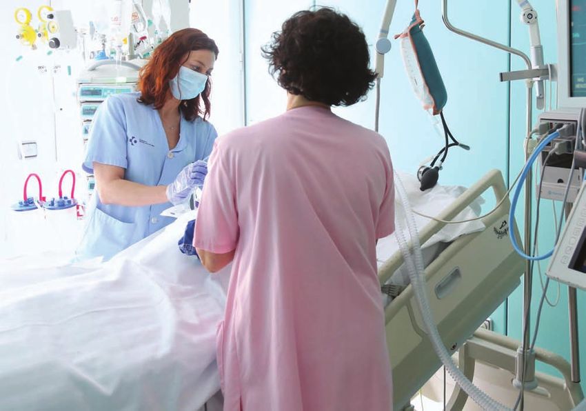
agefotostock / Alamy Stock Photo
MATERNAL SEPSIS: PRESENTATION,
COURSE, TREATMENT, AND OUTCOMES
Courtney Stanley Sundin, MSN, RNC-OB, C-EFM, Kendall Rigg, BSN, RNC-OB, and Kathleen Kistner Ellis, PhD, RN, CCRN-K
Abstract presenting symptom was hypothermia or hyperthermia, followed by
Purpose: The current adult definition of sepsis and septic shock, as tachycardia. Primary laboratory results included above and below
developed in 1992, does not adequately define sepsis in the pregnant normal white blood cell count and elevated lactate levels. Sixty-four
and peripartum women due to the alteration of sepsis presentation in percent of patients diagnosed with sepsis were readmitted postpar-
the maternal population. The purpose of this study was to determine tum and 41% gave birth via cesarean.
potential causative factors for sepsis with the aim of prevention and Clinical Implications: Findings are applicable for nursing care and
reducing morbidity and mortality. maternal sepsis protocol development. Early identification of mothers at
Study Design and Methods: A descriptive observational design via risk for maternal sepsis and tool development for early diagnosis would
a retrospective medical record review was used with a convenience be beneficial to support the ongoing work on decreasing maternal
sample of 22 women who were identified after admission as having morbidity and mortality that have a devastating effect on women, their
sepsis. The setting was the labor and delivery unit of a large urban families, and their health care team. Early warning signs of sepsis can
hospital in Fort Worth, Texas, with over 5,000 births per year. be shared by nurses with new mothers and their families as part of
Results: The most common diagnoses related to maternal sepsis routine postpartum discharge teaching so they know when to call their
included urinary tract infections, endometritis, chorioamnionitis, and primary health care provider and when to seek care in person.
wound infections. Main causative agents identified were predomi-
nantly Escherichia coli (E. coli), followed by group B streptococcus Key words: Chorioamnionitis; Maternal sepsis; Puerperal infection;
(GBS), and group A streptococcus (group A strep). The most prevalent Pyelonephritis.
May/June 2021 MCN 155
Copyright © 2021 Wolters Kluwer Health, Inc. All rights reserved.epsis is identified by organ dysfunction leading to
S a high rate of mortality and morbidity and can
result from a multitude of infections (Novosad et
al., 2016). Organ and tissue damage occur with
sepsis as a reaction to the initial infection. Managing
sepsis starts with understanding that sepsis is a medical
Sepsis is the second leading cause of
maternal mortality and makes up 5% of
maternal ICU admissions in the United States.
emergency requiring prompt assessment, fluid resuscita-
tion, source control, and evaluation of hemodynamic sta-
tus (Levy et al., 2018). Sepsis is the second leading cause
of maternal mortality, accounting for 12.7% of maternal 100,000 births (Bonet et al., 2017). Evidence has shown
deaths in the United States (Centers for Disease Control that maternal sepsis mortality rate is 8%; cases that prog-
and Prevention [CDC], 2020b). Maternal sepsis contrib- ress to septic shock increase the mortality rate to between
utes to 5% of maternal ICU admissions (Parfitt et al., 20% and 28% (Bonet et al.). There is a link between organ
2017a). During pregnancy, signs and symptoms of sepsis system failure and mortality rate (Bonet et al.; Parfitt et al.,
may be increasingly difficult to distinguish due to mater- 2017c). In the United States, there has been an approxi-
nal adaptations to pregnancy, effects of interventions re- mate increase of maternal sepsis by 50% in 2003 (Acosta
quired during labor, maternal pushing efforts, and blood et al., 2014). According to the California Maternal Qual-
loss after birth (Bonet et al., 2017). ity Care Collaborative, “for each maternal death, there are
Physiological adaptations during pregnancy include 50 women who experience life-threatening morbidity
increased circulation, tachycardia, decreased blood pres- from sepsis” (Gibbs et al., 2019). Of the deaths associated
sure, diminished oxygen reserve, and increased suscepti- with sepsis, group A streptococcal infection was identified
bility to infection (Bonet et al., 2017; Escobar et al., as the more prevalent infection related to maternal sepsis
2020). Due to an increase in maternal intervascular vol- mortality, whereas E. coli has been shown to be the more
ume during pregnancy, there is a subsequent increase in prevalent cause of sepsis (Acosta et al.).
maternal heart rate by 10 to 20 beats per minute (bpm), The two greatest sources of infection in the maternal
along with a compensatory decrease in blood pressure population are urinary tract and genital infections in-
due to vasodilation, mimicking signs of sepsis (Escobar et cluding chorioamnionitis and endometritis (Bonet et al.,
al.; Gibbs et al., 2019). Respiratory changes during preg- 2017). In the maternal population, infection can rapidly
nancy include an increase in tidal volume and a decrease progress in less than 24 hours from the first symptom to
in the residual volume and functional residual capacity the diagnosis of sepsis. In women with group A strep in-
(Olvera & Dutra, 2016). This causes a reduction in oxy- fection, infection was found to progress in as little as 9
gen reserve thereby affecting the mother’s ability to com- hours from onset to diagnosis, highlighting importance
pensate when a severe infection potentially leads to meta- of initiating high-dose intravenous antibiotics within 1
bolic acidosis (Olvera & Dutra). Immunologic hour of onset (Acosta et al., 2014). A high number of
adaptations during pregnancy include a decrease in in- women develop sepsis after hospital discharge; therefore,
flammatory response and cell-mediated immunity caus- discharge teaching of signs and symptoms of infection is
ing a predisposition to infection (Parfitt et al., 2017b). To critical (Gibbs et al., 2019).
protect the fetus, this immune response occurs to allow Another study identified a link between antibiotic
tolerance of fetal antigens (Gibbs et al.). The increased treatment during the perinatal period and a greater likeli-
susceptibility to infection and invasion of microorgan- hood of the development of maternal sepsis (Acosta et
isms in the genitourinary tract is likely to cause ascending al., 2014). This implies that there is a progression of the
infections such as pyelonephritis and chorioamnionitis infection even after initiation of antibiotic treatment sug-
(Parfitt et al., 2017b). Cesarean birth increases risk for gesting improper diagnosis, treatment, and follow-up
development of a surgical site and endometrial infection (Acosta et al.). A significant risk factor was identified as
(Olvera & Dutra). These immunologic changes increase an unscheduled cesarean birth that increases the risk for
risk of development of acute respiratory distress syndrome sepsis 5- to 20-fold when compared with a vaginal birth
during pregnancy and postpartum (Olvera & Dutra). The (Parfitt et al., 2017b). Other risk factors that increase a
physiologic and immunologic adaptations that occur may mother’s probability for development of sepsis are also
mask initial signs and symptoms of infection that can re- poorly understood and include obesity, chronic hyperten-
sult in a delay in recognition and treatment (Escobar et sion, anemia, poor nutrition, history of GBS infection,
al.). lack of prenatal care, and nonwhite ethnicity (Parfitt et
The current adult definition of sepsis and septic shock al., 2017b).
as developed in 1992 (Bone et al.) does not adequately The Surviving Sepsis Campaign suggests that to de-
define sepsis in pregnant and peripartum women due to crease sepsis mortality and morbidity there has to be
the altered presentation of sepsis in this population (Acos- awareness, recognition, appropriate treatment, health
ta et al., 2014). Despite improvements in medical care, in- care education, guidelines of care, post intensive unit
cidence of maternal sepsis continues to rise globally (Acos- care, and quality improvement programs (De Backer &
ta et al., 2016). Current incidence of maternal sepsis in Dorman, 2017). The World Health Organization encour-
developed countries is estimated to be from 9 to 49 per ages use of specific maternal guidelines to best recognize
156 volume 46 | number 3 May/June 2021
Copyright © 2021 Wolters Kluwer Health, Inc. All rights reserved.and treat maternal sepsis (Bonet et al., 2017). There is TABLE 1. MATERNAL SEPSIS OUTCOMES
need to understand progression of maternal sepsis to gain
knowledge of treatment and infection control (Acosta et Average Range
al., 2014). Generally, intensivists, general practitioners, Demographics and Pregnancy Outcomes
or emergency room providers have the most sepsis man- Age 27 years 16–40 years
agement education and knowledge in the adult popula-
tion. However, they have less understanding of maternal Gravidity 2.27 1–5
presentation and the rate at which infection rapidly pro- Parity 1.8 1–4
gresses (Acosta et al.). When group A strep infection is Gestation 36.8 weeks 23.1–41.1 weeks
confirmed, providers should treat and manage infection
Length of membrane 31.9 hr 1 min–21 days
as a maternal emergency.
rupture time to birth
Maternal adaptations to pregnancy conceal and mimic
signs and symptoms of maternal sepsis. To better under- Maternal Vital Signs
stand the infectious process, retrospective medical record Temperature 101.8 °F 95.8–103.7 °F
review of cases of mothers with a confirmed sepsis diagno-
Heart rate 125 bpm 84–175 bpm
sis was analyzed. Medical records were evaluated for ma-
ternal presenting signs and symptoms, laboratory and as- Respiratory rate 21.8 bpm 16–48 bpm
sessment findings, and progression of maternal sepsis. The Blood pressure 105/61 mmHg 71/43–140/78 mmHg
main purpose of this study was to determine factors asso- Infant Vital Signs
ciated with sepsis cases with the aim of generating knowl-
edge to help reduce morbidity and mortality in maternity Temperature 99.5 °F 97.8–102.1 °F
patients by early recognition. We also sought to identify Heart rate 164.1 bpm 140–190 bpm
obstacles in recognition of sepsis in this population. Respiratory rate 55.5 bpm 38–80 bpm
Study Design and Methods criteria. All data were collected on a shared, password-
We used a descriptive observational design through a ret- protected electronic file that was accessible only to the
rospective medical record review of cases of maternal sep- members of the research team. Patient medical infor-
sis with the following research question. Among maternity mation included: patient demographics, gravidity and
patients who are subsequently diagnosed with maternal parity, gestational age at birth, type of birth, pertinent
sepsis, what concerning criteria including vital signs, labo- medical and prenatal history, reason for admission, mem-
ratory results, and presenting symptoms are found on a brane status (including time of rupture, meconium stain-
retrospective medical record review? ing), vital signs (infant and mother), pertinent laboratory
results, capillary refill, urinary output, blood cultures,
Sample 12-lead electrocardiogram, treatment information (oxy-
The sample consisted of women who were identified after gen administration, antibiotics, fluid resuscitation), and
admission as having maternal sepsis. The research team infant disposition.
obtained a list from health information management of
patients using discharge and readmission coding for sep- Results
sis and sepsis-related terms: puerperal sepsis, maternal Demographics
sepsis. Inclusion criteria were records of patients who Twenty-four cases of maternal sepsis were identified from
gave birth at the study hospital or who were admitted 2014 to 2019 using the puerperal sepsis diagnosis code.
during the postpartum period, up to 6 weeks after birth Of these, two were excluded due to not meeting specified
with suspected or confirmed sepsis. Exclusion criteria criteria. Average maternal age at time of diagnosis was
were patients who did not give birth at a hospital and 27 years with a range of 16 to 40 (Table 1). Fifty-nine
postpartum patients who did not have coding for sepsis percent were Caucasian, 19% were Hispanic, 13% Afri-
or sepsis-related diagnoses. can American, and 9% were Asian.
Setting Pregnancy and Birth Outcomes
The setting was the labor and delivery unit of Baylor Mothers
Scott & White All Saints Medical Center-Fort Worth, a Average gravidity was 2.27 and average parity was 1.8.
538-bed hospital with a level III neonatal intensive care Average gestation at time of birth was 36.8 weeks. Fifty-
unit and a level III trauma center. The labor and delivery nine percent (14) gave birth vaginally, whereas 41% (9)
unit has 19 labor-delivery-recovery rooms, an obstetric had a cesarean birth. Of the nine cesareans, two were
triage unit, and averages over 5,000 births per year. scheduled repeat cases and seven were unplanned for in-
dications including: failure to progress (4), fetal “distress”
Study Procedures (1), severe intrauterine growth restriction and preeclamp-
After obtaining institutional review board approval, re- sia (1), and twin pregnancy with preterm spontaneous
searchers obtained a list of patients who met inclusion rupture of membranes (1). Meconium-stained fluid was
May/June 2021 MCN 157
Copyright © 2021 Wolters Kluwer Health, Inc. All rights reserved.noted in 9% of cases (2). Average length of time from 1), and shortness of breath (5%, 1). Urinary cultures re-
membrane rupture to birth was 31.9 hours (range: 1 min- sulted in Escherichia coli (E. coli) (12), respiratory cul-
ute to 21 days) (Table 1). Sixty-four percent (14) were ture showed macrobacterium (1), two wound cultures
GBS negative, 23% (5) had unknown status, and 13% resulted in GBS (1) and Vividans strep (1), blood cultures
(3) were GBS positive. There were no maternal deaths. resulted in extended spectrum beta-lactamases (1), E. coli
(1), and group A strep (1).
Babies
Newborn and fetal findings included a fetal tachycardia Laboratory and Assessment Results
in 13% (3) of the cases averaging 173 bpm (120–190 Average diagnostic vital signs included maternal tem-
bpm), average infant weight 3,196.9 g (536 to 4,022 perature of 101.8 °F (min 95.4, max 103.7), heart rate
g), and admittance of 36% (8) of the infants to the neo- 125 bpm (min 84, max 175), respiratory rate 21.8 bpm
natal intensive care unit (NICU). Seventy-five percent (min 16, max 48), and blood pressure 105/61 mmHg
(6) of infants who required NICU admissions resulted (min 71/43, max 140/78). Of the vital sign changes,
from a maternal diagnosis of chorioamnionitis and the most prevalent change was hyperthermia (86%,
hospital policy for NICU observation, whereas the re- 19) and hypothermia (4%, 1) followed by maternal
maining 25% (2) due to prematurity. Average infant tachycardia (77%, 17), and tachypnea (27%, 6). Hy-
vital signs at birth included temperature of 99.5 °F potension (18%, 4) was not as predominant in these
(min 97.8, max 102.1), heart rate of 164.1 bpm (min maternal cases as related to the nonpregnant adult signs
140, max 190), and a respiratory rate of 55.5 breaths and symptoms.
per minute (bpm) (min 38, max 80) (Table 1). Due to Laboratory data included white blood cell (WBC) count of
micro prematurity (23-week twin gestation), two infant 13.45 mcL (min 1.4, max 23.4) of which 36% (8) had bands
deaths had occurred. present. WBCs were elevated in 50% (11) of women, 23%
Of the babies of the women who were readmitted, we (5) were in normal range, 9% (2) were below normal, and
found that infants’ initial vital signs at birth were abnor- 18% (4) were within normal limits but had bands present.
mal including infant tachycardia with an average heart Other laboratory data included timed lactic acid results of
rate of 164 bpm, an average temperature of 99.9 °F, and 2.3/2.0/1.6 mmol/L, 28.9 IU/L; aspartate transaminase (AST)
an average respiratory rate of 56 bpm. 28.9 IU/L and alanine transaminase (ALT) 33 IU/L; random
glucose 98.6 mg/d; platelets 269.1 mcL; creatinine clearance
Maternal Sepsis Source and Trigger 0.79 mL/min; and bilirubin 0.58 µmol/L (Table 2). One
Sixty-four percent (14) of patients did not develop woman had a mental status change, 18% (4) required supple-
maternal sepsis symptoms during their initial birth hospi- mental oxygen, and 13% (3) had reduced urine output (Discussion SUGGESTED CLINICAL IMPLICATIONS
Cesarean Birth
There appeared to be an increased susceptibility to devel- • Due to maternal adaptations of pregnancy, signs and
oping maternal sepsis when birth is by cesarean, as evi- symptoms of sepsis may be masked. The bedside nurse
denced by 41% of our cases. During the study period, should be able to recognize alterations from baseline,
average cesarean birth rate was 30% at our facility. and use clinical assessment skills and critical judgment
for optimal patient outcomes for mothers and babies.
Readmissions • Understanding primary maternal triggers such as
The majority (64%) of patients diagnosed with sepsis hypothermia and hyperthermia, tachycardia, and
were readmitted. These findings were similar to Gibbs et tachypnea warrant further investigation and prompt
al. (2019) and further solidifies need for patient and fam- response.
ily teaching at discharge. When considering babies of the
• Our findings apply to nursing care and offer information
women who were readmitted, infants’ initial vital signs at
for maternal sepsis protocol development to best
birth were abnormal, potentially signifying early signs of recognize and treat maternal sepsis.
infection. This suggests that the infant’s vital signs at
birth could possibly point to maternal sepsis well before • In efforts to decrease maternal morbidity and mortality
the mother’s first signs and symptoms appear. related to sepsis, nurses and health care providers need
Women and their families must be made aware of to be aware of current guidelines, and emerging re-
early warning signs of sepsis as part of discharge teach- search, and continuously revaluate practice to help in
ing so they know when to call their health care provider prompt recognition and the most effective treatment
measures.
and when to seek care in person. Several well-developed
patient teaching aids are available for this purpose in- • Discharge teaching to patient and family is paramount
cluding the POST-BIRTH warning signs education pro- following birth, once signs and symptoms appear,
gram from Association of Women’s Health, Obstetric, follow-up care should be prioritized.
and Neonatal Nurses (2020), and the Urgent maternal
warning signs project from the Council on Patient Safe- • Consider using one or more of the maternal early warning
signs patient education materials from AWHONN, CDC,
ty in Women’s Health Care (2020). The CDC’s (2020a)
and Council on Patient Safety in Women’s Health Care as
Hear Her program offers information for women, their part of routine hospital discharge teaching.
families, and health care professionals.
Laboratory Results (group A strep). The most prevalent presenting symptom
In our cases of maternal sepsis, the only laboratory values was hypothermia or hyperthermia, followed by tachycar-
that were identified as abnormal during the entire patient dia. Primary laboratory results included above and below
stay included WBC, presence of bands, and an elevated lac- normal white blood cell count and elevated lactate levels.
tic acid level. Initial lactic acid levels were elevated. Platelets, Sixty-four percent of patients diagnosed with sepsis were
AST/ALT, blood sugar, creatinine clearance, and bilirubin readmitted postpartum and 41% gave birth via cesarean.
stayed within normal range. This suggests that a nonpreg- Due to maternal adaptations of pregnancy, signs and symp-
nant screen will not necessarily show early chemistry chang- toms of sepsis may be masked. It is essential that bedside
es in the maternal population. nurses recognize alterations from baseline and use clinical
assessment skills and critical judgment for optimal patient
Vital Signs outcomes. Understanding that primary maternal triggers
Our data identified risk factors, maternal triggers, most such as hypothermia and hyperthermia, tachycardia, and
common sources, and began to fill in the gaps of the differ- tachypnea warrant prompt response and notification of
ences between adult sepsis and maternal sepsis presenta- other members of the perinatal health care team is vital to
tion. Additional study of laboratory results, evaluation of promote safe care. Nurses sharing information about ma-
infant vital signs, and a focus on maternal readmissions ternal sepsis as part of postpartum discharge teaching with
after infant birth need to be examined further. new mother and their families can potentially save lives. ✜
Limitations Acknowledgment
Limitations of this study include the small sample size and The authors acknowledge the support of the Baylor Scott
single study site. More cases could allow a better under- & White Health System, Baylor Scott & White All Saints
standing of maternal sepsis triggers and progression. Medical Center – Fort Worth, and the Baylor Scott & White
Research Institute.
Clinical Implications
The most common diagnoses related to maternal sepsis in- Courtney Stanley Sundin is a Quality and Simulation Spe-
cluded urinary tract infections, endometritis, chorioamnio- cialist, Labor & Delivery, Andrew’s Womens Hospital,
nitis, and wound infections. Main causative agents identi- Baylor Scott & White All Saints Medical Center, Fort
fied were predominantly Escherichia coli (E. coli), followed Worth, TX. The author can be reached via email at Court-
by group B streptococcus (GBS), and group A streptococcus ney.Sundin@bswhealth.org
May/June 2021 MCN 159
Copyright © 2021 Wolters Kluwer Health, Inc. All rights reserved.Kendall Rigg is a Charge Nurse, Labor & Delivery, Council on Patient Safety in Women’s Health Care. (2020). Urgent maternal
Andrew’s Womens Hospital, Baylor Scott & White All warning signs. https://safehealthcareforeverywoman.|org/urgentma-
ternalwarningsigns/
Saints Medical Center, Fort Worth, TX. Cunningham, F. G. (2010). Laboratory values in normal pregnancy. In J.
Dr. Kathleen Kistner Ellis is an Assistant Professor, T. Queenan, J. C. Hobbins, & C. Y. Spong (5th ed.), Protocols for
high-risk pregnancies: An evidence-based approach (pp. 587–595).
Texas Woman’s University, Houston, TX. Blackwell Science Ltd.
The authors declare no conflicts of interest. De Backer, D., & Dorman, T. (2017). Surviving sepsis guidelines: A con-
tinuous move toward better care of patients with sepsis. Journal of
Copyright © 2021 Wolters Kluwer Health, Inc. All rights the American Medical Association, 317(8), 807–808. https://doi.
reserved. org/10.1001/jama.2017.0059
Escobar, M. F., Echavarría, M. P., Zambrano, M. A., Ramos, I., & Kusanovic,
DOI:10.1097/NMC.0000000000000712 J. P. (2020). Maternal sepsis. American Journal of Obstetrics & Gyne-
cology MFM, 2(3), 100149. https://doi.org/10.1016/j.ajogmf.2020.100149
References Gibbs, R., Bauer, M., Olvera, L., Sakowski, C., Cape, V., & Main, E.
Acosta, C. D., Harrison, D. A., Rowan, K., Lucas, D. N., Kurinczuk, J. J., (2019). Improving diagnosis and treatment of maternal sepsis: A
& Knight, M. (2016). Maternal morbidity and mortality from severe quality improvement toolkit. California Maternal Quality Care Col-
sepsis: A national cohort study. BMJ Open, 6(8), e012323. https:// laborative.
doi.org/10.1136/bmjopen-2016-012323 Levy, M. M., Evans, L. E., & Rhodes, A. (2018). The surviving sepsis
Acosta, C. D., Kurinczuk, J. J., Lucas, D. N., Tuffnell, D. J., Sellers, S., & campaign bundle: 2018 update. Intensive Care Medicine, 44(6),
Knight, M. (2014). Severe maternal sepsis in the UK, 2011-2012: A 925–928. https://doi.org/10.1007/s00134-018-5085-0
national case-control study. PLoS Medicine, 11(7), e1001672. https:// Novosad, S. A., Sapiano, M. R. P., Grigg, C., Lake, J., Robyn, M., Du-
doi.org/10.1371/journal.pmed.1001672 myati, G., Felsen, C., Blog, D., Dufort, E., Zansky, S., Wiedeman, K.,
Association of Women’s Health, Obstetric, and Neonatal Nurses. (2020). Avery, L., Dantes, R. B., Jernigan, J. A., Magill, S. S., Fiore, A., &
POST-BIRTH warning signs education program. https://awhonn. Epstein, L. (2016). Vital signs: Epidemiology of sepsis: Prevalence of
org/education/hospital-products/post-birth-warning-signs-educa- health care factors and opportunities for prevention. MMWR. Mor-
tion-program/ bidity and Mortality Weekly Report, 65(33), 864–869. https://www.
Bone, R. C., Balk, R. A., Cerra, F. B., Dellinger, R. P., Fein, A. M., Knaus, W. cdc.gov/mmwr/volumes/65/wr/mm6533e1.htm
A., Schein, R. M., & Sibbald, W. J. (1992). Definitions for sepsis and Olvera, L., & Dutra, D. (2016). Early recognition and management of
organ failure and guidelines for the use of innovative therapies in maternal sepsis. Nursing for Women’s Health, 20(2), 182–196.
sepsis. The ACCP/SCCM Consensus Conference Committee. Ameri- https://doi.org/10.1016/j.nwh.2016.02.003
can College of Chest Physicians/Society of Critical Care Medicine. Parfitt, S. E., Bogat, M. L., Hering, S. L., & Roth, C. (2017a). Sepsis in ob-
Chest, 101(6), 1644–1655. https://doi.org/10.1378/chest.101.6.1644 stetrics: Pathophysiology and diagnostic definitions. MCN, The
Bonet, M., Nogueira Pileggi, V., Rijken, M. J., Coomarasamy, A., Lis- American Journal of Maternal Child Nursing, 42(4), 194–198. https://
sauer, D., Souza, J. P., & Gülmezoglu, A. M. (2017). Towards a con- doi.org/10.1097/nmc.0000000000000339
sensus definition of maternal sepsis: Results of a systematic review Parfitt, S. E., Bogat, M. L., Hering, S. L., Ottley, C., & Roth, C. (2017b).
and expert consultation. Reproductive Health, 14(1), 67. https://doi. Sepsis in obstetrics: Clinical features and early warning tools. MCN,
org/10.1186/s12978-017-0321-6 The American Journal of Maternal Child Nursing, 42(4), 199–205.
Centers for Disease Control and Prevention. (2020a). Hear her cam- https://doi.org/10.1097/nmc.0000000000000340
paign. https://www.cdc.gov/hearher/ Parfitt, S. E., Bogat, M. L., & Roth, C. (2017c). Sepsis in obstetrics: Treat-
Centers for Disease Control and Prevention. (2020b). Pregnancy mor- ment, prognosis, and prevention. MCN, The American Journal of
tality surveillance system. https://www.cdc.gov/reproductivehealth/ Maternal Child Nursing, 42(4), 206–209. https://doi.org/10.1097/
maternal-mortality/pregnancy-mortality-surveillance-system.htm nmc.0000000000000341
For additional Nursing Continuing Professional Development (NCPD) activities related to
maternal child nursing, go to nursingcenter.com/ce/mcn.
NCPD Nursing Continuing
Professional Development
INSTRUCTIONS
Maternal Sepsis: Presentation, Course, Treatment, and Outcomes
TEST INSTRUCTIONS PROVIDER ACCREDITATION
• Read the article. The test for this nursing continuing professional Lippincott Professional Development will award 2.0 contact hours
development (NCPD) activity is to be taken online at www.nursing for this nursing continuing professional development activity.
center.com/CE/MCN. Tests can no longer be mailed or faxed. Lippincott Professional Development is accredited as a provider
• You'll need to create an account (it's free!) and log in to access My of nursing continuing professional development by the American
Planner before taking online tests. Your planner will keep track of all Nurses Credentialing Center's Commission on Accreditation.
your Lippincott Professional Development online NCPD activities for This activity is also provider approved by the California Board
you. of Registered Nursing, Provider Number CEP 11749 for 2.0 contact
• There's only one correct answer for each question. A passing score hours. Lippincott Professional Development is also an approved
for this test is 7 correct answers. If you pass, you can print your provider of continuing nursing education by the District of Columbia,
certificate of earned contact hours and access the answer key. If you Georgia, and Florida, CE Broker #50-1223. Your certificate is valid in
fail, you have the option of taking the test again at no additional cost. all states.
• For questions, contact Lippincott Professional Development: Disclosure: The authors and planners have disclosed no potential
1-800-787-8985. conflicts of interest, financial or otherwise.
• Registration deadline is June 2, 2023. Payment: The registration fee for this test is $21.95.
160 volume 46 | number 3 May/June 2021
Copyright © 2021 Wolters Kluwer Health, Inc. All rights reserved.You can also read