Meditation experience is associated with increased cortical thickness
←
→
Page content transcription
If your browser does not render page correctly, please read the page content below
AGEING                                                                                                                              NEUROREPORT

 Meditation experience is associated with increased
                cortical thickness
   Sara W. Lazara, Catherine E. Kerrb, Rachel H. Wassermana,b, Jeremy R. Grayc, Douglas N. Greved,
    Michael T. Treadwaya, Metta McGarveye, Brian T. Quinnd, Je¡ery A. Dusekf,g, Herbert Bensonf,g,
                      Scott L. Raucha, Christopher I. Mooreh,i and Bruce Fischld,j

    a
      Psychiatric Neuroimaging Research Program, Massachusetts General Hospital, bOsher Institute, Harvard Medical School, Boston, Massachusetts
       c
        Department of Psychology,Yale University, New Haven, Connecticut, dAthinoula A. Martinos Center for Biomedical Imaging, Massachusetts
          General Hospital, Boston, eGraduate School of Education, Harvard University, Cambridge, fMind/Body Medical Institute, Chestnut Hill,
    g
     Department of Medicine, Beth Israel Deaconess Medical Center, Harvard Medical School, Boston, hDepartment of Brain and Cognitive Sciences,
                 Massachusetts Institute of Technology, iMcGovern Institute for Brain Research and jComputer Science and AI Lab (CSAIL),
                                          Massachusetts Institute of Technology, Cambridge, Massachusetts, USA.

                  Correspondence and requests for reprints to Sara W. Lazar, PhD, Room 2609,14913th St. Charlestown, MA 02129, USA
                                   Tel: + 1 617 724 7108; fax: + 1617 726 4078; e-mail: lazar@nmr.mgh.harvard.edu

 Sponsorship: This work was supported by NIH/NCCAM K01AT00694 - 01, NCRR (P41RR14075), the MIND Institute, and CDC Grants H75/CCH119124 and
                                         H75/CCH123424. C.K. was supported by Grant R21AT002860 - 02.

                                   Received 26 August 2005; revised16 September 2005; accepted19 September 2005

  Previous research indicates that long-term meditation practice is         in meditation participants than matched controls, including the
  associated with altered resting electroencephalogram patterns,            prefrontal cortex and right anterior insula. Between-group di¡er-
  suggestive of long lasting changes in brain activity. We hypothe-         ences in prefrontal cortical thickness were most pronounced
  sized that meditation practice might also be associated with              in older participants, suggesting that meditation might o¡set
  changes in the brain’s physical structure. Magnetic resonance             age-related cortical thinning. Finally, the thickness of two regions
  imaging was used to assess cortical thickness in 20 participants          correlated with meditation experience. These data provide the
  with extensive Insight meditation experience, which involves              ¢rst structural evidence for experience-dependent cortical plasticity
  focused attention to internal experiences. Brain regions associated       associated with meditation practice. NeuroReport 16:1893^1897
  with attention, interoception and sensory processing were thicker         c 2005 Lippincott Williams & Wilkins.

  Keywords: insula, meditation, plasticity, prefrontal cortex

 Introduction                                                               sensory processing, thereby showing evidence of cortical
 Meditation is a form of mental exercise that has become a                  plasticity.
 popular US health practice. Regular practice of meditation is
 reported to produce changes in mental state and resting
 electroencephalogram patterns that persist beyond the time-                Participants and methods
 period of active practice [1]. We hypothesized that regular                Twenty participants with extensive training in Insight
 meditation practice should also result in significant changes              meditation were recruited from local meditation commu-
 in the cortical structure in regions that are routinely engaged            nities. These participants were not monks, but rather typical
 during this mental exercise. To test this hypothesis, we used              Western meditation practitioners who incorporate their
 magnetic resonance imaging to visualize differences in the                 practice into a daily routine involving career, family, friends
 thickness of the cerebral cortex of experienced Buddhist                   and outside interests. Two participants were full-time
 Insight meditation practitioners. This form of meditation                  meditation teachers, three were part-time yoga or medita-
 does not utilize mantra or chanting. Rather, the main focus                tion teachers and the rest meditated an average of once a
 of Insight meditation is the cultivation of attention and a                day for 40 min, while pursuing traditional careers in fields
 mental capacity termed ‘mindfulness’, which is a specific                  such as healthcare and law. On average, participants had
 nonjudgmental awareness of present-moment stimuli with-                    9.177.1 years of meditation experience and practiced
 out cognitive elaboration [2]. Formal practice involves                    6.274.0 h per week. Participants were required to have
 sustained mindful attention to internal and external sensory               participated in at least 1 week-long Insight meditation
 stimuli. Thus, we tested the hypothesis that between-group                 retreat, which entails approximately 10 h of meditation per
 and experience-dependent differences in cortical thickness                 day. Fifteen control participants with no meditation or yoga
 would be found in brain regions involved in attention and                  experience were also recruited. The meditation and control

 0959- 4965
            c Lippincott Williams & Wilkins                       Vol 16 No 17 28 November 2005 18 93
Copyright © Lippincott Williams & Wilkins. Unauthorized reproduction of this article is prohibited.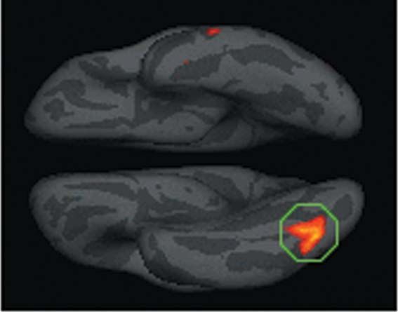
NEUROREPORT                                                                                                                                        LAZAR ETAL.

  participants were matched for sex (meditators 65% male,
                                                                                   (a)
  controls 67%), age (meditators 38.2 years old, controls 36.8
  years old), race (both groups 100% Caucasian) and years of
  education (meditators 17.3 years, controls 17.4 years). All
  participants were physically and psychologically healthy.
  Two meditation participants were left-handed; exclusion of
  the left-handed participants did not significantly alter                                                                           4
  results. All participants provided written, informed consent
  and the study was approved by the Institutional Review
  Board at the Massachusetts General Hospital.
     The present methods utilized a well-validated computa-
  tional approach to measure the thickness of the cerebral
  cortex [3,4]. Cortical thickness was estimated from two
  magnetization prepared rapid gradient echo (MPRAGE)
  structural images collected from each participant that were
  then motion-corrected and averaged together to form a
  single high-resolution image [3–5]. An initial estimate of the
  gray/white matter boundary was constructed by classifying
  all white matter voxels in a magnetic resonance imaging                          (b)
  volume using a combination of geometric and intensity-
                                                                                                                 3
  based information. A surface-deformation procedure was
  then used to obtain subvoxel resolution in the gray/white
  boundary and in the pial surface using a combination of
  smoothness constraints and intensity terms. The resulting
  cortical surface models for all participants were aligned to
  an atlas of cortical folding patterns using a high-dimen-                                                                                 1            2
  sional nonlinear registration technique.

  Results
  The mean thickness across the entire cortex did not differ
  significantly between the groups for either hemisphere
  (P40.10), indicating that it was not the case that the cortex
  of meditators is nonspecifically thicker everywhere. Statis-
  tical thickness-difference maps constructed using the
  Kolmogorov–Smirnoff          statistics  (one-tailed,    a-level                                                                              P
MEDITATION AND CORTICALTHICKNESS NEUROREPORT
between meditation practitioners, using the total number of ling for individual right-hemisphere mean thickness (as a
years of practice is not a sensitive metric of experience. One measure of nonspecific effects on cortical thickness), partial
effect of regular meditation practice is a significant drop in r(17)¼0.73, Po0.001, and still further when controlling for
respiration rate during formal practice [7,8]. We therefore age, partial r(16)¼0.75, Po0.001. When controlling for age
tested whether changes in respiration rate between rest and and individual right-hemisphere average thickness, a partial
meditation could serve as an objective measure of medita- correlation between thickness in this region and years of
tion experience. The change in mean respiration rate from a experience remained significant [partial r(15)¼0.627,
6-min baseline period to the first 6 min of the meditation P¼0.007]. These findings are consistent with the hypothesis
period was calculated for each participant and then that meditation practice promoted thickening in this region.
correlated with the self-reported total number of hours of The most experienced participants were also among the
formal sitting meditation over the participant’s lifetime oldest. As age-related decreases in cortical thickness are
(r¼0.75, Po0.001). The correlation between respiration greatest in frontal regions [5], it is possible that the effect of
rate and total number of years the participant had been age may obscure the modest effects of meditation practice in
practicing was also significant (r¼0.57, P¼0.009); however, these areas. The Pearson correlations between respiration
the correlation with total hours of formal sitting practice rate and cortical thickness in the insula and BA 9/10 were
resulted in a higher coefficient. not significant (r¼0.36, P¼0.12 and r¼0.23, P¼0.33,
To directly test for cumulative effects of meditation respectively), although they became so in the insula after
experience on brain structure, a correlation was performed controlling for age [partial r(17)¼0.48, P¼0.04]. The correla-
between cortical thickness and change in respiration rate. tion between these parameters for the BA 9/10 region was
After correcting for multiple comparisons using a false essentially unchanged [partial r(17)¼0.25, P¼0.30].
discovery rate associated with a P¼0.05, the analysis
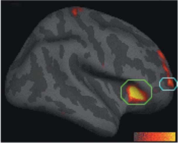
revealed one significant region within the inferior occipito-
temporal visual cortex (Fig. 2). Among the meditation Discussion
group, the zero-order correlation between thickness in this Our data indicate that regular practice of meditation is
region and change in respiration rate was r(18)¼0.72, associated with increased thickness in a subset of cortical
Po0.001, which was effectively unchanged when control- regions related to somatosensory, auditory, visual and
interoceptive processing. Further, regular meditation prac-
tice may slow age-related thinning of the frontal cortex.
(a) Previous studies of cortical plasticity in animals and
humans have shown that when a task requires that attention
be consistently directed towards a behaviorally relevant
sensory stimulus (e.g. a somatosensory [9] or auditory
stimulus [10]) over repeated practice sessions [11], robust
changes in sensory cortical maps result ([12] and Kerr CE,
Wasserman RH and Moore CI. Cortical plasticity as a
therapeutic mechanism for touch healing, under reveiw).
Additional studies suggest that relaxation facilitates the
learning-based process that underlies such cortical plasticity
[13]. It may be useful to conceptualize meditation practice as
engaging in an analogous set of cortical remodeling
processes: namely, directing attention towards behaviorally
relevant sensory stimuli within a relaxing setting over
repeated practice sessions [2,7]. Increased cortical thickness
could be due to greater arborization per neuron, increased
(b) 2.3
glial volume or increased regional vasculature. The methods
employed do not distinguish between these possibilities;
however, each of these mechanisms is supportive of
2.1 increased neural function.
Thickness (mm)
We hypothesized that meditation practice should promote
neural plasticity in regions that are routinely engaged
1.9
during formal practice. Many factors including age, sex,
genetics, neuropathology and psychopathology [4,5,14,15],
1.7 however, influence the thickness of cortex nonspecifically,
confounding these analyses. Perhaps the largest of these
confounds is the effect of age. The rate of age-dependent
1.5 thinning is highly variable across the cortical surface [5].
− 10 −5 0 5
Change in breathing rate (bpm)
Meditation-related effects on thickness may have been
counterbalanced by the effects of age on cortical thinning,
Fig. 2 Visual area correlated with meditation experience. (a) Statistical thereby minimizing our ability to detect significant correla-
map depicting cortical thickness correlated with change in respiration tions. Thinning is most pronounced in the frontal lobe, and
rate. (b) Scatter plot of mean cortical thickness of each participant from
indeed there were many regions in the parietal, temporal
the circled region within the inferior occipitotemporal lobe plotted
versus change in respiration rate. Note: negative change in breathing rate and occipital lobe where there was little if any difference in
(left side) is associated with more hours of meditation experience and a the average thickness in our older and younger participants
thicker cortex. (data not shown). Such age-related effects may account for
Vol 16 No17 28 November 2005 18 9 5
Copyright © Lippincott Williams & Wilkins. Unauthorized reproduction of this article is prohibited.NEUROREPORT LAZAR ETAL.
the fact that the strongest correlation with experience was Insight meditation. The largest between-group difference
found in the occipitotemporal region, while other regions of was in the thickness of right anterior insula. Functional
interest, which all lie in frontal regions, had only low imaging and electrophysiological studies in humans and
correlation with experience. Interestingly, despite the effects monkeys have implicated the right anterior insula in tasks
of aging on the prefrontal cortex, in one focal region of BA related to bodily attention and increased visceral awareness
9/10 the average cortical thickness of the 40–50-year-old [20,21]. Structural measures of gray matter volume of the
meditation participants was similar to the average thickness right anterior insula predict accuracy of objective measures
of the 20–30-year-old meditators and controls, suggesting of interoceptive performance, as well as subjective ratings of
that regular practice of meditation may slow the rate of global visceral awareness [21]. The differential thickness
neural degeneration at this specific locus. Future long- between groups in this region is consistent with increased
itudinal studies will be required to verify this finding. capacity for awareness of internal states by meditators,
Another factor possibly confounding our ability to detect particularly awareness of breathing sensations. Right BA
correlations between thickness and experience is hetero- 9/10 has been shown to be involved in the integration of
geneity in the specific mental exercises that Insight practi- emotion and cognition [22]. It has been hypothesized that by
tioners engage in over time. Beginners are taught to becoming increasingly more aware of sensory stimuli
maintain focused awareness on interoceptive stimuli and during formal practice, the meditation practitioner is
then are gradually taught to expand their awareness to gradually able to use this self-awareness to more success-
focus on thoughts, emotions and external stimuli fully navigate through potentially stressful encounters that
such as sounds, although there is no prescribed schedule arise throughout the day [2,23]. This eastern philosophy of
or order in which these practices are taught. Correspond- emotion dovetails with Damasio’s theory that connections
ingly, the insula, an area associated with the interoceptive between sensory cortices and emotion cortices play a crucial
processes and breath awareness techniques common to role in processing of emotionally salient material and
beginning and experienced meditators, had the largest and adaptive decision making [24].
most significant between-group difference, while unimodal Other forms of yoga and meditation will likely have a
sensory areas, which may be associated with more similar impact on cortical structure, although each tradition
advanced and heterogeneous practices, had less significant would be expected to have a slightly different pattern of
differences. cortical thickening based on the specific mental exercises
As a result of the cross-sectional nature of the study, the involved [7,8,25]. Although numerous studies have
findings are necessarily correlational, and a causal relation- shown that indices of cortical size can decrease as a result
ship between cortical thickness and meditation cannot be of aging and pathology (e.g. [4,5]), there are limited data
inferred. For example, it is possible that people with thicker indicating mechanisms that promote cortical thickening
sensory cortex are for some reason drawn to meditation. [16–18]. Our findings suggest that cortical plasticity can
Several factors, however, suggest that these findings relate occur, in adults, in areas important for cognitive and
to the meditative practice itself. First, although there were emotional processing.
significant ‘regional’ differences in thickness between
groups, there was no between-group difference in ‘global’
mean cortical thickness, indicating that these findings are Conclusion
unlikely to be due to spurious between-group differences Our initial results suggest that meditation may be associated
that might impact cortical structure nonspecifically. Second, with structural changes in areas of the brain that are
the regions of cortical thickening correspond well to the important for sensory, cognitive and emotional processing.
specific activities that practitioners of Insight repeatedly The data further suggest that meditation may impact age-
engage in over time – paying attention to breathing related declines in cortical structure.
sensations and sensory stimuli. It is unlikely that nonspe-
cific lifestyle effects such as diet would be associated with
the specific pattern of differences found. The most plausible
Acknowledgements
explanation for the specific pattern observed is experience-
We thank R. Gollub, D. Salat, M. Bar, G. Kuperberg and
dependent cortical plasticity.
S. Stufflebeam for helpful discussions. We also thank
Finally, both years of practice and change in respiration
I. Rosman for technical assistance, J. Zaki for manuscript
rate (a physiological measure of cumulative meditation
editing, and D. Salat and D. Rosas for access to data.
experience) were correlated with cortical thickness in two
regions, the inferior occipitotemporal visual cortex and right
anterior insula. These findings are consistent with other
cross-sectional reports of experience-dependent differences References
in neural volume [16,17]. In addition, a longitudinal study 1. Lutz A, Greischar LL, Rawlings NB, Ricard M, Davidson RJ. Long-term
meditators self-induce high-amplitude gamma synchrony during mental
[18] has demonstrated that learning to juggle is associated
practice. Proc Natl Acad Sci USA 2004; 101:16369–16373.
with increases in visual motion cortical areas. Our finding of 2. Goldstein J, Kornfield J. Seeking the heart of wisdom: The path of Insight
a correlation between the thickness in two regions and Meditation. Boston: Shambhala Publications; 1987.
amount of experience lends support to the hypothesis that 3. Fischl B, Dale AM. Measuring the thickness of the human cerebral cortex
the observed differences are acquired through extensive from magnetic resonance images. Proc Natl Acad Sci USA 2000; 97:
practice of meditation, and are not simply due to pre- 11050–11055.
4. Rosas HD, Liu AK, Hersch S, Glessner M, Ferrante RJ, Salat DH, et al.
existing or incidental between-group differences.
Regional and progressive thinning of the cortical ribbon in Huntington’s
Most of the regions identified in this study were found in disease. Neurology 2002; 58:695–701.
the right hemisphere. The right hemisphere is essential for 5. Salat DH, Buckner RL, Snyder AZ, Greve DN, Desikan RSR, Busa E et al.
sustaining attention [19], which is a central practice of Thinning of the cerebral cortex in aging. Cereb Cortex 2004; 14:721–730.
18 9 6 Vol 16 No 17 28 November 2005
Copyright © Lippincott Williams & Wilkins. Unauthorized reproduction of this article is prohibited.MEDITATION AND CORTICALTHICKNESS NEUROREPORT
6. Genovese CR, Lazar NA, Nichols TE. Thresholding of statistical maps in 16. Maguire EA, Gadian DG, Johnsrude IS, Good CD, Ashburner J,
functional neuroimaging using the false discovery rate. Neuroimage 2002; Frackowiak RSJ, Frith CD. Navigation-related structural change in the
15:870–878. hippocampi of taxi drivers. Proc Natl Acad Sci USA 2000; 97:4398–4403.
7. Wallace RK, Benson H, Wilson AF. A wakeful hypometabolic 17. Mechelli A, Crinion JT, Noppeney U, O’Doherty J, Ashburner J,
physiological state. Am J Physiol 1971; 221:795–799. Frackowiak RS, Price CJ. Neurolinguistics: structural plasticity in the
8. Lehrer P, Sasaki Y, Sauti Y. Zazen and cardiac variability. Psychosom Med bilingual brain. Proficiency in a second language and age at acquisition
1999; 61:812–821. affect grey-matter density. Nature 2004; 431.
9. Recanzone GH, Merzenich MM, Jenkins WM, Grajski KA, Dinse HR. 18. Draganski B, Gaser C, Busch V, Schuierer G, Bogdahn U, May A. Changes
Topographic reorganization of the hand representation in cortical area 3b in grey matter induced by training. Nature 2004; 427:311–312.
owl monkeys trained in a frequency-discrimination task. J Neurophys 19. Posner MI, Peterson SE. The attention system of the human brain. Annu
1992; 67:1031–1056. Rev Neurosci 1990; 13:25–42.
10. Bao S, Chang EF, Woods J, Merzenich MM. Temporal plasticity in the 20. Craig AD. Interoception: the sense of the physiological condition of the
primary auditory cortex induced by operant perceptual learning. Nat body. Curr Opin Neurobiol 2003; 13:500–505.
Neurosci 2004; 7:974–981. 21. Critchley HG, Wiens S, Rotshtein P, Ohman A, Dolan RJ. Neural systems
11. Bangert M, Altenmuller EO. Mapping perception to action in piano supporting interoceptive awareness. Nat Neurosci 2004; 7:189–195.
practice: a longitudinal DC-EEG study. BMC Neurosci 2003; 4:26. 22. Gray JR, Braver TS, Raichle ME. Integration of emotion and cognition in
12. Merzenich MM, DeCharms RC. Neural representations, experience and the lateral prefrontal cortex. Proc Natl Acad Sci USA 2002; 99:
change. Boston: MIT Press; 1996. 4115–4120.
13. Gottselig JM, Hofer-Tinguely G, Borbely AA, Regel SJ, Landolt HP, Retey 23. Segal ZV, Williams JMG, Teasdale JD. Mindfulness-based cognitive therapy
JV, Achermann P. Sleep and rest facilitate auditory learning. Neurosci for depression: a new approach to preventing relapse. New York: Guilford
2004; 127:557–561. Press; 2002.
14. Kuperberg GR, Broome MR, McGuire PK, David AS, Eddy M, Ozawa F, 24. Damasio AR. The somatic marker hypothesis and the possible functions
et al. Regionally localized thinning of the cerebral cortex in schizophrenia. of the prefrontal cortex. Philos Trans R Soc Lond B Biol Sci 1996; 351:
Arch Gen Psychiatry 2003; 60:878–888. 1413–1420.
15. Rauch SL, Wright CI, Martis B, Busa E, McMullin KG, Shin LM, et al. 25. Lazar SW, Bush G, Gollub RL, Fricchione GL, Khalsa G, Benson H.
A magnetic resonance imaging study of cortical thickness in animal Functional brain mapping of the relaxation response and meditation.
phobia. Biol Psychiatry 2004; 55:946–952. Neuroreport 2000; 11:1581–1585.
Vol 16 No17 28 November 2005 18 9 7
Copyright © Lippincott Williams & Wilkins. Unauthorized reproduction of this article is prohibited.You can also read