Neonatal Anemia and Jaundice - J.Manna Hematopathology - bcsls
←
→
Page content transcription
If your browser does not render page correctly, please read the page content below
Neonatal Anemia and
     Jaundice

                             J.Manna
                      Hematopathology
                                 2015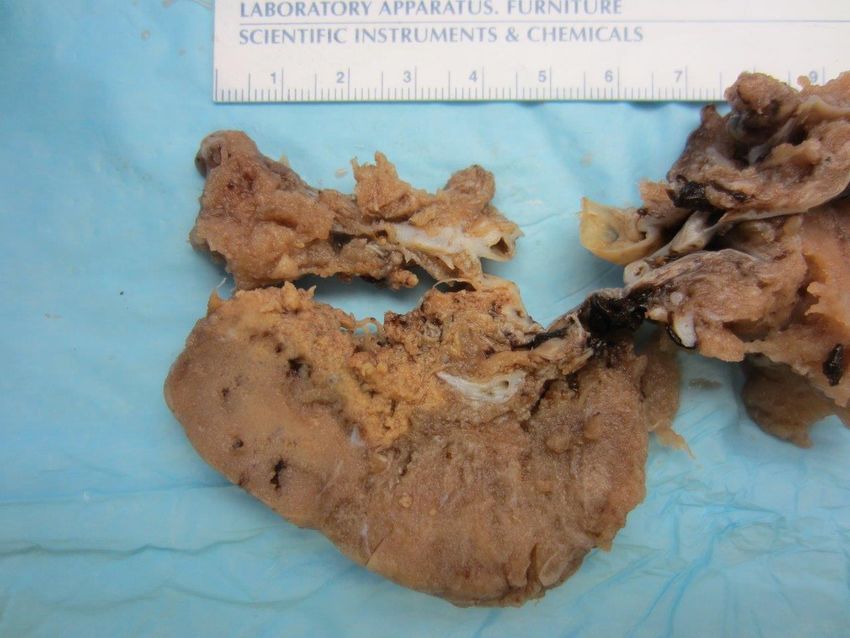
OVERVIEW

1. “Normal” neonatal hematology

2. Principles of Anemia

3. Principles of Jaundice

4. Tests for Assessing Newborn Anemia/Jaundice

5. Unusual cases from 2015
“Normal” Neonatal Hematology

- Newborn =       Small Adult

- Normal range for Hb, MCV and neutrophils different

- At birth, mostly Hb F (2 alpha and 2 gamma globins)

- By 6 months of age, there adult levels of Hb A1
       (2alpha and 2 beta globins)

     Terminology: Neonate = up to 4 weeks of age.
Normal Neonatal Peripheral Smear

-   Mild anisopoikilocytosis
-   +/- Howell-Jolly bodies
-   A few nRBCs for about first 5 days post partum
Blood Loss:

         Hemolysis =
         Decrease RBC lifespan:

Anemia

         Decrease Production:

         Physiologic Anemia:      - at about 2 months of age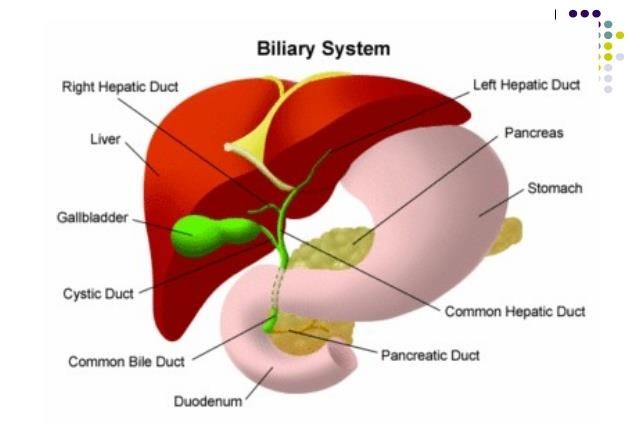
Blood Loss

Common
Causes of
Anemia
At birth
            Hemolysis =
            Decrease RBC lifespan: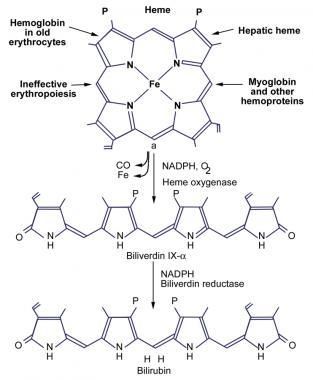
Blood Loss: - Fetal-Maternal Hemorrhage (FMH)
- Twin – Twin transfusion
Hemolysis =
Decrease RBC lifespan: -Immune -Alloiommune
eg. HDN
-Hereditary -Hemoglobinopathy
-Membranopathy
Anemia -Enzymopathy
At birth
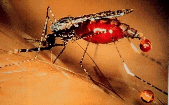
-Infection -Direct eg. malaria
-Indirect eg. DIC, toxin
Decrease Production: - Nutritional -eg. maternal B12
deficiency
- Marrow problem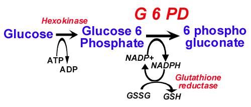
ABOUT JAUNDICE…

Definition = -   Excess bilirubin which manifests as ‘yellow’ skin and/or eyes.

Physiology = -   RBC ‘born’ in marrow, lives 120 days, and ‘dies’ in spleen

             A. Bilirubin formed as a byproduct of RBC breakdown in spleen
             B. Liver enzymes conjugate bilirubin so it is H2O soluble
             C. And can be excreted via gallbladder/biliary tracts into GI system
 A.

                                         B.

                                                                         C.
ABOUT JAUNDICE…
Definition = - Excess bilirubin which manifests as ‘yellow’ skin and/or eyes.
Physiology = - RBC ‘born’ in marrow, lives 120 days, and ‘dies’ in spleen
A. Bilirubin formed as a byproduct of RBC breakdown
B. Liver enzymes conjugate bilirubin so it is H2O soluble
C. And can be excreted via gallbladder/biliary tracts into GI system
Causes of Neonatal Jaundice =
- Physiologic
vs.
- Pathologic Excess Unconjugated Bilirubin
Excess Conjugated BilirubinPhysiologic Jaundice : occurs > 24 hrs post partum
Neonatal
JAUNDICE
Pathologic Jaundice: for sure pathologic if < 24 post partum
Unconjugated bilirubin KERNICTERUS
- Hemolysis
- Crigler-Najjar syndrome – severe UDPGT defect
(- Gilbert’s syndrome – mild UDPGT defect)
Conjugated bilirubin:
- Biliary tract: - atresia, cysts, stones, CA
- Intrahepatic: - hepatitis
- hereditary (alpha 1 tryptase
deficiency, trisomy 18, metabolic, etc)OVERVIEW 1. “Normal” neonatal hematology 2. Principles of Anemia 3. Principles of Jaundice 4. Tests for Assessing Newborn Anemia/Jaundice 5. Unusual cases from 2015
LAB TESTS

           Tests to help answer these questions:

              1. Is there Blood Loss? Usually fetal-maternal
                 hemorrhage (FMH).
ANEMIA

              2. a. Is there Hemolysis?
                 b. If hemolytic anemia (HA) is present,
                         what is the cause?
JAUNDICE
Blood Loss: - Fetal-Maternal Hemorrhage (FMH)
- Twin – Twin transfusion
Hemolysis =
Decrease RBC lifespan: -Immune -Alloiommune
eg. HDN
-Hereditary -Hemoglobinopathy
-Membranopathy
Anemia -Enzymopathy
At birth
-Infection -Direct eg. malaria
-Indirect eg. DIC, toxin
Decrease Production: - Nutritional -eg. maternal B12
deficiency
- Marrow problemLAB TESTS
∙FMH Tests - Kleihauer-Betke test
- HbF Via flow cytometry
•Hemolytic Tests - CBC
- Reticulocyte count
- Bilirubin
- (LDH)
- (Haptoglobin)
•Etiologic HA tests Immune vs. NonimmuneETIOLOGIC H.A. LAB TESTS
HDN (Immune) Testing:
- Baby’s blood = Baby’s Group, DAT, Peripheral smear (PS)
for spherocytes
- Mom blood = Group/screen
Hereditary Hemolytic Tests:
- Enzymopathies = PS, Functional, Molecular tests
- Membranopathies = PS, E5M via Flow Cytometry
- Hemoglobinopathies = PS, HPLC, H-bodies,
Hb Gel Electropheresis, Molecular
Peripheral Smear :
- Spherocytes (HDN, HS, HPP) -Bite/blister cells (G6PD, unstable Hb)
- Schistocytes (DIC, usually none) -Sputnik cells (PKD, usually not present)
-Marked anisopoikilocytosis5 Unusual 2015 Cases
1. Hyperbilirubinemia – at birth , 35 week premature boy
2. A. -Hyperbilirubinemia - 5 days post-partum , 29 weeks premature boy
B. -Acute anemia– 2 yo girl
3. Anemia in-utero – therefore Emergency C-section at 36 weeks
4. Anemia at birth - Emergency C-section at 39 weeks for
decreased fetal heart rate.Case # 1
Hyperbilirubinemia – at birth , 35 week premature boy
- Critically high unconjugated bilirubin
- Exchange transfusion requested STAT due to possible
kernicterus
Evaluating the high unconjugated bilirubin:
- occurred < 24 hrs post partum; therefore pathologic
- Hemolysis vs. inherent liver disease?
- Tests to assess possible hemolysis:
-CBC – initially normal Hb
-HDN tests normal: -Baby = O Rh +, DAT neg,
-Mom = O Rh +, screen neg.
-PSCase # 1 - “RBC Potpourri” = increased anisopoikilocytosis No sepsis - Increased nRBCs and polychromasia No hypoxia
Case # 1

Likely hemolysis, but what is the cause???

      -   Not immune, ie not HDN

      -   Hereditary?
Case # 1 - Hemoglobinopathies: HPLC and H-body = normal/neg - Membranopathies: E5M via flow cytometry = normal - Enzymopathies: G6PD normal - Parents’ CBC and PS = unremarkable - No family history of hemolysis/gallstones - First Nations, siblings healthy -- 4 boys and 1 girl
Case # 1

Hyperbilirubinemia – at birth , 35 week premature boy

        - Critically high unconjugated bilirubin
        - Double exchange transfusion due to possible
           kernicterus

        -Hb continued drifting downwards post exchange
        -required pRBCs at about 2 weeks of age and
           subsequently.

        -Turned to ‘bird seed’ tests on pre-exchange sample:

                - Macleod’s phenotype: not present
                - Macleod’s phenotype =
                  lack of RBC Ag ‘K’ precursor
                  resulting in acanthocytosis and
                  neurological problems
Case # 1
Hyperbilirubinemia – at birth , 35 week premature boy
- Critically high unconjugated bilirubin
- Double exchange transfusion due to possible
kernicterus
- Requiring regular simple transfusions
- Further Tests? Little pre-transfusion sample,
not fresh, ‘bird seed rare tests’ are send outs…
- Molecular studies for enzyme mutations – not
affected by transfusions.
Diagnosis?
Pyruvate Kinase DeficiencyAbout PKD…Understanding the Biochemistry
90 % Glycolysis
Glucose
10 % HMPS
(Hexose monophosphate shunt)Glycolysis----AHHHHHHHH!
About PKD…
  90%               Glucose
  Glycolysis
                                          10%
                    G6P
                                          HMPS   G6PD

2,3-DPG                       2 NADH

                              Net 2 ATP

                    Pep
      PK---------
                    Pyr
      LDH-------
                    Lactate
About Pyruvate Kinase Deficiency. . .
Definition: -Most common glycolytic enz. deficiency
-Most common cause of infant chronic
hemolysis
Epidemiology: - 51 per million in Caucasians
- more prevalent in Europe & Japan
- Autosomal Recessive, males = females
2 Genes Encode 4 Isozymes:
R-type = RBC
L-type = Liver
M1 = Muscle, Brain
M2 = WBCs, all fetal tissueEtiology / Pathology of PKD
Point Mutation, AR
Pyruvate Kinase Deficiency
Chronic HA= HNSHA
MECHANISM OF HEMOLYSIS??
Cell Lysis
PKD ATP Na/K Pump
Increased Volume
Splenic Sequestration / DestructionPKD Lab Tests
1. Peripheral Smear:
-No characteristic morphology - although
‘Sputnik cells’ described but often
not present.
2. Enzyme Function Tests:
-Depletion of NADH
3. Molecular Testing:
-More complex than G6PD due to 2 genes
encoding 4 isozymesEnzyme Function Tests
PEP + ADP PK Pyruvate + ATP
Pyruvate + NADH + H LDH Lactate + NAD
Measures consumption of added NADH
1. Screening Test = UV Fluorescence of NADH
2. Diagnostic Test = Spectrophotometrically measure
light absorption of NADH @ 340nmWhat are 3 reasons for false-negative results
from a PKD screen??
1. Increased reticulocytes
2. Leukocytosis
3. pRBC Transfusion5 Unusual 2015 Cases
1. Hyperbilirubinemia – at birth , 35 week premature boy
2. A. -Hyperbilirubinemia - 5 days post-partum , 29 weeks premature boy
B. -Acute anemia– 2 yo girl
3. Anemia in-utero – therefore Emergency C-section at 36 weeks
4. Anemia at birth - Emergency C-section at 39 weeks for
decreased fetal heart rate.Case #2a
-Hyperbilirubinemia - 5 days post-partum , 29 weeks premature boy
- Critically high unconjugated bilirubin (~ 360)
- Phototherapy helped
Evaluating the high unconjugated bilirubin:
- occurred > 24 hrs post partum; therefore
physiologic or pathologic
- Bili quite high for usual physiologic jaundice – assess for hemolysis:
-CBC – normal Hb at birth, acute anemia at 12 days of age
-HDN tests normal: -Baby = A Rh +, DAT neg,
-Mom = A Rh +, screen neg.
-PS at 5 days old = unremarkable
-PS at 12 days old =Case # 2a
Bite Cell
Bite Cell
Blister CellCase #2a
-Hyperbilirubinemia - 5 days post-partum , 29 weeks premature boy
- Critically high unconjugated bilirubin (~ 360)
- Phototherapy helped
- Bite/blister cells
- East Indian ethnicity
- No evidence of membranopathy or hemoglobinopathy
- Enzyme function tests completed since there was no transfusion
Diagnosis?
G6PD DeficiencyAbout G6PD…
90% Glucose
Glycolysis
10%
G6P
HMPS G6PD
2,3-DPG 2 NADH
Net 2 ATP
Pep
PK---------
Pyr
LDH-------
LactateThe Hexose Monophosphate Shunt
G6PD acts as a “sweeper” of damaging oxidative radicals
Glycolysis Ribose-5-PAbout G6PD Deficiency. . .
Definition: -Most common enzyme deficiency
-Usually asymptomatic to mild
-Usually triggered episodic hemolysis
Prevalence:
Types:About G6PD Deficiency. . .
Definition: -Most common enzyme deficiency
-Usually asymptomatic to mild
-Usually triggered episodic hemolysis
Epidemiology: - mutation is relatively common
- ~ 400 million affected worldwide
- Affects 1/10 Africans, also common
in Asia and the Mediterranean
- X-linked recessive, Males > Females
Types:Selective Advantage to G6PDD? Resistance to Malaria
What are other disorders that may confer resistance
                   to malaria?

        - Hemoglobinopathies---Sickle Cell Trait
        - Thalassemias
        - Membranopathies---SAO
        - No Duffy a/b antigen on RBC
About G6PD Deficiency. . .
Definition: -Most common enzyme deficiency
-Usually asymptomatic to mild
-Usually triggered episodic hemolysis
Prevalence: - mutation is relatively common
- ~ 400 million affected worldwide
- Affects 1/10 Africans, also common
in Asia and the Mediterranean
- X-linked recessive, Males > Females
Types: Normal B G6PD
A + G6PD = 20 % Africans
Abnormal A- = 11% Africans
Mediterranean = Severe
Canton = SevereEtiology / Pathology / Clinical Aspects of G6PDD
Point Mutation, X-Linked recessive
G6PD due to decreased stability
RBC susceptibility to oxidative damage
Neonatal Jaundice Precipitated HA Chronic HA = HNSHA
-Minimal to no hemolysis - Drugs - Very Rare
- Infection
-Due to combined: - Food
G6PD + immature liver
- Diabetic ketoacidosis
enzymesPrecipitated HA in G6PDD
- Drugs: primaquine, dapsone, sulfa, nitrofurantoin, methylene blue
- Infection: pneumonia, typhoid, rocky mountain spotted fever
- Food: fava beans, unripe peach, red suya
- Diabetic Ketoacidosis: ??
Increased Oxidants + Decreased Ability to “mop”
Damage to:
1. Membrane Bite / Blister cells I/V + E/V HA
2. Hb Heinz Bodies E/V HAG6PDD Lab Tests
1. Peripheral Smear:
-Bite/Blister cells
(2. Supravital Stain: Heinz bodies)
3. Enzyme Function:
-Detect NADPH formation
-Screening and Diagnostic Function Tests
4. MolecularEnzyme Function Tests
G6PD
G6P + NADP 6-PG + NADPH + H
GR
NADPH + GSSG NADP + GSH
Measures production (+/- consumption) of NADPH
1. Screening Test = UV fluorescence of NADPH
2. Diagnostic Test = Spectrophotometrically measure
light absorption of NADPH @ 340 nmG6PD UV Fluorescent Screening Test
What are 3 reasons for false-negative results
                from a G6PD screen?

1. Increased reticulocytes --- contain normal levels of
   G6PD in G6PD A – variant

2. pRBC Transfusion

3. Heterozygote woman – ie. a ‘carrier’ of the G6PD
                              mutation
5 Unusual 2015 Cases
1. Hyperbilirubinemia – at birth , 35 week premature boy
2. A. -Hyperbilirubinemia - 5 days post-partum , 29 weeks premature boy
B. -Acute anemia– 2 yo girl
3. Anemia in-utero – therefore Emergency C-section at 36 weeks
4. Anemia at birth - Emergency C-section at 39 weeks for
decreased fetal heart rate.Case #2b
-Acute anemia in a 2 yo African girl
- in ER for infection/fever
- Hb = 80
- Anemia resolved on its own
Evaluating the acute anemia:
- Common pediatric causes = nutritional deficiencies and infection
- Uncommon pediatric causes = non-traumatic blood loss, hemolysisCase #2b
-Acute anemia in a 2 yo African girl
- in ER for infection/fever
- Hb = 80
- Anemia resolved on its own
Evaluating the acute anemia:
- Common pediatric causes = nutritional deficiencies and infection
- Uncommon pediatric causes = non-traumatic blood loss, hemolysis
- Blood loss, nutritional deficiencies and malaria excluded
- CBC = normocytic anemia, mild neutrophilia and monocytosis
- Retic count = normal
- PS = possible spherocytes
- Hemolysis? -- RBC changes, increased LDH but normal bili
- No membranopathy
- one deletion alpha thalassemia detected – red herring?
- Enzyme function studies confirmed……..Case #2b
-Acute anemia in a 2 yo African girl
- in ER for infection/fever
- Hb = 80
- Anemia resolved on its own
G6PD deficiencyCase #2b
-Because G6PDD is X-linked, usually females are asymptomatic carriers
-How can females become symptomatic as in this case?
1. Turners syndrome, XO
2. Homozygous mutation
3. Unfavourable Lyonization: ie. more of the normal, unmutated
X chromosomes are inactivated at the embryonic stage
4. Uniparental disomy5 Unusual 2015 Cases
1. Hyperbilirubinemia – at birth , 35 week premature boy
2. A. -Hyperbilirubinemia - 5 days post-partum , 29 weeks premature boy
B. -Acute anemia– 2 yo girl
3. Anemia in-utero – therefore Emergency C-section at 36 weeks
4. Anemia at birth - Emergency C-section at 39 weeks for
decreased fetal heart rate.Case # 3
Anemia in-utero – therefore Emergency C-section at 36 weeks
- at birth, Hb critically low at 96
Evaluating the Anemia:
- Common causes in neonate = blood loss and hemolysis
- Kleihaur-Betke test normal = blood loss unlikely
- Hemolysis? Initial tests = CBC, bilirubin, PSCase # 3
Case # 3

Anemia in-utero – therefore Emergency C-section at 36 weeks

                 - at birth, Hb critically low at 96

Evaluating the Anemia:

        - Common causes in neonate = blood loss and hemolysis

        - Kleihaur-Betke test normal = blood loss unlikely

        - Hemolysis?      -Hb = 96, increasing bilirubin
                          -PS = spherocytes + increased polychromasia
                                   & nRBCs for age

        - Most common cause of neonatal hemolysis?
Case # 3
Anemia in-utero – therefore Emergency C-section at 36 weeks
- at birth, Hb critically low at 96
Evaluating the Anemia:
- Hemolysis? -Hb = 96, increasing bilirubin
-PS = spherocytes + increased polychromasia
& nRBCs for age
-Most common cause of neonatal hemolysis?
Immune Hemolysis = HDN (hemolytic disease of the newborn)Case # 3
Anemia in-utero – therefore Emergency C-section at 36 weeks
- at birth, Hb critically low at 96
Evaluating the Anemia:
- Hemolysis? -Hb = 96, increasing bilirubin
-PS = spherocytes + increased polychromasia
& nRBCs for age
- HDN tests:
- Baby = O Rh pos, DAT is strongly positive, PS = see above
- Mom = B Rh neg, screen (IDAT) shows
allo-anti - D and anti-E present.
- Treatment = IVIG + exchange transfusionCase # 3
Anemia in-utero – therefore Emergency C-section at 36 weeks
- at birth, Hb critically low at 96
Evaluating the Anemia:
- HDN tests:
- Baby = O Rh pos, DAT is strongly positive, PS = spherocytes + nRBCs/poly
- Mom = B Rh neg, screen (IDAT) shows
allo-anti - D and anti-E present.
- Mom is G2P1, had RhIg in previous pregnancy
- This pregnancy ------ - Allo- anti-D and anti-E detected
- Anti-D and anti-E titers monitored
- Monitored baby via doppler US of MCA to
assess for anemia
-Paternal tests showed D+ and E+ phenotypeAbout HDN:
Etiology & Pathophysiology
Fetal Ag pos RBC into maternal blood
Ag neg Mom produces alloantibody
+/- Amnestic response upon SUBSEQUENT pregnancy
IgG alloantibody crosses placenta & binds to Ag
Extravascular hemolysis results in fetusAbout HDN:
Epidemiology
• Mom = RBC antigen negative
• Fetus = RBC antigen positive
• Most common = ABO, IgG ---- 1 / 150
• Most potent = Rh (anti-D) ---- 1 / 1000
• Other less common
But potentially significant =
- Kell, Kidd, Duffy, C,c,E,e…About HDN:
ABO vs Rh HDN
ABO Rh
-Most common: 1/150 -Less common: 1/1000
-Asymptomatic to mild -Symptomatic
-3 to 5 dys post-partum -In-utero or ex-utero
-Not amnestic -AmnesticAbout HDN:
ABO vs Rh HDN
-Prevention of Rh HDN = Rh Ig given to Rh neg mom
-HDN better understood in the 50’s
-RhIg became more routine in the 70’s
-RhIg significantly decreased Rh HDN in the 80’s and afterAbout HDN:
ABO vs Rh HDN
-ABO HDN = typically asymptomatic to mild due to:
1. to antigen not fully expressed at birth
2. Antibody neutralized by A & B Ag in fluid and tissues
-ABO HDN can be severe in Africans, Asians, Arabians, Hispanics:
-In 2014, African-Canadian newborn with severe anti-B
HDN
-exchange transfusion and IVIG
-Rare case studies describing severe anti-B HDN
specifically in the African populationHDN Testing:
- If “HDN testing” or DAT requested, then the following tests are done together:
-Baby’s blood = Group, DAT, peripheral smear
-Mom’s blood = Group and Screen
- If mom has known allo-antibodies, then CBS results summarized:
- Mom’s group, phenotype
- Antibody titers
- Paternal phenotype5 Unusual 2015 Cases
1. Hyperbilirubinemia – at birth , 35 week premature boy
2. A. -Hyperbilirubinemia - 5 days post-partum , 29 weeks premature boy
B. -Acute anemia– 2 yo girl
3. Anemia in-utero – therefore Emergency C-section at 36 weeks
4. Anemia at birth - Emergency C-section at 39 weeks for
decreased fetal heart rate.Case # 4
Anemia at birth - Emergency C-section at 39 weeks for decreased fetal heart rate.
-At birth, Hb markedly decreased at 40!
Evaluating the Anemia:
- Common causes in neonate = blood loss and hemolysis
- Kleihaur-Betke test normal, flow cytometry of HbF was normal
- Hemolysis? - Bilirubin not increased!
- Retic count increased
- Peripheral smearCase # 3:
HDN
Case # 4Case # 4
Anemia at birth - Emergency C-section at 39 weeks for decreased fetal heart rate.
-At birth, Hb markedly decreased at 40!
-Baby also had - cardiomegaly
- hepatomegaly
- absent spleen
- no overt hydrops fetalis (swelling)
- normal 21 week ultrasound
-Mom = - 42 yo
- Normal pregnancy
- one child, previous normal
pregnancy/deliveryWhen things don’t make sense…
- Clerical error? --- No
- Atypical presentation of a common disorder (Anemia)?
-Eg. atypical hemolysis
- Very rare disorder?Case # 4
Anemia at birth - Emergency C-section at 39 weeks for decreased fetal heart rate.
-At birth, Hb markedly decreased at 40 and erythroblastosis!
-Cardiomegaly, hepatomegaly, absent spleen
Evaluating the Anemia:
- Common causes in neonate = blood loss and hemolysis
- Kleihaur-Betke test normal, flow cytometry of HbF was normal
- Hemolysis? -bilirubin not increased!
-PS = increased polychromasia & nRBCs for age
- Etiologic tests for hemolysis:
- not HDN, baby = A pos, DAT neg, mom = A pos, screen neg
-No H-bodies, therefore alpha thalassemia major very unlikely
- Excluded hemoglobinopathies, membranopathies,
& enzymopathiesWhen things don’t make sense…
- Clerical error? --- No
- Atypical presentation of a common disorder (Anemia)?
- Eg. atypical hemolysis --- No
- Eg. Blood loss but not into maternal circulation?
- Very rare disorder?
--- A syndrome including marrow pathology?
-- how would erythroblastosis still occur
in this setting?
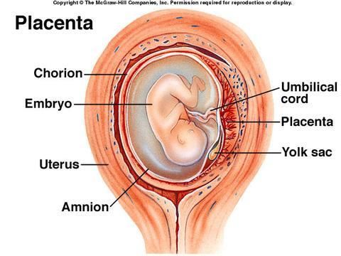
- Need placental examination
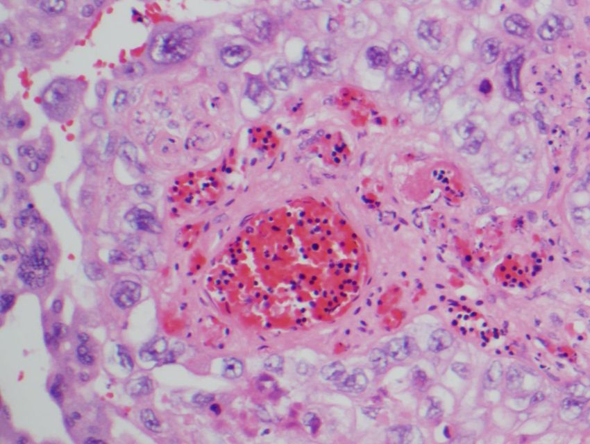
- Bone marrow biopsyCase # 4 - Autopsy declined (baby tragically died) - Placental examination showed …
Case # 4

B

A

    Gross Placental Examination
Case # 4
Diagnosis = Fetal blood loss into intraplacental choriocarcinoma
Common cause of anemia secondary to an extremely rare diseaseAbout Intraplacental
Choriocarcinoma
Definition = extremely rare cancer of the placental, chorionic cellsAbout Intraplacental
Choriocarcinoma
Epidemiology: -Very rare – 0.04% of all gestational trophoblastic diseases (GTDs).
-Most common GTD = hydatidiform mole
Risks: -Occurs in older (> 35 yo) or very young women
-No increased risk of subsequent pregnancies developing this cancer
Pathophysiology
& Clinical: -Aggressive cancer that clinically manifests in the third trimester, at
birth or just after birth.
-Marked fetal/neonatal anemia due to blood loss into placental
malignancy.
Diagnosis: -Placental examination – subtle cancer, so can be missed.
-Clinical information crucial.About Intraplacental
Choriocarcinoma
Management: -Usually emergency C-section due to fetal distress
-Supportive therapy for baby, eg. pRBCs
-Assess for metastasis in baby and mom: hCG levels
and depending on hCG, radiologic scanning may be required.
- If metastasis is present, then chemotherapy is initiated.
Case #4: -Baby unfortunately died
(from heart failure secondary to the severe anemia)
-Mother’s hCG normalized, no metastasis therefore no
chemotherapy was requiredAcknowledgments
BCCH, Hematopathology Department
-Thank you to the hematopathologists and lab technologists
re: discussions and conducting G6PD diagnostic testing.
Hamilton Laboratory Staff
-Thanks for conducting diagnostic molecular PK testing
Victoria, Anatomic Pathology Department
-Thank you to Dr. N. Van der Westhuizen and lab technologists
for discussions, articles, images and diagnosing intraplacental choriocarcinoma
Victoria, Hematopathology Department
-Thank you to special hematology, core lab and transfusion
medicine for their collaborationReferences 1. Alarcon, Pedro de & Werner, Eric. Neonatal Hematology. 2005. Cambridge University Press. 2. Beutler, Ernest. G6PD Deficiency. Blood 1994 84:3613-3636. 3. Drabik-Clary, K. et al, Severe hemolytic disease of the newborn in a group B African-American infant delivered by a group O mother. Ann Clin Lab Sci Spring 2006 vol.36 no.2, 205-207. 4. Henningsen, A.A. et al. Massive fetomaternal hemorrhage caused by an intraplacental choriocarcinoma: a case report. Case Reports in Medicine Volume 2010, Article ID 767218, 3 pages. 5. Jeong, J.E. et al. Fetomaternal hemorrhage caused by an intraplacental choriocarcinoma. 6. Kauchansky, K. Et al. William Hematology 8th edition. McGraw-Hill Companies, Inc. 2010. 7. Murray, N.A .& Roberts, I.A.G., Haemolytic disease of the newborn. Arch Dis Child Fetal Neonatal Ed. 2007 March; 92 (2): F83- F88. 8. Zanella, A. et al. Red cell pyruvate kinase deficiency: molecular and clinical aspects. British Journal of Haematology, 2005, 130, 11-25.
You can also read