Neurovascular evidence for a co-occurrence of teeth and baleen in an Oligocene mysticete and the transition to filter-feeding in baleen whales ...
←
→
Page content transcription
If your browser does not render page correctly, please read the page content below
Zoological Journal of the Linnean Society, 2021, XX, 1–21. With 5 figures.

Neurovascular evidence for a co-occurrence of teeth and
baleen in an Oligocene mysticete and the transition to

                                                                                                                                   Downloaded from https://academic.oup.com/zoolinnean/advance-article/doi/10.1093/zoolinnean/zlab017/6278618 by guest on 10 October 2021
filter-feeding in baleen whales
ERIC G. EKDALE1,2,* and THOMAS A. DEMÉRÉ2
Department of Biology, San Diego State University, 5500 Campanile Drive, San Diego, CA 92182 USA
1

Department of Paleontology, San Diego Natural History Museum, 1788 El Prado, San Diego, CA, 92101,
2

USA

Received 29 October 2020; revised 11 February 2021; accepted for publication 5 March 2021

Extant baleen whales (Mysticeti) have a deciduous foetal dentition, but are edentulous at birth. Fossils reveal that
the earliest mysticetes possessed an adult dentition. Aetiocetids, a diverse clade of Oligocene toothed mysticetes,
have a series of small palatal foramina and associated sulci medial to the postcanine dentition. The openings
have been homologized with lateral palatal foramina that transmit neurovascular structures to baleen in extant
mysticetes, thereby implying a co-occurrence of teeth and baleen in aetiocetids. However, homology of the foramina
and sulci have been questioned. Using CT-imaging, we report that the lateral palatal foramina of Aetiocetus weltoni
are connected internally to the superior alveolar canal, which transmits neurovascular structures to baleen in extant
mysticetes and to teeth in extant odontocetes. Furthermore, the lateral palatal foramina of Aetiocetus are separate
from the more medially positioned canals for the greater palatine arterial system. These results provide critical
evidence to support the hypothesis that the superior alveolar neurovasculature was co-opted in aetiocetids and later
diverging mysticetes to serve a new function associated with baleen. We evaluate competing hypotheses for the
transition from teeth to baleen, and explore the transition from raptorial feeding in early mysticetes to filter-feeding
in extant species.

ADDITIONAL KEYWORDS: anatomy – comparative anatomy – cranium – morphology – Oligocene – osteological
correlate – skull anatomy – soft-tissue reconstruction – vertebrate palaeontology.

                    INTRODUCTION                                    and certainly most diverse clade of Oligocene toothed
                                                                    mysticetes, is Aetiocetidae, members of which retained
Extant baleen whales (Mysticeti) are edentulous at
                                                                    a full dentition into adulthood. Besides possessing
birth, but possess a deciduous foetal dentition early
                                                                    an adult dentition, aetiocetid fossils also preserve
in development. This ontogenetic pattern reflects
                                                                    a series of palatal foramina and sulci positioned on
a toothed ancestry for mysticetes that is strongly
                                                                    the lateral portion (alveolar process) of the maxilla
supported by a rapidly improving fossil record. Extinct
                                                                    just medial to the toothrow, hereafter referred to as
latest-Eocene and Oligocene basal stem mysticetes
                                                                    lateral palatal foramina (Deméré & Berta, 2008).
retained a full complement of functional postnatal teeth
                                                                    These features are hypothesized to be homologous
(Fitzgerald, 2006; Fordyce & Marx, 2010, 2018; Geisler
                                                                    with the neomorphic bony conduits for neurovascular
et al., 2017; Lambert et al., 2017), while later diverging
                                                                    structures that nourish and innervate the baleen
Oligocene stem mysticetes displayed a variety of dental
                                                                    apparatus that characterizes extant edentulous
patterns (e.g. closely spaced, multicusped, postcanine
                                                                    mysticetes (Deméré et al., 2008). The implication of this
teeth; widely spaced, simple-cusped, postcanine
                                                                    hypothesis is a co-occurrence of teeth and some form
teeth) (Deméré et al., 2008; Boessenecker & Fordyce,
                                                                    of ‘proto-baleen’ in aetiocetids (Fig. 1; Deméré et al.,
2015a; Marx et al., 2016). Perhaps the best-known,
                                                                    2008), with an underlying prediction that the lateral
                                                                    palatal foramina in aetiocetids connect internally to
*Corresponding author. E-mail: eekdale@sdsu.edu                     the canal for the superior alveolar blood vessels and

© 2021 The Linnean Society of London, Zoological Journal of the Linnean Society, 2021, XX, 1–21                                1
This is an Open Access article distributed under the terms of the Creative Commons Attribution-NonCommercial License
(http://creativecommons.org/licenses/by-nc/4.0/), which permits non-commercial re-use, distribution, and reproduction in any
medium, provided the original work is properly cited. For commercial re-use, please contact journals.permissions@oup.com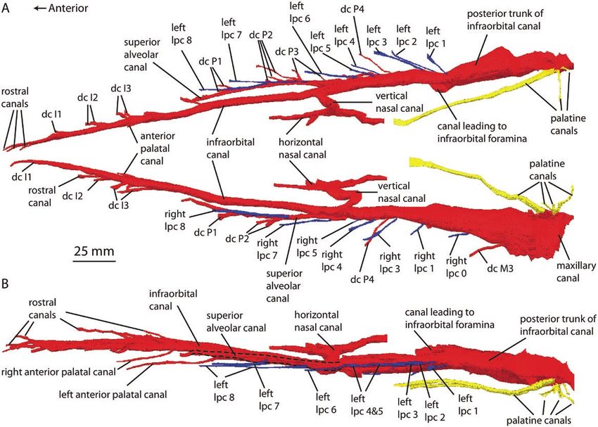
2   E.G. EKDALE and T.A. DEMÉRÉ

                                                                                                                               Downloaded from https://academic.oup.com/zoolinnean/advance-article/doi/10.1093/zoolinnean/zlab017/6278618 by guest on 10 October 2021
Figure 1. Hypothesis of baleen evolution. Relationships based on published phylogenetic analyses (Uhen, 2013; Fordyce
& Marx, 2018; Peredo et al., 2018). Thick bars represent stratigraphic ranges downloaded from the Paleobiology Database
(paleobiodb.org) on 14 February 2020, using the taxonomic name search form for each terminal taxon named on the
cladogram. Red branches indicate presence of lateral palatal foramina.

nerves, which nourish and innervate the maxillary             Fordyce & Marx, 2018). If these alternative hypotheses
dentition in odontocetes and the baleen apparatus in          were true, one might predict that the lateral palatal
extant mysticetes (Ekdale et al., 2015).                      foramina in aetiocetids would connect internally to
  However, the homology of lateral palatal foramina           the canal for palatine vessels and nerves that nourish
and sulci in aetiocetids relative to osteological             and innervate soft tissues covering the hard palate in
structures associated with baleen in living mysticetes        extant mysticetes (Ekdale et al., 2015) and terrestrial
has been questioned (Marx, 2011; Marx et al., 2016;           artiodactyls (O’Brien et al., 2016; O’Brien, 2017). We
Peredo et al., 2017, 2018). In particular, purportedly        contend that acceptance (or falsification) of any of
similar sulci occur in specimens of extinct odontocetes,      these competing hypotheses hinges on the pattern
basilosaurids (Peredo et al., 2018) and in the                of connections of the lateral palatal foramina to the
Late Eocene stem toothed mysticete Llanocetus                 internal rostral canals in aetiocetids, and requires a
denticrenatus Mitchell, 1989 (Fordyce & Marx,                 discussion of the soft-tissue correlates of those canals.
2018). However, we contend that these suggested                 As a test of these competing hypotheses, we present
similarities in other taxa are purely superficial and         new high-resolution X-ray computed tomography (CT)-
do not take into consideration the actual complexity of       based data on the internal neurovascular anatomy of
neurovascular structures of the mysticete palate (see         the holotype skull (UCMP 122900) of Aetiocetus weltoni
Discussion). Further, if the lateral palatal foramina         Barnes & Kimura in Barnes et al., 1994 that focuses on
and sulci in aetiocetids are not homologous with those        the bony canals associated with the maxillary artery
that are associated with baleen in extant mysticetes,         and nerve, and their branches. These observations
then an alternative explanation for these structures in       are then compared to neurovascular structures in the
aetiocetids is necessary. Some workers have suggested         rostra of other cetaceans using extant exemplars – a
that increased palatal vascularization is perhaps             mysticete [Eschrichtius robustus (Lilljeborg, 1861)] and
unrelated to, and preceded, the evolution of baleen,          an odontocete [Tursiops truncatus (Montagu, 1821)].
and instead might be associated with alternative              We go on to discuss the implications of our new findings
structures and functions that are unknown in                  to the larger question of the teeth to baleen transition
extant mysticetes, such as ‘thickened gums’ related           and the various competing hypotheses of mysticete
to enhanced oral suction-feeding (Marx et al., 2016;          form, function and evolution related to this question.

                             © 2021 The Linnean Society of London, Zoological Journal of the Linnean Society, 2021, XX, 1–21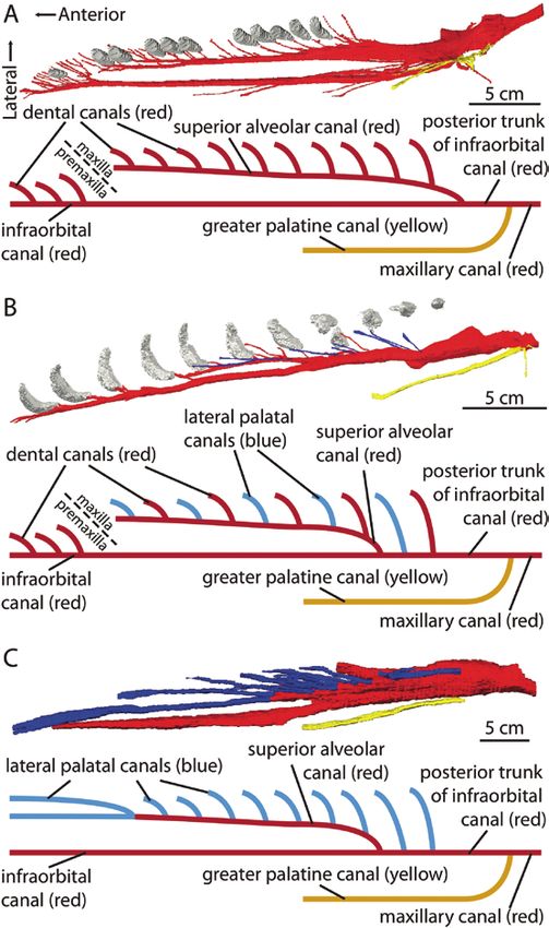
EVIDENCE FOR BALEEN IN AETIOCETUS                     3

            MATERIAL AND METHODS                                 Standring et al., 2008), we identify the medial division
                                                                 as the canal for the palatine artery and nerve, the
          Specimens and data collection
                                                                 intermediate division as the infraorbital canal for the
The skull and partial right dentary (attached to                 infraorbital artery and nerve and their subsequent
rostrum) of the holotype of Aetiocetus weltoni (UCMP             branches, and the lateral division as the canal for the
122900) were CT-scanned by YXLON International in                superior alveolar blood vessels and nerves that nourish

                                                                                                                                Downloaded from https://academic.oup.com/zoolinnean/advance-article/doi/10.1093/zoolinnean/zlab017/6278618 by guest on 10 October 2021
San Jose, CA using a Y.CT Modular cone-beam system.              and innervate the maxillary teeth (Figs 2, 3). It is
A total of 3866 high-resolution slices were acquired             important to emphasize that the anatomy described here
with a slice thickness of 0.27 mm and pixel dimensions           is of the bony canals revealed in CT images (Fig. 4) and
of 0.14 mm by 0.14 mm. In order to better visualize the          not the actual blood vessels and nerves that presumably
internal connections of the lateral palatal foramina             travelled within these canals. As noted by Ekdale et al.
and thereby test hypotheses of homology, the maxillary           (2015), in Eschrichtius, the superior alveolar artery
canal and its subsequent branches were segmented                 branches from the infraorbital artery within the bony
and isolated using Avizo Lite 9.3 (FEI Visualization             infraorbital canal posterior to the divergence of the
Sciences Group, 2017). The original CT data can be               superior alveolar canal. As such, not all branches of
accessed at the Data Dryad open access data repository           the superior alveolar artery that extend to the baleen
(https://doi.org/10.5061/dryad.05qfttf1h).                       aparatus via lateral palatal canals depart from the
   The segmented rostral canals of Aetiocetus weltoni            superior alveolar canal, but rather some of them depart
were compared with published CT data of the skull of             from the posterior trunk of the infraorbital canal. More
an extant grey whale Eschrichtius robustus (Ekdale               detailed anatomical descriptions of rostral canals, along
et al., 2015), as well as new digital segmentations              with slice-by-slice identifications of structures, are
of the rostral canals of an extant bottlenose dolphin            provided in the Supporting Information (Appendices S1,
Tursiops truncatus (SDSNH 21212; using previously                S2; Table S1).
published CT data; Colbert et al., 2005), and published             The canal for the greater palatine vessels (medial
descriptions and digital images of the surface                   division; yellow branches in Figs 2, 3) is the first
morphology of the palate of Llanocetus denticrenatus             (most posterior) to diverge from the main trunk of
Mitchell, 1989 (Fordyce & Marx, 2018) that together              the maxillary canal. Several small canals depart
serve as toothed cetacean outgroups without baleen.              ventromedially to open as minor palatine foramina
                                                                 through the palatine bone along the posterior portion
                                                                 of the palate (small yellow branches in Fig. 2). A larger
            Institutional abbreviations                          canal extends anteromedially and ventrally before
AMP, Ashoro Museum of Paleontology, Ashoro,                      opening on to the palatine process of the maxilla via
Hokkaido, Japan; ChM, Charleston Museum,                         the greater palatine foramen. Externally, an elongate
Charleston, South Carolina, USA; KMNH VP,                        sulcus continues anteriorly from the greater palatine
Kitakyushu Museum of Natural History and Human                   foramen along the surface of the maxilla, well medial
History, Kitakyushu, Fukuoka, Japan; NMP, Museum                 to the tooth row (Fig. 2A). No connections were
Victoria, Melbourne, Australia; SDSNH, San Diego                 observed between the palatine canals and either the
Society of Natural History, San Diego, California,               dental alveoli or the lateral palatal foramina.
USA; UCMP, University of California Museum of                       The maxillary canal (intermediate division) extends
Paleontology, Berkeley, California, USA; USNM,                   anteriorly as the infraorbital canal (red branch in Figs
National Museum of Natural History, Smithsonian                  2, 3; transverse slices in Fig. 4). A large branch extends
Institution, Washington, DC, USA; UWBM, University               dorsally to open as a series of dorsal infraorbital foramina
of Washington, Burke Museum of Natural History and               on the external surface of the maxilla (Fig. 2B). Several
Culture, Seattle, Washington, USA.                               smaller branches extend laterally from the infraorbital
                                                                 canal to join the alveoli of the molars and P4 (small, red
                                                                 branches labelled as dental canals in Figs 2, 3), as well
                        RESULTS                                  as to a series of lateral palatal foramina (small, blue
                                                                 branches labelled as lateral palatal canals in Figs 2, 3;
             General anatomy of canals                           transverse slices in Fig. 4). As noted above, these lateral
The high-resolution CT images of Aetiocetus weltoni              palatal canals that diverge from the posterior trunk of
reveal three primary longitudinal canals within the              the infraorbital canal presumably carried branches of
rostrum that we interpret as major ‘divisions’ (medial,          the superior alveolar artery. The observed number of
intermediate and lateral) of the canal for the maxillary         lateral palatal canals branching from the infraorbital
branch of the external carotid artery. Given the positions       canal differs between the right and left sides of the
and architecture of these canals compared to the rostral         rostrum, as there are five lateral palatal canals and
canals of extant mammals (Davis, 1964; Evans, 1993;              foramina on the right and six on the left. However, the

© 2021 The Linnean Society of London, Zoological Journal of the Linnean Society, 2021, XX, 1–21
4 E.G. EKDALE and T.A. DEMÉRÉ
Downloaded from https://academic.oup.com/zoolinnean/advance-article/doi/10.1093/zoolinnean/zlab017/6278618 by guest on 10 October 2021
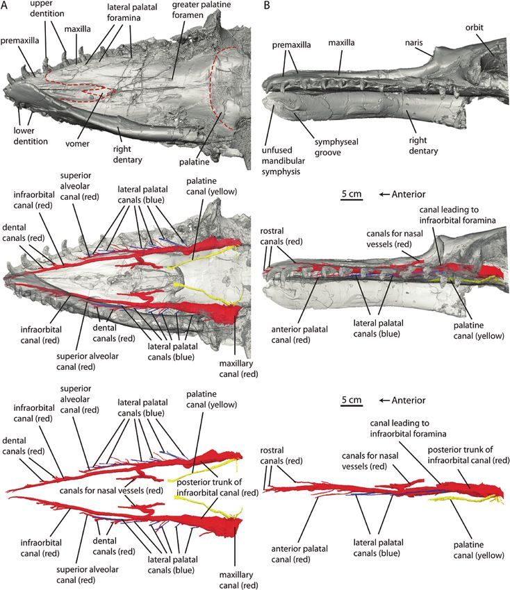
Figure 2. Neurovascular canals through rostrum of Aetiocetus weltoni (UCMP 122900). A, ventral view of 3D rendering
of skull (top), skull rendered semi-transparent to reveal internal canals of rostrum (middle), and digital segmentation of
rostral canals (bottom). B, lateral view of 3D rendering of skull (top), skull rendered semi-transparent to reveal internal
canals of rostrum (middle), and digital segmentation of rostral canals (bottom).
© 2021 The Linnean Society of London, Zoological Journal of the Linnean Society, 2021, XX, 1–21EVIDENCE FOR BALEEN IN AETIOCETUS 5
Downloaded from https://academic.oup.com/zoolinnean/advance-article/doi/10.1093/zoolinnean/zlab017/6278618 by guest on 10 October 2021
Figure 3. Digital segmentation of neurovascular canals through rostrum of Aetiocetus weltoni (UCMP 122900). A, rostral
canals in ventral view. B, rostral canals in lateral view. Abbreviations: dc, dental/alveolar canal; lpc, lateral palatal canal.
exact number may be obscured by damage to both sides the rostrum via a series of premaxillary foramina (red
of the rostrum in the posterior region of the maxillary premaxillary canals in Figs 2B, 3).
canal (see Supporting Information, Appendix S1). The superior alveolar canal (lateral division) diverges
A large canal departs dorsomedially from the from the infraorbital canal and sends branches (dental/
infraorbital canal near the divergence of the superior alveolar canals) to alveoli of the maxillary teeth (C and
alveolar canal (lateral division). This dorsomedial branch P1–3) (small, red branches labelled as dental canals
divides into anterior and posterior extensions that open in Fig. 2), as well as to canals that open on to the
into the narial fossa (red canals for nasal vessels in Figs palate via lateral palatal foramina positioned medial
2, 3). The canal likely carried the sphenopalatine artery to the dentition (small, blue branches labelled as
in life, which in turn is divided into various nasal arteries lateral palatal canals in Figs 2, 3). Two lateral palatal
(Davis, 1964; Evans, 1993). Anterior to the divergence of canals and foramina directly connected to the superior
the superior alveolar canal, an elongate canal departs alveolar canal are observed in this area on both the
ventromedially from the infraorbital canal and extends right and left sides of the rostrum (Deméré et al., 2008:
anteromedially within the suture between the maxilla fig. 1D, E). The superior alveolar canal terminates at
and premaxilla before opening on to the palate well the alveolus of the canine. There are no observable
medial to the tooth row (Supporting Information, Videos anastomoses between the superior alveolar canal and
S1, S2). More distally, the infraorbital canal sends feeder either the infraorbital or greater palatine canals.
canals to alveoli of the premaxillary teeth (small, red, Preliminary segmentation of CT data of the partial
dental canals in Figs 2, 3) and ultimately separates right dentary has revealed the internal structure of
into branches that open on to the external surface of the mandibular canal and subsequent branches. The
© 2021 The Linnean Society of London, Zoological Journal of the Linnean Society, 2021, XX, 1–216 E.G. EKDALE and T.A. DEMÉRÉ
Downloaded from https://academic.oup.com/zoolinnean/advance-article/doi/10.1093/zoolinnean/zlab017/6278618 by guest on 10 October 2021
Figure 4. CT-scan data of Aetiocetus weltoni (UCMP 122900). Slice A–A’ taken oblique to the horizontal plane through the
skull as indicated on the surface medial in lateral view to image course of infraorbital canal. Slices B–B’, C–C’ and D–D’
taken along the transverse plane (original scan axis) at different positions along the rostrum as indicated on the surface
model in dorsal view.
mandibular canal is oval in cross-section and decreases DISCUSSION
in diameter anteriorly (Supporting Information,
Comparisons with extant cetaceans
Appendices S1, S2; Table S1). Numerous small-diameter
dental canals extend from the dorsal edge of the The general branching pattern of the rostral canals in
mandibular canal to connect with the dental alveoli, extinct Aetiocetus (toothed Mysticeti) closely mirrors
which are set in a distinct alveolar groove. In edentulous that observed in both extant Tursiops (toothed
mysticetes with a closed alveolar groove, these canals Odontoceti) and Eschrichtius (toothless Mysticeti). The
have been redirected to emerge on the medial side obvious difference, however, is that the lateral palatal
of the dorsal margin of the dentary via the gingival canals and foramina are absent in Tursiops, while
foramina and transmit neurovascular structures to the dental canals and alveoli with teeth are absent in
the mandibular gingiva. In contrast, lateral branches Eschrichtius (Fig. 5). In the two extant cetaceans, the
separate from the mandibular canal and open via mental medially positioned palatine canal branches from the
foramina on the external surface of the dentary. A total maxillary canal posterior to the other two branches to
of seven mental canals and foramina were observed, the transmit palatine vessels and nerves to the palatine
anteriormost two of which are positioned on the extreme process of the maxilla. The infraorbital canal extends
anterolateral corner of the dentary below the alveolus anteriorly and transmits dental vessels and nerves
of i1 (Supporting Information, Appendices S1, S2; Table to the premaxillary alveoli in Tursiops (Fig. 5A) and
S1). Medial to these foramina on the lingual side of the the distal end of the edentulous rostrum within the
dentary is a smooth symyphseal surface anterior to a premaxilla–maxilla suture in Eschrichtius (Fig. 5C).
sharply defined symphyseal groove (Fig. 2B; Deméré & Importantly, the superior alveolar canal in extant
Berta, 2008). The observed symphyseal morphology and cetaceans diverges from the infraorbital canal anterior
neurovascular anatomy of the dentary are consistent to the branching of the palatine canal and transmits
with the phylogenetic position of aetiocetids as the superior alveolar vessels and nerves to the maxillary
sister-group to all later diverging mysticetes. A more alveoli and teeth via dental/alveolar canals in Tursiops
detailed description and analysis of these structures (Fig. 5A) and to the lateral palatal foramina and baleen
will be presented elsewhere, as the discussions in the apparatus via lateral palatal canals in Eschrichtius
current report focus on the rostrum only. (Fig. 5C). Sawamura (2008a, 2008b) described a similar
© 2021 The Linnean Society of London, Zoological Journal of the Linnean Society, 2021, XX, 1–21EVIDENCE FOR BALEEN IN AETIOCETUS 7
(2017) relied in part on their inability to understand
how the superior alveolar artery and nerve could
simultaneously have played this dual role of nourishing
and innervating both teeth and baleen. As discussed
below, we contend that this anatomical ‘solution’ is a
case of exaptation, wherein the dental/alveolar canals
Downloaded from https://academic.oup.com/zoolinnean/advance-article/doi/10.1093/zoolinnean/zlab017/6278618 by guest on 10 October 2021
of stem toothed mysticetes were co-opted to serve as
the neomorphic lateral palatal canals of aetiocetids and
later diverging edentulous mysticetes.
In offering this hypothesis of homology, we recognize
that there are potential problems stemming from such
a limited sample size (i.e. CT-imaging of the skulls of
one aetiocetid, one extant odontocete and one extant
mysticete). However, the general architecture of the
structures we are imaging is so consistent within
Mammalia (i.e. the positions and successive branching
pattern of the infraorbital canal; Davis, 1964; Evans,
1993; Standring et al., 2008) that we are confident that
our interpretations have not been adversely affected
by sample size. We also recognize the potential
problems related to limited ontogenetic sampling,
especially our reliance on the neurovascular anatomy
of a single grey whale neonate. For mysticetes, access
to specimens and the exponential increase in logistical
issues with increasing body size places obvious
limitations on research, which leaves researchers
with few options. Fortunately, however, the relevant
neurovascular structures and enclosing rostral bones
of the grey whale neonate that we studied (Ekdale
et al., 2015) have passed through a sufficient number
of developmental stages (e.g. resorption of the foetal
dentition, closure of the foetal alveolar groove, and
formation of the lateral palatal canals, foramina and
sulci) that the resulting neonate anatomy is broadly
representative of the juvenile and adult condition
Figure 5. Digital segmentations (top) and graphical (Ridewood, 1923; Lanzetti, 2019).
reconstructions of rostral canals in select cetaceans
(bottom). A, extant toothed odontocete Tursiops truncatus
(SDSNH 21212). B, extinct toothed mysticete Aetiocetus Hypothesis of homology and presence of
weltoni (UCMP 122900). C, extant toothless mysticete baleen in aetiocetids
Eschrichtius robustus (modified from: Ekdale et al., 2015).
Our results establish structural and positional
homology between the lateral palatal canals, foramina
neurovascular anatomy in minke whales (presumably and sulci in the toothed mysticete Aetiocetus weltoni
Balaenoptera bonaerensis Burmeister, 1867) and, like us, (and we contend all aetiocetids) and the lateral
emphasized the importance of distinguishing between palatal canals, foramina and sulci in edentulous
vessels of the greater palatine artery that exit on to the crown mysticetes (i.e. connection of the lateral palatal
palatine process of the maxilla and vessels of the superior foramina directly to the superior alveolar canal and not
alveolar artery that exit on to the alveolar process of to the greater palatine canal). Given the clear pattern
the maxilla. CT-scan results reveal that the condition in extant mysticetes of transmission of superior
in Aetiocetus weltoni is intermediate between that seen alveolar blood vessels and nerves via the lateral
in Tursiops and Eschrichtius in that the lateral division palatal foramina to the ‘root’ of the baleen apparatus,
of the maxillary canal (superior alveolar canal) has the most parsimonious explanation for the pattern
connections that extend to both dental alveoli (dental/ seen in Aetiocetus weltoni is that the lateral palatal
alveolar canals) and lateral palatal foramina (lateral foramina of aetiocetids performed the same function,
palatal canals) (Fig. 5C). In rejecting the hypothesis that namely transmission of superior alveolar blood vessels
aetiocetids possessed teeth and baleen, Peredo et al. and nerves to nourish and innervate baleen. In this
© 2021 The Linnean Society of London, Zoological Journal of the Linnean Society, 2021, XX, 1–218 E.G. EKDALE and T.A. DEMÉRÉ
view, aetiocetids would have possessed both an adult those of extant mysticetes and, therefore, cannot be
dentition and some form of ‘proto-baleen’. Further, the homologous.
retention of connections between the superior alveolar Studies that recognize the presence of lateral
canal/posterior trunk of the infraorbital canal and palatal foramina in Aetiocetus weltoni but reject the
dental alveoli in Aetiocetus weltoni suggests that these hypothesized co-occurrence of teeth and proto-baleen
former alveolar structures were co-opted in aetiocetids in aetiocetids make the following arguments: (1) the
Downloaded from https://academic.oup.com/zoolinnean/advance-article/doi/10.1093/zoolinnean/zlab017/6278618 by guest on 10 October 2021
and all later diverging mysticetes to supply blood and presence of baleen is functionally incompatible with the
nerves to the baleen apparatus. In our view, hypotheses presence of teeth (Marx et al., 2016) and (2) the lateral
of novel functions for unobserved and unknowable palatal foramina of aetiocetids are homologous with
structures in extinct taxa such as ‘enlarged gums’ (e.g. those of extant mysticetes, but the structures serve a
Marx et al., 2016; Peredo et al., 2018; Fordyce & Marx, different function (e.g. innervation and blood supply
2018) overcomplicate the issue. for simple cornified gingival tissues or ‘thickened
gums’) and, therefore, are not directly relevant to the
origin of baleen (Fordyce & Marx, 2018).
Arguments against the hypothesis of baleen in We discuss all of these counter-arguments below.
aetiocetids
Needless to say, the original formulation of the
hypothesis for a co-occurrence of teeth and baleen in Lateral palatal foramina in non-mysticetes?
aetiocetids (Fitzgerald, 2006; Deméré & Berta, 2008; A corollary of the hypothesis for a co-occurrence of
Deméré et al. 2008) has not been widely accepted (for teeth and baleen in aetiocetids is that the osteological
a supportive analysis, see: de Muizon et al., 2019), and correlate for baleen (lateral palatal foramina) should
arguments against the idea have focused on a lack be absent in extinct taxa for which baleen would be
of direct evidence for baleen, questionable homology unlikely to occur from a phylogenetic perspective
of lateral palatal foramina and sulci, and alternative (i.e. hypotheses of multiple origins of baleen). If the
functions for lateral palatal foramina and sulci. hypothesized osteological correlates are present in
Concerning the lack of direct evidence, clearly, the those taxa, then the structures either are not reliable
best evidence to definitively confirm the presence of indicators of baleen, as has been suggested by Fordyce
baleen in a toothed mysticete such as Aetiocetus would & Marx (2018) and Peredo et al. (2018), or else they
be discovery of a fossil with preserved baleen. Although indicate baleen in a taxon for which baleen has not
a few examples of fossilized baleen have been reported been inferred previously. Care must be taken then to
(Esperante et al., 2008; Bisconti, 2012; Gioncada et al., accurately identify the homology of specific palatal
2016; Marx et al., 2017), there are no known aetiocetid foramina in fossil specimens. The original hypothesis
fossils with preserved baleen and such fossils may of baleen in aetiocetids was explicit in stating that the
never be discovered owing to the low preservation osteological correlates are specifically on the lateral
potential of this keratinous structure. This is not an border of the palate and separate from the more
unusual conundrum in paleontology, where it is often medially positioned palatine foramina that penetrate
necessary to establish bony correlates for important the palate of all mammals (Deméré et al., 2008). Extant
soft anatomical structures, in this case, baleen. mysticetes and extinct aetiocetids thereby possess both
Studies that recognize the presence of lateral medial palatal foramina for the descending palatine
palatal foramina in Aetiocetus weltoni but reject the artery and nerve in addition to the lateral palatal
hypothesized homology with similar bony structures in foramina (Deméré et al., 2008), which transmit blood
extant mysticetes (e.g. Peredo et al., 2018) rely on several vessels and nerves for baleen in extant taxa (Ekdale
arguments: (1) palatal foramina are present in taxa for et al., 2015). In other words, there are two primary
which baleen has never been reconstructed, including series of palatal canals and foramina in aetiocetids
certain stem toothed mysticetes, basilosaurids and and later diverging mysticetes.
stem odontocetes; (2) the lateral palatal foramina of In order for lateral palatal foramina connected
aetiocetids are not homologous with those in extant internally to the superior alveolar canal to have
mysticetes because the foramina are not present in predictive power as osteological correlates of baleen,
all aetiocetids and, therefore, are not universal within they must be present only in animals that have
the clade; (3) palatal foramina that connect with the baleen. Therefore, any specimen of a non-baleen
superior alveolar canal are possibly absent in select bearing mammal that possesses both a medial and a
taxa that share a more recent common ancestry with lateral series of palatal foramina, the latter of which
crown Mysticeti than do aetiocetids (e.g. Maiabalaena connects internally to the superior alveolar canal,
nesbittae Peredo, Pyenson, Marshall, Uhen, 2018); would reduce the utility of the lateral palatal foramina
and (4) the lateral palatal foramina of aetiocetids as a correlate. Such a case has been suggested for the
differ in size, orientation and overall morphology from basilosaurid Dorudon atrox Andrews, 1906 (Peredo
© 2021 The Linnean Society of London, Zoological Journal of the Linnean Society, 2021, XX, 1–21EVIDENCE FOR BALEEN IN AETIOCETUS 9
et al., 2018). However, the palatal foramina that and near the midline of the palate medial to P3 of the
have been described for Dorudon were homologized holotype (USNM 256517) (figured but not described
with the medial series for the palatine neurovascular by Peredo et al., 2018: supplemental information,
structures (Uhen, 2004). We agree that these small fig. S3B). These foramina were homologized with the
foramina on the palate of Dorudon are homologous major palatine foramina, while additional smaller
with the anterior palatine foramina of other mammals, palatal foramina on the maxilla were identified as
Downloaded from https://academic.oup.com/zoolinnean/advance-article/doi/10.1093/zoolinnean/zlab017/6278618 by guest on 10 October 2021
rather than the lateral palatal foramina described for minor palatine foramina (Fordyce, 2002: figs 2, 4).
Aetiocetus weltoni and extant mysticetes, given that Thus, the palatal foramina referenced by Peredo
they penetrate the palate near its midline. Dorudon et al. (2018) correspond to openings that most likely
also possesses posterior palatine foramina (Uhen, connect internally to the canal for the palatine artery
2004), in agreement with the position of the same and nerve. Importantly, no separate series of lateral
foramina described for Aetiocetus weltoni here and palatal foramina has been described for Simocetus.
elsewhere (Deméré et al., 2008). Nowhere in Uhen’s In light of this information, the foramina reported in
description of the palate of Dorudon is there mention Eocene basilosaurids and the Oligocene odontocete
of a separate series of lateral palatal foramina Simocetus are most likely homologous with the
(Uhen, 2004). medial series of palatal foramina that are well known
Peredo et al. (2018) have also asserted (but did for most mammals, rather than the neomorphic
not describe) that palatal foramina ‘similar’ to lateral palatal foramina of aetiocetids, eomysticetids
those in Aetiocetus weltoni occur on the maxilla of and crown mysticetes. Additional analyses of critical
the basilosaurids Zygorhiza kochii Carus, 1847 and fossils utilizing CT-scanning are needed to confirm
Basilotritus wardii (Uhen, 1999) (= Platyosphys wardii; this hypothesis.
see: Van Vliet et al., 2020), as well as the Oligocene Hopefully, it has become clear how critical it is
odontocete Simocetus rayi Fordyce, 2002. Given to understand that not all palatal foramina are the
that Zygorhiza is closely related phylogenetically to same in terms of their relevance to the question of
Dorudon (Uhen, 2013), and acknowledging that the the origin/presence of baleen. The presence of palatal
palate of the former taxon has not been adequately foramina is not the question, but rather what their
described in the literature, we provisionally suggest internal connection is with the major bony conduits for
that the configuration and associated internal the neurovasculature of the palate. To be relevant to
anatomy of palatal foramina would be the same in the question of baleen in the context of our findings, a
both basilosaurid taxa and thus not similar to the palatal foramen must connect to the superior alveolar
condition we describe here for Aetiocetus weltoni. canal or to the posterior trunk of the infraorbital
For another basilosaurid Platyosphys wardii, Peredo canal (both of which presumably carried the superior
et al. (2018: fig S3) only provide a photograph of alveolar artery and its branches), which generally
the holotype rostral fragment (USNM 310633) and means that the foramen is positioned on the lateral/
do not describe the purported palatal foramina, alveolar process of the maxilla. If a foramen connects
which appear to penetrate the tip of a narrow and to the greater palatine canal, then in our view it has
attenuated rostral fragment composed solely of the no bearing on whether baleen was present or not. This
premaxillae, not the maxillae as erroneously reported critical distinction has not always been considered
by Peredo et al. (2018). There are several problems in the various arguments surrounding the origin of
with their assertion that these purported palatal baleen, and in many cases these arguments have
foramina are relevant to the discussion of lateral even failed to make the distinction between a given
palatal foramina in aetiocetids: (1) the structures foramen and its associated sulcus or its position on
are in the premaxilla and not the maxilla; and (2) the palate.
the tooth rows are closely adjacent to the midline In our view, it has yet to be demonstrated that any
making it impractical to call a palatal feature medial non-mysticete taxon has foramina along the lateral
or lateral relative to the dental alveoli. We contend border of the maxilla that are connected internally
that since the superior alveolar canal terminates to canals for the superior alveolar blood vessels and
at the canine alveolus and, therefore, does not even nerves separate from the medial palatine series.
extend into the premaxilla in Aetiocetus, Tursiops Thus, the most parsimonious reconstruction for
and Eschrichtius (Fig. 5), the features in Platyosphys the soft tissues associated with the lateral palatal
are anterior palatine foramina and not homologous canals and foramina of Aetiocetus weltoni would be
to the lateral palatal foramina of mysticetes. The blood vessels and nerves that are homologous to the
original description of the simocetid stem odontocete neurovascular structures that nourish and innervate
Simocetus rayi (Fordyce, 2002), noted the occurrence baleen in the sister-group of aetiocetids – the baleen-
of palatal foramina and associated sulci at the bearing chaeomysticetes (eomysticetids plus crown
anterolateral corner of the maxillary–palatine suture mysticetes).
© 2021 The Linnean Society of London, Zoological Journal of the Linnean Society, 2021, XX, 1–2110 E.G. EKDALE and T.A. DEMÉRÉ
Lateral palatal foramina in early diverging and homology with aetiocetids and chaeomysticetes
toothed mysticetes? was established, this would suggest either a
Fordyce & Marx (2018) described the presence of dual origin of baleen, given the basal position of
a network of narrow (~1–2 mm) palatal sulci (not Llanocetus in mysticete phylogenies (Fordyce &
foramina) medial to, and partially surrounding, Marx, 2018; however, for an alternative phylogenetic
the left P1–4 alveoli on the damaged and incomplete hypothesis and discussion of possible palatal
Downloaded from https://academic.oup.com/zoolinnean/advance-article/doi/10.1093/zoolinnean/zlab017/6278618 by guest on 10 October 2021
holotype skull (USNM 183022) of the Late Eocene keratinous ‘appendages’, see: de Muizon et al., 2019)
toothed mysticete Llanocetus denticrenatus. The or else loss of baleen and associated foramina in
delicate sulci of Llanocetus generally occur in the stem toothed mammalodontid mysticetes (Fig.
‘peri-dental bundles’ that converge medially and 1). The latter possibility generally relies on the
posteriorly within a fractured region of the palate once proposed, but now rejected, hypothesis of a
containing longitudinal ‘striations’ that the authors monophyletic mammalodontid + aetiocetid grouping
speculate contained ‘crushed and ventrally eroded (Marx et al. 2016; Boessenecker & Fordyce, 2016;
vascular canals and sulci’ (supplemental information Hocking et al., 2017; de Muizon et al., 2019).
of Fordyce & Marx, 2018). Importantly, the authors
admit that there are no palatal foramina preserved
in association with the observed palatal sulci. In fact, Taxonomic distribution of lateral palatal
throughout their report, the authors focus solely foramina among aetiocetids
on discussions of palatal sulci and are mute on the Peredo et al. (2017, 2018) noted that lateral palatal
structure and importance of lateral palatal foramina. foramina have been described for only three of
Although noting that the configuration of the palatal the nine nominal aetiocetid species [Aetiocetus
sulci in Llanocetus is autapomorphic relative to all cotylalveus Emlong, 1966, Aetiocetus weltoni and
known mysticetes, and in spite of the absence of Fucaia goedertorum (Barnes & Furusawa in Barnes
evidence of associated lateral palatal foramina in et al., 1994)] implying that lateral palatal foramina
this taxon, Fordyce & Marx (2018) speculate that are not characteristic of Aetiocetidae, but rather of
these sulci were part of a well-developed palatal a small subset of the clade. While the statement by
vascular system that nourished ‘enlarged gums’ in Peredo et al. (2017, 2018) is accurate at face value, it
a manner equivalent to that possessed by extant is misleading given that specimens assigned to five
mysticetes, sans the subsequently evolved baleen [Chonecetus sookensis (Russell, 1968); Morawanocetus
apparatus. Missing from their discussion was yabukii Kimura & Barnes in Barnes et al., 1994;
any recognition of the different configuration and Aetiocetus tomitai Kimura & Barnes in Barnes et al.,
components of the neurovascular bony palatal 1994; Fucaia buelli Marx, Tsai & Fordyce, 2015;
features seen in aetiocetids and extinct and extant Salishicetus meadi Peredo & Pyenson, 2018] of the
chaeomysticetes). Whereas Llanocetus possesses other six nominal aetiocetid species lack rostra entirely
bundles of delicate sulci medial to, and partially and, therefore, do not preserve evidence for either the
surrounding, individual premolar alveoli, aetiocetids presence or absence of lateral palatal foramina. The
and most chaeomysticetes typically possess single, one remaining candidate species that does preserve
isolated sulci intimately associated with single, a rostrum, Aetiocetus polydentatus Sawamura in
distinct, lateral palatal foramina, which, in the case Barnes et al., 1994, is based on a specimen (AMP-12)
of aetiocetids, lie entirely medial to the premolar with marked diagenetic deformation of the rostrum
and molar alveoli. We contend that these positional (e.g. transversely oblique en echelon ‘faults’ and
(i.e. lateral on the palate and medial to dental ‘folds’; irregular, fractured and possibly delaminated
alveoli) and structural (i.e. single lateral palatal palatal cortical bone). Thus, the specimen is neither
foramina connected internally via bony canals to the well enough preserved nor currently prepared to an
superior alveolar canal and the posterior trunk of extent that would allow accurate determination of the
the infraorbital canal; Fig. 5) features are critical presence or absence of the salient palatal structures.
to the question of homology and here suggest that Additional preparation and CT-imaging of AMP-12
the bundles of delicate palatal sulci described in could provide an important answer to this question.
Llanocetus may be neomorphic and unrelated to Further, an additional specimen of a new unnamed
the neurovascular anatomy of baleen that first aetiocetid (Marx et al., 2016) has been reported as
evolved in the common ancestor of aetiocetids and lacking lateral palatal foramina (Peredo et al., 2018),
chaeomysticetes. Importantly, if the necessary but the right and left lateral margins of the palate are
positional and structural features were discovered not preserved in that specimen (NMV P252567) and
in the holotype (with additional preparation or thus provide no evidence either way for the presence or
imaging) or recovery of other specimens of Llanocetus absence of these critical neurovascular features.
© 2021 The Linnean Society of London, Zoological Journal of the Linnean Society, 2021, XX, 1–21EVIDENCE FOR BALEEN IN AETIOCETUS 11
Of course, absence of a structure due to poor Teeth are neither preserved nor associated with the
preservation or incomplete study (e.g. lack of high holotype specimen (USNM 314627) and there are no
resolution CT-imaging) is not a valid argument for a clear alveoli along the purportedly ‘nearly complete’
true anatomical absence of the structure and it would right lateral margin of the maxilla nor along the
be just as accurate to state that 100% of aetiocetids dorsal margin of the dentaries (Peredo et al., 2018). It
for which the lateral palatal margin is sufficiently should be noted, however, that the authors mention
Downloaded from https://academic.oup.com/zoolinnean/advance-article/doi/10.1093/zoolinnean/zlab017/6278618 by guest on 10 October 2021
preserved and prepared possess lateral palatal (but do not describe or illustrate) the presence of
foramina. Reinforcing this conclusion is the occurrence three ‘openings’ along the anterior portion of the
of an additional aetiocetid specimen from the Upper left side of the rostrum in Maiabalaena (presumably
Oligocene Morawan Formation of Hokkaido, Japan all in the maxilla), but dismiss these openings as
assigned to the genus Morawanocetus and partially being dental alveoli because the structures open
described by Sawamura (2008a, b). The specimen anteriorly and are oriented horizontally, and are
in question (AMP-14) preserves a series of distinct not mirrored on the more complete right side of the
lateral palatal foramina along the incomplete lateral rostrum. We suggest that the anteriorly facing and
margin of the right maxilla (the lateral margin of the horizontal orientation of the openings does not in
left maxilla is even less complete). A formal description itself disqualify these features as alveoli given that
and analysis of AMP-14 is currently being prepared (T. the incisor alveoli of Aetiocetus weltoni described here
Ando, personal communication). The addition of this (Fig. 2; Supporting Information, Appendix S1), as well
specimen and the genus Morawanocetus to the list as the anterior dental alveoli of Aetiocetus cotylalveus
of known aetiocetids with lateral palatal foramina is (Peredo et al., 2018: fig. 3), also open anteriorly
strong evidence for the general taxonomic distribution and are clearly oriented horizontally within the
of this feature within this relatively diverse clade of rostrum. In addition, specimens of the later diverging
toothed mysticetes. eomysticetids, Waharoa ruwhenua Boessenecker
Although tangential to the question of baleen in & Fordyce, 2015 (see further discussion below) and
aetiocetids, it is noteworthy that all four nominal Yamatocetus canaliculatus Okazaki, 2012, possess
aetiocetid taxa with preserved mandibles (Aetiocetus anterolaterally oriented alveoli within the anterior
polydentatus, A. weltoni, Fucaia goedertorum and portion of the rostrum (Okazaki, 2012; Boessenecker
Salishicetus meadi) possess a smooth, unfused & Fordyce, 2015b).
mandibular symphysis with a longitudinal symphyseal Concerning the assertion that the holotype skull of
groove (Fig. 2B), which represents an apomorphy Maiabalaena lacks features that could be called lateral
shared with all later diverging chaeomysticetes palatal foramina; we contend that the case for this is
(Deméré & Berta, 2008; Fitzgerald, 2012). Viewed also equivocal. Peredo et al. (2018) describe at least
in a phylogenetic context, we suggest that it is not a four and possibly eight palatal foramina on the right
coincidence that the evolution of this key feature of maxilla as ‘shallow, laterally narrow foramen [sic]
bulk filter-feeding (kinetic mandibular symphysis) with anteroposteriorly elongated sulci that are angled
and baleen-bearing mysticetes is first reported in between 20 and 45 degrees from the sagittal plane’
aetiocetids. and add that similar foramina were not observed
on the left maxilla (unpaginated supplemental
information of Peredo et al., 2018. While admitting
Lateral palatal foramina in Maiabalaena that inadequate CT resolution may be responsible, the
nesbittae? authors contend that these foramina are superficial,
A recently described Oligocene mysticete, Maiabalaena penetrate less than 5 mm into the maxilla, and do not
nesbittae, from Washington, USA, which is purported connect internally to the superior alveolar canal (or
to occupy a more crownward phylogenetic position to any other neurovascular canals). Combined, these
than aetiocetids and a more stemward position than interpretations are puzzling at best and suggest that
eomysticetids (Fig. 1), was interpreted as lacking diagenesis is masking (e.g. through fragmentation
both teeth and baleen (Peredo et al., 2018). Such a and crushing of the rostrum) or has removed (through
conclusion could indicate either a dual origin of baleen loss of the lateral margins of the maxillae) critical
in aetiocetids and later diverging mysticetes, a loss of morphology from the holotype. For example, how can
baleen and lateral palatal foramina in Maiabalaena, there only be foramina on the lateral portion of the
or evolution of baleen closer to crown Mysticeti than in right maxilla and not on the left maxilla or palatines?
the ancestor of aetiocetids and extant clades (Fig. 1). Or how can a feature that is determined to be a palatal
However, for various reasons we contend that the foramen not be connected internally to a canal? As with
evidence that Maiabalaena lacked the neurovascular the holotype of Aetiocetus polydentatus, preservational
structures here correlated with the presence of baleen problems with the holotype of Maiabalaena nesbittae
is not as conclusive as originally presented. may have made recognition of unequivocal palatal
© 2021 The Linnean Society of London, Zoological Journal of the Linnean Society, 2021, XX, 1–2112 E.G. EKDALE and T.A. DEMÉRÉ
neurovascular structures difficult, if not impossible, to borders of the palate to a sufficient extent to allow
determine. recognition of the foramina. Indeed, major sections of
Additional general ‘palatal foramina’ are described the lateral border of the palate are missing, but the
for Maiabalaena, but they are not identified as authors contend that a portion of the right side of
lateral palatal foramina specifically (Peredo et al., the palate is nearly complete. It is critical to consider
2018). The authors go on to assert that CT-imaging that further mechanical or chemical preparation of
Downloaded from https://academic.oup.com/zoolinnean/advance-article/doi/10.1093/zoolinnean/zlab017/6278618 by guest on 10 October 2021
reveals that the palatal foramina of Maiabalaena the holotype or higher resolution CT-imaging could
do not connect to the superior alveolar canal, reveal critical features. Either way, concluding that
which implies that they are not homologous with the lateral foramina do not connect to the superior
the lateral palatal foramina of extant mysticetes. alveolar canal in Maiabalaena is premature until
However, the same authors score the character of higher resolution CT-imaging and analysis of the
lateral palatal foramina as present for Maiabalaena holotype are conducted or better-preserved specimens
in their phylogenetic data matrix, and do the same of this species are discovered. In the absence of such
for another Oligocene mysticete from Washington, studies, we remain unconvinced that Maiabalaena
USA, Sitsqwayk cornishorum Peredo & Uhen, 2016. (and Sitsqwayk) lacked both teeth and baleen.
Although Peredo & Uhen (2016) originally described
the holotype skull of Sitsqwayk cornishorum
(UWBM 82916) as possessing palatal ‘nutrient
grooves’ that suggested the presence of baleen, they Lateral palatal foramina in eomysticetids
subsequently reversed their interpretation without Also critical to the evaluation of character evolution
discussion (Peredo et al., 2018) and inferred that and the origin of baleen in mysticetes is the palatal
this species lacked both teeth and baleen, and was morphology of members of the later diverging
the sister-taxon of Maiabalaena in an unnamed Eomysticetidae. Members of this clade were initially
monophyletic grouping that they contend is sister reported to be entirely edentulous given that the
to eomysticetids and crown mysticetes. Given this holotype of Eomysticetus whitmorei Sanders & Barnes,
phylogenetic hypothesis, it is confusing that Peredo 2002 (ChM PV4253) from the Late Oligocene of South
et al., (2018) score lateral palatal foramina as present Carolina, USA preserves no evidence of teeth in the
in Maiabalaena + Sitsqwayk, the same score that incomplete upper or lower jaws (Sanders & Barnes,
they give for aetiocetids, eomysticetids and extant 2002). However, more recent work has suggested that
mysticetes (Peredo & Uhen, 2016). Identical scoring at least some eomysticetids retained a partial anterior
of the character implies homology of those structures dentition that was likely non-functional (in terms of
predicting that aetiocetids and Maiabalaena would, feeding), namely Matapa waihao Boessenecker &
in fact, possess the same bony canals that transmit Fordyce, 2017, Waharoa ruwhenua and Tokarahia
blood vessels and nerves that nourish and innervate sp. cf. T. lophocephalus (Marples, 1956) from the Late
baleen in extant taxa. Our observations of Aetiocetus Oligocene of New Zealand (Boessenecker & Fordyce,
weltoni internal rostral anatomy illustrate the 2015a, 2015b; 2016), and Yamatocetus canaliculatus
conflict between those statements. from the Late Oligocene of Japan (Okazaki, 2012).
Again, the contradiction between hypotheses of Unfortunately, the evidence for teeth in these taxa
homology resulting from study of Maiabalaena may be remains equivocal, since no specimens actually
due to the low-resolution CT-imaging used to analyse preserve in situ dentitions that are directly associated
the holotype (Peredo et al., 2018). However, the authors with the purported upper and lower dental alveoli.
also hypothesize that a lack of connection between the However, an isolated partial tooth discovered during
foramina and the superior alveolar canal may be the preparation of a specimen (OU 22081) provisionally
result of bone remodelling around alveoli in the upper referred to Tokarahia lophocephalus has been
jaw. It seems unlikely that a loss of connection between suggested to represent an anterior vestigial tooth of
the lateral palatal foramina and the superior alveolar this eomysticetid (Boessenecker & Fordyce, 2015a).
canal would be a result of the loss of alveoli, given that This tooth does not preserve any enamel and consists
the lateral palatal foramina clearly connect with the of a linguolabially flattened root that matches the
superior alveolar canal as well as the posterior trunk flattened oval-shaped alveoli found at the anterior
of the infraorbital canal in extant mysticetes, which end of well-preserved eomysticetid rostra. Contrary to
themselves lack distinct alveoli at any point in their suggestions by Peredo et al. (2018) that eomysticetids
development (Ridewood, 1923; Ekdale et al., 2015; potentially also lacked baleen, there are several critical
Lanzetti, 2019). Because the medial series of palatal fossils that strongly suggest otherwise.
foramina for the palatine artery and nerve is present Holotype and paratype skulls of Waharoa ruwhenua
in Maiabalaena, a more likely scenario is that the possess an elongate and flattened rostrum with
holotype specimen does not preserve the crucial lateral 6–13 lateral palatal foramina and sulci preserved
© 2021 The Linnean Society of London, Zoological Journal of the Linnean Society, 2021, XX, 1–21EVIDENCE FOR BALEEN IN AETIOCETUS 13
posteriorly and alveoli preserved anteriorly. The connections between the lateral palatal foramina
latter may have housed a non-functional dentition and neurovascular canals within the rostrum. The
on the premaxilla and anterior maxilla. Dentaries initiation of such studies is strongly encouraged.
preserve an alveolar groove, no gingival foramina (i.e.
neurovascular foramina representing the ontogenetic
remnants of the alveolar groove), and at least three Morphological differences of lateral palatal
foramina between aetiocetids and crown
Downloaded from https://academic.oup.com/zoolinnean/advance-article/doi/10.1093/zoolinnean/zlab017/6278618 by guest on 10 October 2021
alveoli anteriorly, which may have also housed a non-
functional dentition. Boessenecker & Fordyce (2015b) mysticetes
concluded that Waharoa likely possessed a baleen Peredo et al. (2017) assert that the lateral palatal
apparatus, at least on the posterior two-thirds of the foramina in aetiocetids are much smaller, fewer in
palate, as well as a reduced, anterior, non-functional number and exhibit a different distribution and
dentition. As such, they suggest that eomysticetids orientation pattern than seen in crown mysticetes
were a transitional clade that spans the morphological (except, perhaps, species of Balaenoptera). The
gap between aetiocetids with a functional dentition implication is that the lateral palatal foramina of
and probable baleen apparatus, and fully edentulous, aetiocetids do not show positional or structural
baleen-bearing crown mysticetes. They go on to homology with the lateral palatal foramina of
speculate that the baleen ‘plates’ in eomysticetids crown mysticetes and, therefore, do not represent
likely were short because of the flatness of the rostrum, osteological correlates of baleen. However, in making
that there was likely an anterior subrostral gap in this argument the authors also emphasize the ‘widely
the baleen apparatus, that vestigial anterior teeth variable’ pattern of lateral palatal foramina exhibited
were likely present and that the animals practiced a by extant balaenopterids, balaenids, Eschrichtius and
unique type of skim filter-feeding using their short and Caperea marginata (Gray, 1846) (all of which clearly
elongated baleen racks. possess baleen), even going so far as to incorrectly
Likewise, the holotype skull of Yamatocetus state that balaenids entirely lack these foramina.
canaliculatus (KMNH VP 000.017) preserves an Deméré et al. (2008) briefly summarized the pattern
interesting array of palatal features, including a of morphological diversity in palate vascularization of
complex alveolar groove that runs along the lateral extant mysticetes, noting a general dichotomy between
margin of the premaxilla and anterior half of the the pattern of vascularization of the anterior and
maxilla before stepping slightly medially to extend the posterior portions of the maxilla. Anteriorly, the general
remaining length of the maxilla as an open, parasagittal mysticete pattern consists of parasagittally aligned
linear groove positioned approximately 2 cm medial to foramina with prominent, elongate and somewhat en
the lateral rostral margin. Okazaki (2012) illustrated echelon, anteriorly directed sulci; while posteriorly,
five flattened, oval-shaped alveoli in the alveolar groove the pattern is variable. Posteriorly in balaenopterids
on each side of the scalloped anterior margin of the (except Megaptera Gray, 1846), there typically is a
rostrum, two or three in the premaxilla and three or series of curvilinearly aligned foramina with laterally
two in the maxilla. Additional salient palatal features oriented sulci that display a radial orientation
include three lateral palatal sulci on the right maxilla through an arch of ~85 degrees. In balaenids, the
and one lateral palatal sulcus on the left maxilla, as posterior vascularization pattern is characterized by
well as one medially placed palatine sulcus and one a longitudinal maxillary (alveolar) groove that is open
anteriorly placed parasagitally elongate sulcus in each posteriorly to the margin of the infraorbital plate, and
maxilla. Okazaki (2012) made no mention of foramina from which short, curved and often faint sulci extend
associated with the lateral palatal sulci. However, laterally across the narrow surface of the maxilla. In
presumably foramina are present but not visible Caperea Gray, 1864, the general pattern consists of
because of incomplete preparation of the palate and/or a more extensive longitudinal maxillary groove that
lack of CT-imaging. As with Waharoa, it appears that does not reach to the infraorbital plate, but from
Yamatocetus possessed both baleen and teeth, with the which numerous short, transversely oriented sulci
former possibly confined to the posterior two-thirds of extend laterally across the maxilla. In Eschrichtius
the rostrum and the latter found only in the anterior Gray, 1864, the posterior portion of the maxilla has
portion of the rostrum. The dentary of Yamatocetus numerous and variably sized lateral palatal foramina
also has an alveolar groove and no reported gingival that are irregularly arranged in a series of two to four
foramina. roughly linear parasagittal rows, with or without short
Although, lateral palatal foramina have been associated sulci and no maxillary groove.
noted as occurring in all eomysticetid fossils that Extinct species of crown mysticetes preserve
preserve the lateral margins of the palate, there similar and, in some cases, additional patterns of
have not been follow-up CT-imaging studies of these palate vascularization, and it seems clear from this
specimens to document and critically analyse the limited review that pattern disparity does not equate
© 2021 The Linnean Society of London, Zoological Journal of the Linnean Society, 2021, XX, 1–21You can also read