Novel BTK mutation in X-linked agammaglobulinemia: Report of a 17-year-old male
←
→
Page content transcription
If your browser does not render page correctly, please read the page content below
Allergol Immunopathol (Madr). 2021;49(2):80–83 eISSN:1578-1267, pISSN:0301-0546
Allergologia et
immunopathologia
Sociedad Española de Inmunología Clínica,
Alergología y Asma Pediátrica
CODON
P U B L I C A T I O N S
www.all-imm.com
ORIGINAL ARTICLES OPEN ACCESS
Novel BTK mutation in X-linked agammaglobulinemia:
Report of a 17-year-old male
Zoha Shakaa, Helia Mojtabavib,c, Elham Rayzanc,d, Samaneh Zoghie,f,g,h,
Sepideh Shahkaramih,i,i, Jimenez Heredia Raule,f,g,k, Iraj Sedighil, Kaan Boztuge,f,g,k,m,
Nima Rezaeic, d,h,j*
a
School of Medicine, Iran University of Medical Sciences, Tehran, Iran
b
School of Medicine, Tehran University of Medical Sciences, Tehran, Iran
c
Systematic Review and Meta-Analysis Expert Group (SRMEG), Universal Scientific Education and Research Network (USERN),
Tehran, Iran
d
International Hematology/Oncology of Pediatrics Experts (IHOPE), Universal Scientific Education and Research Network (USERN),
Tehran, Iran
e
Ludwig Boltzmann Institute for Rare and Undiagnosed Diseases, Vienna, Austria
f
St. Anna Children’s Cancer Research Institute (CCRI), Vienna, Austria
g
CeMM Research Center for Molecular Medicine of the Austrian Academy of Sciences, Vienna, Austria
h
Research Center for Immunodeficiencies, Children’s Medical Center, Tehran University of Medical Sciences, Tehran, Iran
i
Department of Pediatrics, Dr. Von Hauner Children’s Hospital, University Hospital, Ludwig-Maximilians-Universität München
(LMU), Munich, Germany
j
Medical Genetics Network (Megene), Universal Scientific Education and Research Network (USERN), Tehran, Iran
k
Department of Pediatrics and Adolescent Medicine, Medical University of Vienna, Vienna, Austria
l
Department of Paediatrics, School of Medicine, Hamedan University of Medical Sciences, Hamedan, Iran
m
Department of Paediatrics and Adolescent Medicine, St Anna Children’s Hospital, Medical University of Vienna, Vienna, Austria
Received 23 August 2020; Accepted 11 November 2020
Available online 1 March 2021
KEYWORDS Abstract
agammaglobulinemia; Introduction and objectives: X-linked agammaglobulinemia (XLA), the first known primary
immunodeficiency; immunodeficiency, is caused by rare mutations in Bruton’s tyrosine kinase (BTK) gene.
mutation; tyrosine Mutations in the BTK gene lead to a failure in the development and maturation of B-cell linage.
kinase, X-linked A decreased number of B-cells results in agammaglobulinemia and increased susceptibility to
agammaglobulinemia a variety of infections. Therefore, patients with XLA usually manifest with repetitive bacterial
infections, such as upper respiratory tract infections, septic arthritis, osteomyelitis, and uri-
nary tract infections, since their infancy.
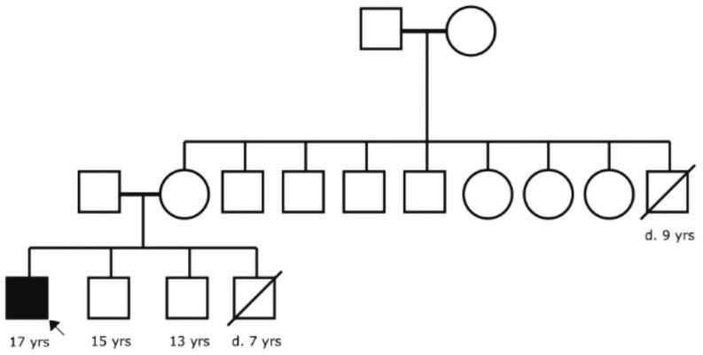
Patients: We report a 17-year-old Iranian boy with XLA, referred to us with a history of severe
and recurrent episodes of bacterial infections for a period of six years.
*Corresponding author: Nima Rezaei, MD, PhD, Children’s Medical Center Hospital, Dr. Qarib St, Keshavarz Blvd, Tehran 14194, Iran.
Email address: rezaei_nima@tums.ac.ir, rezaei_nima@yahoo.com
https://doi.org/10.15586/aei.v49i2.62
Copyright: Shaka Z, et al.
License: This open access article is licensed under Creative Commons Attribution 4.0 International (CC BY 4.0). http://creativecommons.org/
Novel BTK mutation in XLA                                                                                                    81

Results: Genetic analysis using the whole Exome sequencing revealed a hemizygous missense
mutation in the BTK gene (c.428 A > T, p.His143Leu).
Conclusion: To our knowledge, c.428 A > T has not been reported in the BTK gene.
© 2021 Codon Publications. Published by Codon Publications.

Introduction                                                       Case presentation

X-linked agammaglobulinemia (XLA) is an inherited disorder         A 17-year-old Iranian boy was referred due to recurrent
characterized by a profound deficiency of all isotypes of          infections to the Pediatrics Center of Excellence of the
immunoglobulins (Ig) with a significant reduction in mature        Children’s Medical Center Hospital, Tehran, Iran. His symp-
B-cell counts (less than 1%) in the peripheral circulation.1       toms started with a fever and chronic productive cough at
This disease belongs to a broader group of rare genetic dis-       the age of four, and was hospitalized for recurrent respi-
orders called primary immunodeficiency diseases (PIDs).            ratory tract infections such as pneumonia and sinusitis at
XLA, after years of study and gaining a better insight about       the age of five. Later, he experienced several episodes of
PIDs, can be described as human inborn errors of immu-             fever and swelling in his right shoulder joint and right knee,
nity.2 PID affects different components of innate and adap-        which were diagnosed as recurrent osteomyelitis and septic
tive immune system at various levels of development and            arthritis.
maturation. Up to date, most reported cases of PID in chil-            He was the second child of non-consanguineous par-
dren are that of XLA as it is known as the most common             ents who had lost their first male child at the age of seven
inherited antibody deficiency.1–4                                  because of severe pneumonia. The other two male siblings,
     In 1993, Bruton tyrosine kinase (BTK) gene located on X       aged 15 and 13 years, were asymptomatic at the time of
chromosome was found to be the responsible gene defect             the study. Apart from his brother, in their extended family
for XLA. It was named after pediatrician Ogden Bruton,             history, his mother had lost a male sibling in early child-
who first discovered the disease in 1952.3 All the blood cell      hood due to central nervous system (CNS) infection. The
lineages derived from hematopoietic stem cells can express         other seven siblings of his mother were healthy and totally
BTK, anon-receptor protein tyrosine kinase, except T-cells         complaint-free. The family pedigree in detail is illustrated
and natural killer cells. In B-cell linage, BTK is involved as     in Figure 1.
a key regulator of different signals, transduction pathways,           Our patient was born with a favorable perinatal and
and plays an essential role in the differentiation and mat-        birth history, manifested normal growth indices, and
uration of premature B-lymphocytes. Therefore, mutations           followed through normal developmental milestones.
in the BTK gene result in developmental arrest from the            Considering the patient’s medical history of severe recur-
pro-B-cell to pre-B-cell stage.5                                   rent infections and family history of deaths of their male
     Owing to the lack of sufficient humoral immunity              relatives at a young age because of infections, immuno-
and failure in the production of immunoglobulins, XLA              deficiency disorders were under consideration. At the age
patients generally suffer from the most severe and recur-          of 13, an antibody profile study was performed for the
rent infections. Symptoms typically appear after the age of        patient using the enzyme-linked-immunosorbent serologic
six months when the protective trans-placental maternal            assay (ELISA) method, which revealed a total decrease in
Immunoglobulin G (IgG) antibodies decrease and patients            serum immunoglobulins (serum level of IgG 131.1 mg/dL
are unable to produce antibodies on their own.4 The most           [range: 667–1464 mg/dL] and that of IgM 39.1 mg/dL (range:
common clinical manifestations in XLA patients are upper           49–261 mg/dL]).
and lower respiratory tract infections, such as otitis media,
sinusitis, bronchitis, and pneumonia. Severe infections,
including septicemia, meningitis, osteomyelitis and septic
arthritis, and gastrointestinal tract infections, may also
occur in XLA patients.2–6
     Since 1952, immunoglobulin replacement therapy
and prophylactic antibiotic therapy are the cornerstone
treatments for XLA patients.7 In immunoglobulin replace-
ment therapy, a collection of pooled antibodies (>90% IgG
and
82 Shaka Z et al.
Table 1 Laboratory findings.
2014 (age: 13 years) 2017 (age: 15 years) 2019 (age: 17 years)
Immunoglobulin Reference Immunoglobulin Reference Immunoglobulin Reference
profile range profile range profile range
IgG 131.1 mg/dL 667–1464 mg/dL IgG 259 mg/dL 630–1300 mg/dL IgG 115 mg/dL 639–1349 mg/dL
IgM 39.1 mg/dL 49–261 mg/dL IgM 3 mg/dL 40–270 mg/dL IgM Undetectable 37–286 mg/dL
IgA 37.7 mg/dL 77–219 mg/dL IgA 9 mg/dL 60–300 mg/dL IgA Undetectable 60–337 mg/dL
IgE 31.970 IU/ml T,
leukocytes count was 5.58 × 10³ (granulocytes 54%, mono- p.His143Leu).
cytes 9%, and lymphocytes 37%), which led us to perform Even though the genetic analysis is a powerful tool for
flowcytometric study of his blood sample. Flowcytometric diagnosing XLA, it cannot determine the prognosis and
analysis was done on 20,000 WBC/µL; it revealed 90% CD3+ severity of the disease. In fact, the genotype–phenotype
lymphocytes (in total: 32%), CD19+ lymphocyte count was correlation has not been entirely understood yet. Some
0.5% (in total: 0.1%), and undetectable CD20+. studies suggest that the severity of XLA disease can be
The result of genetic analysis by whole exome sequenc- influenced by specific mutations, while several others have
ing (WES) confirmed the novel hemizygous missense muta- concluded that the correlation between genotype and phe-
tion (ENST00000308731.7:c.428A>T, p.His143Leu) in the BTK notype in XLA is not significant.6,14,15
gene; hence, XLA diagnosis was confirmed for the patient. The main treatment for this disease is the lifelong
The variant was not found in public databases such as 1000 immunoglobulin replacement therapy for every three to
genome project or GnomAD and had a damaging prediction four weeks, in addition to the use of prophylactic or inten-
score (v1.4 CADD score of 25.8).10,11 sive antibiotic treatment in case of more infections. The
IVIG therapy is an effective method to decrease the risk of
infection in XLA patients.6,14 As a matter of fact, our case
Discussion did not experience the pre-mentioned infection episodes
yet after receiving the IVIG therapy.
XLA is a rare disease, so it is difficult to determine the Over the past two decades, the prognosis of XLA has
exact number of its prevalence and frequency; however, improved significantly and patients can survive into the
it has been reported that the prevalence of this disease is adulthood as the result of early diagnosis of XLA, immuno-
around 0.0004% (1 in 250,000) of live births and the disease globulin replacement therapy, and better management in
frequency is approximately 0.001% (1 case in 100,000) in treating infections. It has been reported in some previous
male newborns.6,9 studies that the annual mortality rates of XLA have beenNovel BTK mutation in XLA 83
dropped from 17–25% to 1%. However, chronic lung diseases 5. Hendriks RW, Bredius RG, Pike-Overzet K, Staal FJJ Eoott.
and sepsis are still the most common cause of death in Biology and novel treatment options for XLA, the most com-
these patients, and because of lung complications, more mon monogenetic immunodeficiency in man. Expert Opin
than half of the patients die before reaching the age of Ther Targets. 2011;15(8):1003–21. https://doi.org/10.1517/1472
8222.2011.585971
45.5,7,16
6. Lougaris V, Soresina A, Baronio M, Montin D, Martino S, Signa S,
Even though the immunoglobulin replacement therapy et al. Long term follow-up of 168 patients with X-linked agam-
has improved life expectancy and quality of life in XLA maglobulinemia reveals increased morbidity and mortality.
patients, it is not a curative and final treatment for XLA JAllergy Clin Immunol. 2020 Aug;146(2):429–437 2020. https://
and has some limitations. For instance, the high cost of doi.org/10.1016/j.jaci.2020.03.001
IVIG in most countries often combined with their limited 7. Shillitoe B, Gennery AJCI. X-linked agammaglobulinaemia:
resources, a lifelong course of follow-up, and failing against Outcomes in the modern era. Clin Immunol; 2017;183:54–62.
some pathogens because of the lack of IgM and IgA are https://doi.org/10.1016/j.clim.2017.07.008
some of the limitations of this therapy. 8. Pyne D, Ehrenstein M, Morris VJR. The therapeutic uses of
Other treatment options for XLA patients are hemato- intravenous immunoglobulins in autoimmune rheumatic dis-
eases. Rheumatology (Oxford) 2002;41(4):367–74. https://doi.
poietic stem cell transplantation (HSCT) and stem cell gene
org/10.1093/rheumatology/41.4.367
therapy by viral vectors. These treatments are still under 9. Mazhar M, Waseem M. Agammaglobulinemia. [Internet].
study and need to be improved, but the gene therapy has Treasure Island (FL): StatPearls Publishing; 2020 Jan.
the potential to be the future ideal treatment for XLA, 10. Karczewski KJ, Francioli LC, Tiao G, Cummings BB, Alföldi J,
which can cure the defected BTK gene for life.5,7,9 Wang Q, et al. The mutational constraint spectrum quantified
from variation in 141,456 humans. Nature. 2020;581(7809):434–
43. https://doi.org/10.1530/ey.17.14.3
Ethical disclosure 11. Kircher M, Witten DM, Jain P, O’Roak BJ, Cooper GM,
Shendure JJ Ng. A general framework for estimating the rel-
After describing the novelty of genomic mutation caus- ative pathogenicity of human genetic variants. Nat Genet.
2014;46(3):310–5. https://doi.org/10.1038/ng.2892
ing the disease to the patient’s family, they orally con-
12. Carrillo-Tapia E, García-García E, Herrera-González NE,
sented to the authors to use patient’s medical records for Yamazaki-Nakashimada MA, Staines-Boone AT, Segura-
publication. Mendez NH, et al. Delayed diagnosis in X-linked agam-
maglobulinemia and its relationship to the occurrence of
mutations in BTK non-kinase domains. Expert Rev Clin
Immunol. 2018;14(1):83–93. https://doi.org/10.1080/17446
References 66X.2018.1413349
13. Lee J, Rhee M, Min TK, Bang HI, Jang M-A, Kang E-S, et al.
1. Suri D, Rawat A, Singh Surjit. X-linked Agammaglobulinemia. A novel BTK gene mutation, c. 82delC (p. Arg28 Alafs* 5), in
Indian J Pediatr. 2016;83(4):331–7. https://doi.org/10.1007/ a Korean family with X-linked agammaglobulinemia. Korean
s12098-015-2024-8 J Pediatr. 2016;59(Suppl 1):S49. https://doi.org/10.3345/
2. Winkelstein JA, Marino MC, Johnston RB, Jr, Boyle J, Curnutte J, kjp.2016.59.11.S49
Gallin JI, et al. Chronic granulomatous disease: Report on 14. Han S-P, Lin Y-F, Weng H-Y, Tsai S-F, Fu L-SJFii. A novel BTK
a national registry of 368 patients. Medicine (Baltimore) gene mutation in a child with atypical X-linked agammaglob-
2000;79(3):155–69. https://doi.org/10.1097/00005792-200005000- ulinemia and recurrent hemophagocytosis: A case report.
00003 Front Immunol. 2019;10:1953. https://doi.org/10.3389/fimmu.
3. Vihinen M, Kwan SP, Lester T, Ochs HD, Resnick I, Väliaho 2019.01953
J, et al. Mutations of the human BTK gene coding for bru- 15. Abolhassani H, Vitali M, Lougaris V, Giliani S, Parvaneh N,
ton tyrosine kinase in X-linked agammaglobulinemia. Hum Parvaneh L, et al. Cohort of Iranian patients with congeni-
Mutat. 1999;13(4):280–5. https://doi.org/10.1002/(SICI)1098- tal agammaglobulinemia: Mutation analysis and novel gene
1004(1999)13:43.0.CO;2-L defects. Expert Rev Clin Immunol. 2016;12(4):479–86. https://
4. Kanegane H, Futatani T, Wang Y, Nomura K, Shinozaki K, doi.org/10.1586/1744666X.2016.1139451
Matsukura H, et al. Clinical and mutational characteristics 16. El-Sayed ZA, Abramova I, Aldave JC, Al-Herz W, Bezrodnik
of X-linked agammaglobulinemia and its carrier identified by L, Boukari R, et al. X-linked agammaglobulinemia (XLA):
flow cytometric assessment combined with genetic analy- Phenotype, diagnosis, and therapeutic challenges around the
sis. J Allergy Clin Immunol. 2001;108(6):1012–20. https://doi. world. World Allergy Organ J. 2019;12(3):100018. https://doi.
org/10.1067/mai.2001.120133 org/10.1016/j.waojou.2019.100018You can also read