Successful pregnancies after transplantation of ovarian tissue retrieved and cryopreserved at time of childhood acute lymphoblastic leukemia - a ...
←
→
Page content transcription
If your browser does not render page correctly, please read the page content below
Successful pregnancies after transplantation of ovarian tissue
retrieved and cryopreserved at time of childhood acute lymphoblastic
leukemia – a case report

by Kenny A. Rodriguez-Wallberg, Milan Milenkovic, Kiriaki Papaikonomou, Victoria Keros,
Britt Gustafsson, Fotios Sergouniotis, Ida Wikander, Ronak Perot, Birgit Borgström,
Per Ljungman, and Gisela Barbany

Haematologica 2021 [Epub ahead of print]

Citation: Kenny A. Rodriguez-Wallberg, Milan Milenkovic, Kiriaki Papaikonomou, Victoria Keros,
Britt Gustafsson, Fotios Sergouniotis, Ida Wikander, Ronak Perot, Birgit Borgström, Per Ljungman,
and Gisela Barbany. Successful pregnancies after transplantation of ovarian tissue retrieved and
cryopreserved at time of childhood acute lymphoblastic leukemia – a case report.
Haematologica. 2021; 106:xxx
doi:10.3324/haematol.2021.278828

Publisher's Disclaimer.
E-publishing ahead of print is increasingly important for the rapid dissemination of science.
Haematologica is, therefore, E-publishing PDF files of an early version of manuscripts that
have completed a regular peer review and have been accepted for publication. E-publishing
of this PDF file has been approved by the authors. After having E-published Ahead of Print,
manuscripts will then undergo technical and English editing, typesetting, proof correction and
be presented for the authors' final approval; the final version of the manuscript will then
appear in print on a regular issue of the journal. All legal disclaimers that apply to the
journal also pertain to this production process.
Successful pregnancies after transplantation of ovarian tissue retrieved and

     cryopreserved at time of childhood acute lymphoblastic leukemia – A case

                                              report

    Kenny A. Rodriguez-Wallberg1, 2, Milan Milenkovic1, 2, Kiriaki Papaikonomou3, Victoria

      Keros2, Britt Gustafsson4, Fotios Sergouniotis1, 2, Ida Wikander2, Ronak Perot5, Birgit

                         Borgström1, Per Ljungman6 & Gisela Barbany7

1
    Department of Oncology-Pathology, Karolinska Institutet, Stockholm, Sweden
2
    Department of Reproductive Medicine, Division of Gynecology and Reproduction, Karolinska

University Hospital, Stockholm, Sweden
3
    Department of Women’s and Children’s Health, Division of Obstetrics and Gynecology,

Karolinska Institutet, Stockholm, Sweden
4
    Department of Women’s and Children’s Health, Division of Pediatric Oncology, Karolinska

Institutet, Stockholm, Sweden
5
    Department of Gynecology, Division of Gynecology and Reproduction, Karolinska University

Hospital, Stockholm, Sweden
6
    Dept. of Cellular Therapy and Allogeneic Stem Cell Transplantation, Karolinska University

Hospital and Division of Hematology, Department of Medicine Huddinge, Karolinska Institutet,

Stockholm, Sweden.
7
    Department of Molecular Medicine and Surgery, Karolinska Institutet and Department of

Clinical Genetics, Laboratory Division Karolinska University Hospital, Stockholm, Sweden.

                                                                                                1
Corresponding author: Kenny A. Rodriguez-Wallberg, Department of Reproductive Medicine,
Division of Gynecology and Reproduction, Karolinska University Hospital, 141 86 Stockholm,
SwedenE-mail: kenny.rodriguez-wallberg@ki.se Telephone number: +46 8585 800 00
Running heads: Pregnancies after ovarian transplant
Word count text: 1520
Trial registration: ClinicalTrials.gov Identifier: NCT04602962.
ACKNOWLEDGEMENTS: We are grateful to Hanna Nilsson at Karolinska Institutet for
expert assistance in the preparation of the manuscript. We kindly acknowledge Katja
Pokrovskaja Tamm for her expert assistance at the childhood leukemia biobank of Karolinska
Institutet.
FUNDING: This study was supported by research grants from the Swedish Childhood Cancer
Fund (Grant number KP2016-0031, PR2016-0115), The Swedish Cancer Society (Grant number
CAN 2017/704), Radiumhemmets Research Funds (Grant number 201313, 181272), the
Stockholm county council (Grant number 20160626) and Karolinska Institutet (Grant number
2020-00339) (to KARW). The funding sources have played no role in the research design,
analysis or reporting.
2AUTHORSHIP CONTRIBUTION: KARW designed the study, performed surgery and
assisted reproductive treatments, collected and analyzed data, and wrote the manuscript together
with MM and GB; MM, KP, RP performed surgery; FS, IW and MM performed assisted
reproductive treatments; VK performed cryopreservation and thawing of ovarian tissue. BG, BB
and PL collected data on clinical follow-up. GB performed the molecular investigations for
evaluation of ovarian tissue. All authors approved the last version of the manuscript.
CONFLICT OF INTEREST:
The authors declare that they have no competing financial interests.
3CASE REPORT
Fertility preservation (FP) has gained recognition as an integral part of cancer-treatment in young
patients and the interest in developing methods that can also be offered to children is increasing
(1). Ovarian tissue cryopreservation (OTC) is the only option for FP for prepubertal girls, and
also a frequently offered method to young female adolescents (2). Retrieval of the tissue is
usually performed through laparoscopy and can be planned without significant delay and with
low postoperative risks, as reported in large series (2).
The outcome for pediatric patients suffering from acute leukemia has improved dramatically
over the last decades and OTC aimed at FP is becoming an established routine procedure in
many cancer centers. However, fundamental questions remain, especially with regard to the
potential of reseeding of malignant cells, as well as the future functionality of OTC if the patients
have already received several chemotherapy rounds. At present, only a few cases of ovarian
tissue transplantation (OTT) in survivors of adult acute leukemia have been reported (3-5), but
no cases of OTC at time of childhood leukemia have been reported.
The case hereby described refers to a female pediatric patient, 14 years of age, diagnosed in 2001
with Philadelphia chromosome positive, high risk, acute lymphoblastic leukemia (Ph+ ALL).
Induction treatment according to the protocol of Nordic Society Pediatric Hematology and
Oncology, NOPHO ALL 2000 high risk protocol (Vincristine, Doxorubicine, Cytarabine and
Prednisolone plus Methotrexate), was initiated immediately upon diagnosis. After consolidation
the patient achieved complete remission and she was planned for allogenic stem cell
transplantation (HSCT), as stipulated by the protocol. Bone marrow flow-cytometry analysis
showed cells with the leukemia-associated immunophenotype below 0,1% at that timepoint. Due
4to the high gonadotoxicity of the conditioning regime (3Gy x 4 total body irradiation combined
with high dose cyclophosphamide) the patient was referred for FP counselling (Figure 1).
OTC was considered appropriate due to the confirmed remission status. An ovarian biopsy of 1/2
the right ovary was obtained by laparoscopy and cryopreserved in pieces of about 2mm x 4mm
size, using a propanediol-based slow-freezing protocol (6). Following HSCT-conditioning the
patient developed permanent ovarian failure (Figure 1).
In 2016, 15 years after remission and OTC, the patient consulted the fertility clinic expressing a
wish to retransplant her ovarian tissue. The serum gonadotropin levels after interruption of
hormonal substitution were in postmenopausal range (Figure 1). The ovaries were small and
appeared inactive on ultrasound scans.
Although the ovarian tissue had been harvested after several courses of chemotherapy and during
confirmed remission, an additional molecular analysis to further evaluate the safety of the
ovarian tissue to be transplanted was discussed with the patient in 2017. No molecular
investigations had been performed at the time of leukemia-diagnosis; however, frozen blood
cells taken at diagnosis in 2001 were available at the Karolinska Institute’s biobank and were
used to establish that the patient’s leukemic cells harboured the BCR-ABL minor fusion
transcript. Subsequently, the presence of the BCR-ABL transcript was investigated in the
patients’ cryopreserved ovarian tissue. Eight ovarian tissue pieces each approximately 1-2mm x
4 mm, comprising approximately 15% of the cryopreserved tissue (the rest was eventually
transplanted), were randomly selected and processed in parallel using the EZ1 RNATM tissue
mini kit (Qiagen, Sweden) in 2017. To maximize sensitivity, we used all the RNA extracted from
the ovarian tissue and reverse transcribed it using the SuperscriptTM VILOTM kit (ThermoFischer,
5Sweden). The presence of the BCR-ABL transcript was investigated in a total of 80 independent
PCR reactions (7). GUS transcript was used to measure RNA integrity and quantity. No BCR-
ABL transcript was detected in any of the real-time PCR reactions and we could estimate a
detection level corresponding to 1/105 malignant to non-malignant cells. As our investigations
showed a reassuringly low likelihood for leukemic cell contamination, this supported the
decision to proceed to transplantation of the ovarian tissue with reproductive purposes.
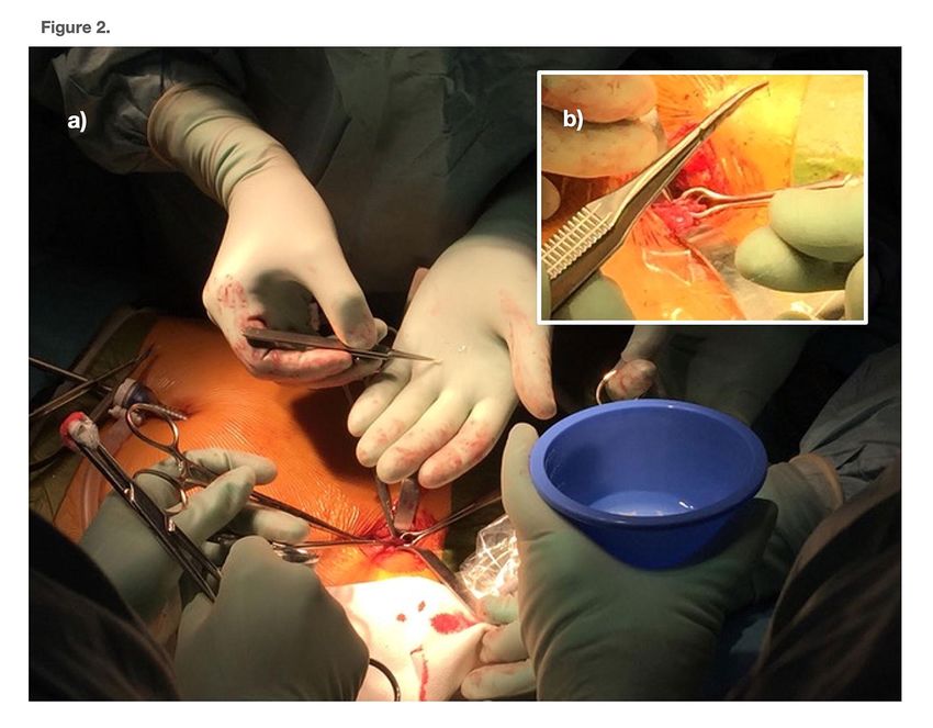
Twenty-seven ovarian tissue pieces were thawed and transplanted in November 2017 through a
modified laparoscopic technique that allowed the ovary to be exteriorized facilitating the
insertion of the ovarian tissue transplants in ovarian subcortical pockets (Figure 2).
The patient suspended hormonal substitution therapy the day before surgery but gonadotropins
still indicated postmenopausal levels one-month post-surgery. Ovarian engraftment was
followed-up through monitoring of clinical signs of estrogen secretion, climacteric symptoms,
and serum levels of gonadotropins, estrogen and Anti-Mullerian Hormone (AMH). The
gonadotropins returned to pre-menopausal levels 85 days after transplantation, increasing serum
estradiol levels were demonstrated, and climacteric symptoms improved (Figure 1). Antral
follicles were visualized on ultrasound and controlled ovarian stimulation with gonadotropins
(COS) using an antagonist protocol aiming at in vitro fertilization (IVF) was initiated. Four
attempts of IVF failed due to poor response, with only one oocyte retrieved that was not
fertilized. A second transplantation was therefore performed in November 2018 using the
remaining ovarian tissue that included 19 thawed pieces; of those 7 pieces had been previously
thawed in 2017 but not used for the molecular analysis and re-cryopreserved. Spontaneous
menstruation occurred 86 days following transplantation. On ultrasound scans the size of the
6uterus and the ovaries was significantly enlarged from baseline estimations, but the serum AMH
levels were low, 0.15μg/l, indicative of a much-reduced ovarian reserve. A new attempt at IVF
was initiated when gonadotropins returned to premenopausal levels and this treatment resulted in
one oocyte retrieved and fertilized by standard IVF technique. Embryo morphological
development was normal and transferred was at 4-cell stage. Pregnancy was established and the
patient delivered a healthy baby boy at 35+5 gestational weeks in November 2019. No
complications associated with prematurity occurred. Breast feeding was possible, but successful
only for three months and thereafter suspended. Menstrual cycles resumed three months after
delivery and continued for nine months until the woman achieved a natural conception
confirmed incidentally in November 2020 at time of consultation to our fertility center. On
ultrasonography, the pregnancy was confirmed as ongoing at gestational week 8. At the time of
this report the pregnancy is progressing into week 35th.
Although OTC have been reported in women and girls with leukemia in large series of fertility
preservation (2, 8), only isolated cases of ovarian tissue transplantation (OTT) in survivors of
hematological malignancies have been described to date (4, 8, 9). The majority of the reported
OTT for hematological malignancies concern lymphomas, while only a handful cases of OTT in
survivors of adult acute leukemia have been reported (Table 1) (3-5, 8).
Residual disease is often detected when the tissue is investigated with sensitive molecular
methods targeting leukemia-specific genetic markers and OTC has been performed before the
patient has achieved molecular remission (10-12). Also, xenografts of cryopreserved OT from
women diagnosed with CML and ALL into SCID mice could transfer leukemic cells,
demonstrating that viable malignant cells may be present in the grafts. However, only the
7xenografts with detectable leukemic markers, as determined by sensitive molecular methods,
showed leukemic cell transfer together with the xenografted OT (10). Importantly, studies have
shown that OTC retrieved after the patient has achieved complete remission in the bone marrow
in most cases also is negative for leukemia markers in the cryopreserved ovarian tissue (8, 13,
14) and OTCs that scored negative for molecular leukemic markers did not transfer leukemia to
the recipient mice (13). In a recent study Chevillon and coworkers investigated the presence of
minimal residual disease (MRD) in OTC harvested from acute leukemia patients that achieved
complete remission and showed significant concordance between bone marrow and OT;
however, 4/9 discordant patients had undetectable MRD in the bone marrow while positive in the
OT (15).
Taken together, these studies support that OTC harvested once the patient has achieved
remission in the bone marrow has a very low likelihood to transmit leukemia provided the OTC
is negative for the molecular marker present in the blast cells.
The present case shares several important features with the published reports of live births
following OTT in patients cured from acute leukemia that provide details on treatment preceding
the OTC (Table 1). In all cases the OTC was performed after induction and consolidation therapy
when the patient had achieved complete remission in the bone marrow (3-5). One important
common fact regarding OTT patients previously reported and our patient is that they had all
received allogeneic HSCT as part of the leukemia treatment and graft vs leukemia effect may
contribute to eliminate small amounts of leukemic cells potentially introduced with the OTT (3,
5). Another important aspect in common is the availability of a fusion transcript unique to the
malignant blasts that could be investigated in the OTT with very sensitive methods (3, 5).
8To the best of our knowledge three live births have been reported to date after ovarian tissue
transplantation in patients with adult acute leukemia (3-5). The present case is the third reported
of a live birth after OTT in a patient with acute leukemia and the first one in pediatric ALL, as
well as the first case where spontaneous pregnancy has additionally occurred after ovarian tissue
transplantation in a survivor from pediatric ALL that recovered fertility following the
transplantation procedures. The patient has been disease-free for 20 years after the diagnosis of
ALL, and at the time of this report 43 and 31 months have elapsed from the first and second
transplantations, respectively. Our results suggest that OT harvested in remission after several
courses of chemotherapy and cryopreserved may be a reproductive option for young women and
girls treated for ALL. However, one should be cautious and carefully inform the patients about
the limitations of the available methods to exclude persistence of leukemic cells in the ovarian
tissue. We believe important aspects to sustain a future plausible transplantation of the ovarian
tissue include the performance of OTC at a confirmed remission stage, as well as the availability
of using a relatively large portion of the OTC for safety assessments. Only if these assessments
point towards a low likelihood of risks can a decision to transplant ovarian tissue back to a
patient cured from cancer been made when she aims at regaining fertility.
Ethical approval:
Ethical approval for the study of children undergoing gonadal tissue cryopreservation aiming at
fertility preservation and for the follow-up of patients to adulthood was granted by the Ethical
Review Board of Karolinska University Hospital (Dnr. 427/03) and the Regional Ethics
Committee of Stockholm (Dnr. 2011/1158-31/2, 2014/470-32, 2016/2530-32 and 2018/2255-32).
9The patient has given her written informed consent to publish and discuss the case and the
pictures here included. The patient has also read and approved the submission of this
manuscript.
10REFERENCES
1. Rodriguez-Wallberg KA, Tanbo T, Tinkanen H, et al. Ovarian tissue cryopreservation and
transplantation among alternatives for fertility preservation in the Nordic countries - compilation
of 20 years of multicenter experience. Acta Obstet Gynecol Scand. 2016;95(9):1015-1026.
2. Rodriguez-Wallberg KA, Marklund A, Lundberg F, et al. A prospective study of women and
girls undergoing fertility preservation due to oncologic and non-oncologic indications in
Sweden-Trends in patients' choices and benefit of the chosen methods after long-term follow up.
Acta Obstet Gynecol Scand. 2019;98(5):604-615.
3. Shapira M, Raanani H, Barshack I, et al. First delivery in a leukemia survivor after
transplantation of cryopreserved ovarian tissue, evaluated for leukemia cells contamination.
Fertil Steril. 2018;109(1):48-53.
4. Silber SJ, DeRosa M, Goldsmith S, Fan Y, Castleman L, Melnick J. Cryopreservation and
transplantation of ovarian tissue: results from one center in the USA. J Assist Reprod Genet.
2018;35(12):2205-2213.
5. Sonmezer M, Ozkavukcu S, Sukur YE, Kankaya D, Arslan O. First pregnancy and live birth in
Turkey following frozen-thawed ovarian tissue transplantation in a patient with acute
lymphoblastic leukemia who underwent cord blood transplantation. J Assist Reprod Genet.
2020;37(8):2033-2043.
6. Keros V, Xella S, Hultenby K, et al. Vitrification versus controlled-rate freezing in
cryopreservation of human ovarian tissue. Hum Reprod. 2009;24(7):1670-1683.
7. Gabert J, Beillard E, van der Velden VH, et al. Standardization and quality control studies of
'real-time' quantitative reverse transcriptase polymerase chain reaction of fusion gene transcripts
for residual disease detection in leukemia - a Europe Against Cancer program. Leukemia.
2003;17(12):2318-2357.
8. Poirot C, Fortin A, Dhedin N, et al. Post-transplant outcome of ovarian tissue cryopreserved
after chemotherapy in hematologic malignancies. Haematologica. 2019;104(8):e360-e363.
9. Meirow D, Ra'anani H, Shapira M, et al.Transplantations of frozen-thawed ovarian tissue
demonstrate high reproductive performance and the need to revise restrictive criteria. Fertil
Steril. 2016;106(2):467-474.
10. Dolmans MM, Marinescu C, Saussoy P, Van Langendonckt A, Amorim C, Donnez J.
Reimplantation of cryopreserved ovarian tissue from patients with acute lymphoblastic leukemia
is potentially unsafe. Blood. 2010;116(16):2908-2914.
11. Meirow D, Hardan I, Dor J, et al. Searching for evidence of disease and malignant cell
contamination in ovarian tissue stored from hematologic cancer patients. Hum Reprod.
2008;23(5):1007-1013.
1112. Rosendahl M, Andersen MT, Ralfkiaer E, Kjeldsen L, Andersen MK, Andersen CY.
Evidence of residual disease in cryopreserved ovarian cortex from female patients with
leukemia. Fertil Steril. 2010;94(6):2186-2190.
13. Greve T, Clasen-Linde E, Andersen MT, et al. Cryopreserved ovarian cortex from patients
with leukemia in complete remission contains no apparent viable malignant cells. Blood.
2012;120(22):4311-4316.
14. Jahnukainen K, Tinkanen H, Wikstrom A, et al. Bone marrow remission status predicts
leukemia contamination in ovarian biopsies collected for fertility preservation. Leukemia.
2013;27(5):1183-1185.
15. Chevillon F, Clappier E, Arfeuille C, et al. Minimal residual disease quantification in ovarian
tissue collected from patients in complete remission of acute leukemia. Blood.
2021;137(12):1697-1701.
12Table 1: Outcomes of ovarian tissue transplantation in survivors of adult acute leukemia
Diagnosis Age Age Chemotherapy MRD Treatment Evaluation OTC Outcome Reference
OTC OTT prior to OTC bone after OTC MRD
marrow
at OTC
AML 26 37 2 x ADE CR Allogeneic Molecular ROF 8
HSCT (TBI*) detection of
leukemia-
specific marker
AML 19 27 1 x MAC, 1 x CR Allogeneic Molecular ROF 8
HDAC+ASPA HSCT detection of
leukemia-
specific marker
AML 19 32 Ara- CR Allogeneic Molecular 1 LB 3
C+Daunorubicin HSCT detection of
fusion transcript
+ xenograft
ALL 24 39 no information CR no no information 1 LB 4
information
AML 25 28 no information CR no no information ROF 4
information
ALL 19 26 VD+ASPA+MTX CR Allogeneic Molecular 1 LB 5
HSCT detection of
FluCy-TBI fusion transcript
+ IHC
OTC: ovarian tissue cryopreservation; OTT: ovarian tissue transplantation; MRD: minimal residual
disease; AML: acute myeloid leukemia; ALL: acute lymphoblastic leukemia; ROF: re-establishment
of ovarian function; ADE: Ara-C (Cytarabine), Daunorubicin, Etoposide; MAC: Myeloablative
conditioning Aracytine Mitoxantrone; HDAC+ASPA: Aracytine, L-Asparaginase; VD+ASPA+MTX:
Vincristine, Daunorubicin, L-Asparaginase, Methotrexate; CR: complete remission; HSCT:
hematopoietic stem cell transplantation, TBI: total body irradiation; FluCy: Fludarabine
cyclophosphamide; IHC: immunohistochemistry; LB: live birth
13FIGURE LEGENDS
Figure 1. Timeline from diagnosis of Acute Lymphoblastic Leukemia (ALL) to first
pregnancy
Abbreviations: p.o: per os; i.v: intravenously; i.th: intrathecal injection; TBI: Total Body
Irradiation; FSH: Follicle Stimulating Hormone; hMG: human menopausal gonadotropin; COS:
Controlled Ovarian Stimulation using gonadotropins for treatment involving assisted
reproduction; IVF: In vitro fertilization; IGF-1: Insulin-like growth factor 1; GH: Growth
Hormone; POF: Premature Ovarian Failure.
Laboratory reference for Serum-FSH: Postmenopausal >26 IU/L.
Serum AMH: 1 µg/L = 7,14 pmol/L
Figure 2. Transplantation of ovarian tissue
a) The first transplantation was conducted in November 2017. In total 27 ovarian tissue
pieces of size 1-2x4 mm were thawed and transplanted. The majority of ovarian pieces
were implanted in subcortical pockets using microsurgical instruments (10 in the right
ovary, and five in the left ovary) and 12 pieces in peritoneal pockets in the mesosalpinx.
All pockets were closed by non-absorbable sutures 6-0. The surgery and postoperative
period were uneventful. The second transplantation included 19 thawed pieces. Same
technique as previous transplant.
b) A modified laparoscopic technique was used, to allow the ovary being grasped
14with Babcock forceps and exteriorized through the anterior abdominal wall using a 3 cm
incision to facilitate inserting the tissue pieces.
15You can also read