Observations of coral and cryptobenthic sponge fluorescence and recruitment on autonomous reef monitoring structures - (ARMS)
←
→
Page content transcription
If your browser does not render page correctly, please read the page content below
Coral Reefs (2022) 41:877–883
https://doi.org/10.1007/s00338-022-02283-2

    NOTE

Observations of coral and cryptobenthic sponge fluorescence
and recruitment on autonomous reef monitoring structures
(ARMS)
Margaux Steyaert1,2 · Andrew Mogg3 ·
Nicholas Dunn2,4 · Rosalie Dowell2,4 ·
Catherine E. I. Head1,2

Received: 22 September 2021 / Accepted: 2 June 2022 / Published online: 28 June 2022
© The Author(s) 2022

Abstract Fluorescence imaging of benthic communities                   Introduction
is a widely used tool for determining the rate of hard coral
recruitment in tropical reefs. Whilst fluorescent proteins are         Recording the success and rate of the recruitment of reef
well-studied in scleractinian corals, less is understood about         benthic organisms can give us vital insights into coral reef
their distribution and function in other sessile reef inver-           health, recovery, and diversity. With high mortality bleach-
tebrates. This short study examines fluorescence images                ing events increasing in frequency across tropical reefs, and
of benthic communities on Autonomous Reef Monitoring                   in the face of bleak climate predictions (IPCC 2021), it is
Structures (ARMS) from a remote and protected Indian                   necessary to monitor recruitment patterns if we are to predict
Ocean reef system. We compare the abundance of adult and               and understand the state of future reefs and implement use-
juvenile hard corals across three sites and between the top-           ful management plans. This is especially true of reefs which
side and underside of ARMS recruitment plates. We also                 have experienced severe bleaching events in the past and
discuss observations of skeletal fluorescence in sponges,              can now be said to be at high risk from further large-scale
as well as uneven green fluorescent protein (GFP) concen-              climate-induced mortality events (Sheppard et al. 2020).
trations across adult coral colonies. Our findings provide                Reef sessile invertebrates, such as hard corals, soft cor-
an insight into the recovery of shallow reefs previously hit           als, sponges, ascidians, tube-forming worms and bivalve
by severe bleaching events and highlight the potential of              molluscs, shape or are anchored to the reef matrix. Cryptic
ARMS fluorescence imaging for the analysis of cryptoben-               surfaces and crevices within the reef matrix often harbour
thic communities.                                                      highly diverse communities of non-hard coral invertebrates
                                                                       and provide shelter for young coral recruits (Kornder et al.
Keywords Fluorescence · Recruitment · Coral · Sponge ·                 2021). Studying the recruitment of both hard corals and non-
GFP · Autonomous reef monitoring structures                            coral invertebrate recruitment in-situ can be complicated, as
                                                                       cryptic spaces are often inaccessible for sampling or pho-
                                                                       tography. Autonomous Reef Monitoring Structures (ARMS)
                                                                       are artificial recruitment devices used for the collection
Topic Editor: Anastazia Teresa Banaszak                                and study of diversity usually found in cryptic reef spaces
                                                                       (Carvalho et al. 2019; Pearman et al. 2020). Composed of
* Margaux Steyaert                                                     9 stacked PVC plates, with alternating gaps between each
  margaux.steyaert@zoo.ox.ac.uk                                        layer, each ARMS provides recruitment surfaces and varied
1
     Department of Zoology, University of Oxford,                      microhabitats for cryptobenthic fauna. These devices are
     Oxford OX1 3SZ, UK                                                now employed around the world to study these communi-
2
     Institute of Zoology, Zoological Society of London,               ties using a mix of standardised genetic and image analyses.
     London NW1 4RY, UK                                                   Fluorescence imaging is a popular census technique for
3
     Tritonia Scientific Ltd., Oban, Argyll PA37 1QA, UK               identifying coral recruits on artificial tiles or in-situ reef sur-
4
     Department of Life Sciences, Imperial College London,             faces (Baird et al. 2006; Zweifler et al. 2017). This method
     Silwood Park Campus, Ascot SL5 7PY, UK                            allows the capture of fluorescent pigments within organisms

                                                                                                                              13
                                                                                                                        Vol.:(0123456789)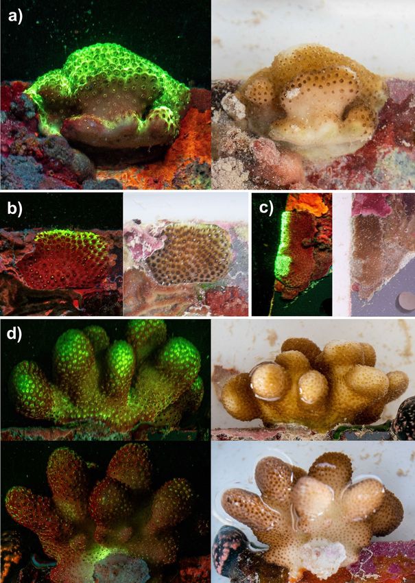
878 Coral Reefs (2022) 41:877–883
such as green fluorescent proteins (GFPs), other fluorescent Photographs were taken at night inside a shaded bin with
proteins (FPs) and photosynthetic pigments such as chloro- filtered seawater. Some plates were photographed multiple
phyll. Fluorescence imaging of hard coral recruits on arti- times to enhance resolution.
ficial recruitment tiles has been widely used, but no study Images were processed using Adobe Lightroom Classic
has yet used it to look at sessile communities on ARMS (for cropping, merging and enhancing brightness and con-
devices. In this study, we present results from high-resolu- trast). The number of hard corals was counted in each image;
tion fluorescence images of ARMS devices deployed across individuals smaller than 15 mm were recorded as juveniles,
the Chagos Archipelago, a remote and protected Indian whilst larger individuals were recorded as adults (Sheppard
Ocean reef system. We record abundances of hard coral et al. 2017).
adult colonies and juvenile recruits from sites previously Counts of corals were then plotted using the ‘ggplot2’
impacted by severe bleaching events (Head et al. 2019) and package in R (v3.3.5) and negative binomial generalised
present observations of the distribution of coral fluorescent linear models were used to test for differences in abundance
pigments across ARMS. Observations of fluorescence from between sites and ARMS (as the data is count-based and
the skeletal elements of several sponge specimens are also does not follow a normal distribution), using the ‘MASS’ and
presented and discussed. ‘vegan’ packages (v7.3.54 and v2.5.7, respectively)(Vena-
bles & Ripley, 2002; Wickham, 2016; Oksanen et al. 2020).
Materials and methods
Results and discussion
Fluorescence images were taken of Autonomous Reef
Monitoring Structures (ARMS) in April 2021 in the Cha- Hard coral recruitment across sampling sites
gos Archipelago Marine Protected Area (MPA) as part of a and ARMS microhabitats
wider research project on shallow reef benthic communities.
Triplicate ARMS devices were retrieved from a depth of A total of 268 hard corals were counted on ARMS devices
approximately 5–12 m across three sites across the northern across the three sampling sites, with an average of 35 indi-
atolls, including two exposed ocean-facing reefs (Ile Ang- viduals per m2. Juvenile recruits (< 15 mm) were consist-
laise, 5°20′04.7"S 72°12′48.5"E, and Moresby, 5°14′00.3"S ently more abundant than adult colonies, with 57 juveniles
71°49′50.3"E) and one sheltered lagoonal reef (Ile du Coin, and 34 adults found on average on each ARMS unit, and
5º27′04.5"S 71º46′30.8"E). ARMS plates photographed for a density of 22 juvenile and 13 adult individuals per m2
this article were retrieved and processed following stand- (Fig. 2). No significant differences in the abundance of adult
ardised Global ARMS NOAA protocols after a 36-month or juvenile corals were observed between sampling sites,
deployment (Leray et al. 2015). suggesting uniform recruitment patterns across sampled
A Sony RX100 MkII camera with a Nightsea 450 nm bar- reefs.
rier filter and two Inon Z240 UV strobe lights with Nightsea The abundance of juvenile corals was found to be equal
fluorescence excitation filters were used to capture sessile between the underside and topside of ARMS plates across
fluorescence on 153 plate faces across 9 ARMS devices (17 all sites. Adult coral abundance was also equal between plate
plate faces per device), from three shallow reef sites (Fig. 1). faces in Ile Anglaise and Moresby but was significantly
Fig. 1 Fluorescence images of
two Autonomous Reef Monitor-
ing Structure (ARMS) recruit-
ment plates (23 cm × 23 cm).
Red boxes highlight scleractin-
ian coral adult colonies and
juvenile recruits. The left-hand
image is of a ‘closed’ surface,
where PVC bars were placed
across this plate and its adjoin-
ing neighbour to create four
distinct recruitment surfaces,
and the right-hand image is the
equivalent with no PVC bars
13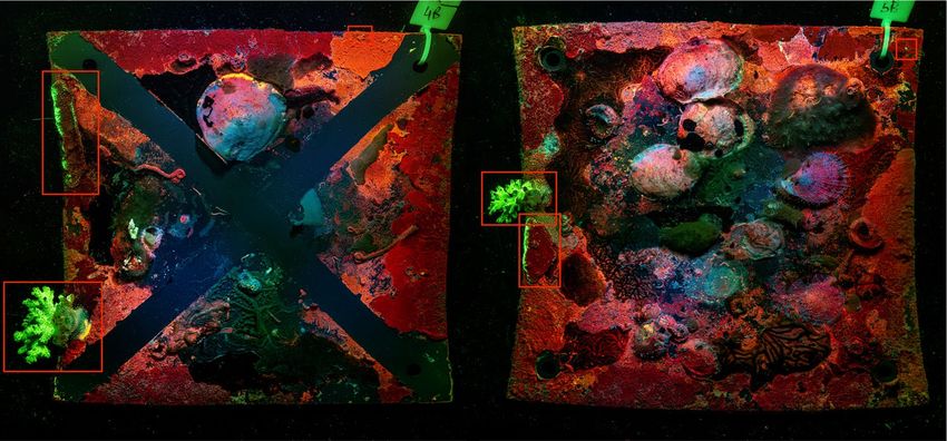
Coral Reefs (2022) 41:877–883                                                                                                            879

Fig. 2  Boxplots displaying count abundances of (a) all hard corals across top and bottom ARMS plate face images and of (b) juvenile recruits
and adult coral colonies on ARMS devices across sampling sites

higher on plate undersides in Ile du Coin (p < 0.001***).                  The highly standardised format of ARMS makes them
This reef site is sheltered from prevailing currents and higher         an advantageous tool for studying coral recruitment with
sedimentation has been observed in-situ compared to the two             minimal disturbance to the natural matrix, as well as moni-
exposed reef sites. Juvenile recruit survival may be lower              toring in-situ coral density of cryptic surfaces. However,
on the topside of ARMS plates at this site due to sediment              whilst previous work has shown ARMS-based communi-
loading.                                                                ties are comparable to those found across dead coral heads
   The surface area of cryptic reef cavities has been shown             (Plaisance et al. 2011), further work is now required to deter-
to exceed that of exposed benthos by up to a factor of eight            mine whether ARMS’s PVC plates bias the attachment and
(Scheffers et al. 2010), meaning exposed surfaces represent             survival of hard coral juveniles compared to exposed natu-
only a portion of available recruitment space on the reef               ral reef surfaces. Fluorescence imaging allows for a quick
matrix. ARMS provide ideal refugia for non-photosynthe-                 scan of adult and juvenile recruits across ARMS plates but
sising cryptobenthic invertebrates but inadvertently also pro-          may overestimate counts due to increased signal to noise
vide a desirable settlement surface for hard corals. Similar            ratio or underestimate them due to genetic variability or the
patterns of coral recruitment have been observed in other               fact that shaded ARMS surfaces may lead to minimal or
studies using artificial settlement tiles, with higher juvenile         absent recruit fluorescence. Further work investigating coral
coral recruitment recorded in cryptic and grooved ridges                recruitment on ARMS could include both daylight and fluo-
than on flat exposed surfaces (Mallela 2018). Tight gaps                rescence counts to allow for meaningful comparisons with
between ARMS plates likely provide protection from graz-                similar studies.
ers and physical damage (e.g. from loose rubble), resulting
in coral colonies growing across sampling sites. Our results            Observations of fluorescence concentration
provide new insights into coral recruitment across reefs                across colonies
which are still recovering from back-to-back high-mortal-
ity bleaching events (Sheppard et al. 2020) and highlight               Fluorescent proteins are ubiquitous in scleractinian corals,
the importance of investigating both exposed and hidden                 but their functional role has often been a highly debated
surfaces when assessing coral recruitment rates.                        topic. Green fluorescent proteins (GFP) have been suggested

                                                                                                                                 13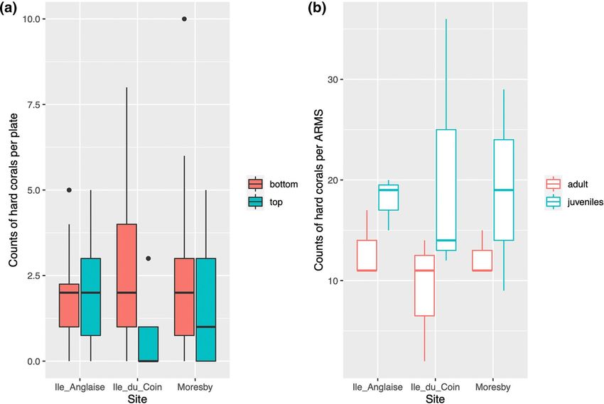
880                                                                                               Coral Reefs (2022) 41:877–883

to play multiple roles including photoprotection (Smith et al.   polyps along the topside of an adult Pocillopora sp. colony
2013), internal light regulation in mesophotic corals (Smith     growing off the side of an ARMS plate (i.e. facing towards
et al. 2017), immune response regulation (Palmer et al.          surface light) were shown to emit brighter fluorescence than
2009), and to attract symbionts (Field et al. 2006; Aihara       polyps found on the underside of this colony (i.e. facing reef
et al. 2019).                                                    benthos) (Fig. 3d).
   Several adult hard coral colonies displayed uneven con-          ARMS plates are stacked closely together with only
centrations of green fluorescence across ARMS plates             1-2 cm gaps between each plate face; this likely blocks a
(Fig. 3). Encrusting colonies were observed to emit the          large amount of daylight from reaching the centre of each
brightest fluorescence closer to and along the exposed edges     device. Patterns of fluorescence concentration across hard
of ARMS recruitment plates (Fig. 3a, b and c). Furthermore,      coral colonies were consistent with areas of ARMS plates

Fig. 3  Images of the same scle-
ractinian coral colonies under
UV light and daylight, where
differences in GFP concentra-
tion can be observed across
colonies close to ARMS plate
edges (a, b and c) and between
the topside (top left and right-
hand images) and underside
(bottom left and right-hand
images) of the same Pocillopora
spp. colony (d)

13
Coral Reefs (2022) 41:877–883 881
most exposed to direct sunlight (i.e. the outer edge of plates). This in turn likely benefits the metabolic activity of pho-
Our results support similar findings from other studies ana- totrophic organisms and their associated sponge host. We
lysing coral fluorescence patterns in response to controlled hypothesise that fluorescence observed here likely originates
(D’Angelo et al. 2008; Smith et al. 2013) or in-situ (Bollati from algae which developed in close association with sponge
et al., 2020) light conditions. skeletal elements, and ongoing genetic and microscopy work
will determine specimen taxonomy. Sponge fluorescence
Fluorescence patterns in sponges and other on coral reefs is poorly documented or understood and has
cryptobenthic taxa not previously been reported from sponge specimens in the
Chagos Archipelago. Furthermore, to our knowledge, in-situ
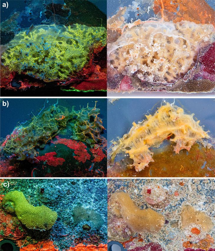
Green and orange fluorescence was also observed across the images of fluorescing tropical reef sponges of this kind have
skeletal elements of a few sponge specimens, from ARMS not previously been published.
in the exposed Ile Anglaise site (Fig. 4). Deep-sea glass Fluorescence was also observed from other ses-
sponge spicules have been shown to have fibre-optical fea- sile invertebrates across ARMS plates, such as solitary
tures (Sundar et al. 2003), and the close association between ascidians (yellow-green fluorescence), limpets (red fluo-
green algae and the siliceous spicules of demosponge Tethya rescence), and from the opercula of serpulid calcareous
seychellensis has been hypothesised, and since confirmed, to worms (green fluorescence). Almost all research on fluo-
serve as a natural pipeline for light (Gaino and Sara 1994). rescence from coral reef benthic communities focuses on
Fig. 4 Images of sponge
specimens on ARMS recruit-
ment plates under both UV light
(left-hand side) and daylight
(right-hand side)
13882 Coral Reefs (2022) 41:877–883
hard corals, and little is known about the presence and role Carvalho S, Aylagas E, Villalobos R, Kattan Y, Berumen M, Pearman
of fluorescent proteins in non-coral sessile reef inverte- JK (2019) Beyond the visual: using metabarcoding to character-
ize the hidden reef cryptobiome. Proc Biol Sci 286:20182697
brates (Zawada and Mazel 2014). Multicolour fluorescence D’Angelo C, Denzel A, Vogt A, Matz MV, Oswald F, Salih A, Nien-
is observed across ARMS plates (Fig. 1), and we recom- haus GU, Wiedenmann J (2008) Blue light regulation of host
mend that future ARMS studies could use fluorescence pigment in reef-building corals. Mar Ecol Prog Ser 364:97–106
imaging to extract quantitative information from crypto- David R, Uyarra MC, Carvalho S, Anlauf H, Borja A, Cahill AE,
Carugati L, Danovaro R, De Jode A, Feral J, Guillemain D,
benthic communities. Martire ML, D’Avray LT, Ville De, Pearman JK, Chenuil A
Benthic communities found on ARMS devices have been (2019) Lessons from photo analyses of autonomous reef moni-
shown to be highly diverse (Carvalho et al. 2019; Pearman toring structures as tools to detect (bio-)geographical, spatial,
et al. 2020), with both genetic and image analysis methods and environmental effects. Mar Pollut Bull 141:420–429
Field SF, Bulina MY, Kelmanson IV, Bielawski JP, Matz MV (2006)
required to determine community composition and diver- Adaptive evolution of multicolored fluorescent proteins in reef-
sity patterns (Pearman et al. 2016). Analysis of ARMS plate building corals. J Mol Evol 62:332–339
images has so far been conducted under white light, followed Gaino E, Sara M (1994) Siliceous spicules of Tethya seychellensis
by a random point count approach to determine the recruit- (Porifera) support the growth of a green alga: a possible light
conducting system. Mar Ecol Prog Ser 108:147–152
ment cover and composition of sessile communities (David Head CEI, Bayley DTI, Rowlands G, Roche RC, Tickler DM, Rog-
et al. 2019). This approach is ideal for determining overall ers AD, Koldewey H, Turner JR, Andradi-Brown D (2019)
functional composition but is likely inaccurate for recording Coral bleaching impacts from back-to-back 2015–2016 ther-
coral recruit abundance. Our study of ARMS demonstrates mal anomalies in the remote central Indian Ocean. Coral Reefs
38:605–618
how fluorescence imaging of these devices could be an ideal IPCC (2021) Climate change 2021: the physical science basis. Con-
standardised tool for studying the in-situ recruitment of hard tribution of working Group I to the sixth assessment report of
corals as well as the presence and patterns of fluorescent the Intergovernmental Panel on Climate Change. Cambridge
proteins in cryptobenthic invertebrates. University Press. In press
Kornder N, Cappelletto J, Mueller B, Zalm M, Martinez SJ, Vermeij
M, Huisman J, De Goeij J (2021) Implications of 2D versus 3D
Acknowledgements This work was funded by the Bertarelli Founda- surveys to measure the abundance and composition of benthic
tion as part of the Bertarelli Marine Science Programme. coral reef communities. Coral Reefs 40:1137–1153
Leray M, Knowlton N (2015) DNA barcoding and metabarcoding of
Declarations standardized samples reveal patterns of marine benthic diver-
sity. Proc Natl Acad Sci USA 112:2076
Conflict of interest On behalf of all authors, the corresponding au- Mallela J (2018) The influence of micro-topography and external
thor states that there is no conflict of interest. bioerosion on coral-reef-building organisms: recruitment, com-
munity composition and carbonate production over time. Coral
Open Access This article is licensed under a Creative Commons Reefs 37:227–237
Attribution 4.0 International License, which permits use, sharing, adap- Oksanen J, Blanchet G, Friendly M, Kindt R, Legendre P, McGlinn
tation, distribution and reproduction in any medium or format, as long D, Minchin PR, O’Hara RB, Simpson GL, Solymos P, Henry
as you give appropriate credit to the original author(s) and the source, M, Stevens H, Szoecs E, Wagner H (2020). Vegan: community
provide a link to the Creative Commons licence, and indicate if changes ecology package. R package version 2.5–7. https://CRAN.R-
were made. The images or other third party material in this article are project.org/package=vegan
included in the article’s Creative Commons licence, unless indicated Palmer CV, Modi CK, Mydlarz LD (2009) Coral fluorescent proteins
otherwise in a credit line to the material. If material is not included in as antioxidants. PLoS ONE 4:e7298
the article’s Creative Commons licence and your intended use is not Pearman JK, Anlauf H, Irigoien X, Carvalho S (2016) Please mind
permitted by statutory regulation or exceeds the permitted use, you will the gap – Visual census and cryptic biodiversity assessment at
need to obtain permission directly from the copyright holder. To view a central Red Sea coral reefs. Mar Environ Res 118:20–30
copy of this licence, visit http://creativecommons.org/licenses/by/4.0/. Pearman JK, Chust G, Aylagas E, Villarino E, Watson JR, Chenuil A,
Borja A et al (2020) Pan-regional marine benthic cryptobiome
biodiversity patterns revealed by metabarcoding Autonomous
Reef Monitoring Structures. Mol Ecol 29:4882–4897
Plaisance L, Caley MJ, Brainard RE, Knowlton N (2011) The diver-
References sity of coral reefs: what are we missing? PLoS ONE 6:e25026
Scheffers S, van Soest R, Nieuwland G, Bak R (2010) Coral reef
framework cavities: is functional similarity reflected in com-
Aihara Y, Maruyama S, Baird AH, Iguchi A, Takahashi S, Minagawa position of the cryptic macrofaunal community? Atoll Res Bull
J (2019) Green fluorescence from cnidarian hosts attracts sym- 583:1–24
biotic algae. PNAS 116:2118–2123 Sheppard C, Sheppard A, Mogg A, Bayley D, Dempsey AC, Roche
Baird AH, Salih A, Trevor-Jones A (2006) Fluorescence census R, Turner J, Purkis S (2017) Coral bleaching and mortality in
techniques for the early detection of coral recruits. Coral Reefs the Chagos Archipelago. Atoll Res Bull 613:1–26
25:73–76 Sheppard C, Sheppard A, Fenner D (2020) Coral mass mortalities
Bollati E, D’Angelo C, Alderdice R, Pratchett M, Ziegler M, Wieden- in the Chagos Archipelago over 40 years: regional species and
mann J (2020) Optical feedback loop involving dinoflagellate assemblage extinctions and indications of positive feedbacks.
symbiont and scleractinian host drives colorful coral bleaching. Mar Pollut Bull 154:111075
Curr Biol 30:2433–2445
13Coral Reefs (2022) 41:877–883 883
Smith EG, D’Angelo C, Salih A, Wiedenmann J (2013) Screening Zawada DG, Mazel CH (2014) Fluorescence-based classification
by coral green fluorescent protein (GFP)-like chromoproteins of Caribbean coral reef organisms and substrates. PLoS ONE
supports a role in photoprotection of zooxanthellae. Coral Reefs 9:e84570
32:463–474 Zweifler A, Akkaynak D, Mass T, Treibitz T (2017) In situ analysis of
Smith EG, D’Angelo C, Sharon Y, Tchernov D, Wiedenmann J coral recruits using fluorescence imaging. Front Mar Sci 4:273
(2017) Acclimatization of symbiotic corals to mesophotic light
environments through wavelength transformation by fluorescent Publisher’s Note Springer Nature remains neutral with regard to
protein pigments. Proc r Soc b: Biol Sci 284:20170320 jurisdictional claims in published maps and institutional affiliations.
Sundar VC, Yablon AD, Grazul JL, Ilan M, Aizenberg J (2003)
Fibre-optical features of a glass sponge. Nature 424:899–900
Venables WN, Ripley BD (2002) Modern applied statistics with S,
4th edn. Springer, New York
Wickham H (2016) ggplot2: elegant graphics for data analysis.
Springer-Verlag, New York
13You can also read


