Ophthalmology Department of - Our Third Century of Leadership in Education - New York Eye and Ear Infirmary
←
→
Page content transcription
If your browser does not render page correctly, please read the page content below
Department of
                         Ophthalmology
                         Staying Focused:
                         Our Third Century of
                         Leadership in Education

SPECIALT Y REP ORT   |    FALL 202 1   |   W W W.NYEE .EDU
CONTENTS
                                  4    Message From the Department of Ophthalmology Leadership

                                  6    Reimagining NYEE for the Next Century of Growth

                                  10   Oldest Eye Hospital in America Is Hitting a New Stride With Its Training Program
                                       ⚫   Recognizing a Legacy of Passion and Dedication to Microsurgery and Education

                                  14   For One Researcher, the Fight to Cure Blindness Is Personal

           ring Our                    ⚫   A Promising New Pathway Toward Retinal Cell Replacement Therapy

         no         P                  ⚫   Racing Against Time to Discover a Gateway to COVID-19 Transmission

                           as
                                  20   An Innovative New Role for OCT Angiography in the Battle Against Sickle Cell Retinopathy
    Ho

                             t
                                  22   A Novel Gene Therapy Approach to Help Prevent Retinal Vision Loss

                                  24   Going Deeper Into the Microworld of Ocular Disease

                                  26   Coffee Alert for Those at High Risk of Glaucoma

                                  27   Reassurance for Patients With Glaucoma Drainage Implants
    En

                             re
                                  28   Cataract Surgery in the Tightest of Spaces
            1820 - 2020
     sio
                           tu
    vi

                             u
                                  30   When a ‘Less Is More’ Approach Is Best Medicine for a Newborn

           n in g       ur F      33   A Severe Infection Leads to a ‘Huge Save’ for Surgeon and Her Young Patient

                    O             36   A New Application of Orbital Brachytherapy Helps a Patient Keep Her Eye and Her Vision

                                  38   Innovative New Eye Stroke Program Proves a ‘Godsend’ for a Patient

                                  41   The Surgical Robot Is Learning New Skills

                                  45   Department of Ophthalmology at a Glance

                                  46   Accolades and Retirees

                                  48   Faculty News

2
MESSAGE              FUTURE            TRAINING PROGRAM               RESEARCH           PATIENT STORIES            CLINICAL PROGRAMS                  NYEE STATS   ACCOLADES AND RETIREES   FACULTY NEWS

Message From the Department
Of Ophthalmology Leadership
Fresh off our bicentennial             • Our researchers uncovered a        we do at NYEE. That dynamic
year, we immediately began               novel gene therapy approach to     will intensify as we draw on
writing the next chapter of our          save retinal ganglion cells and    our rich capabilities in the                    James C. Tsai, MD, MBA
storied history with a narrative         preserve vision in the treatment   areas of imaging, genomics,              President, New York Eye and Ear Infirmary
                                         of severe injuries involving       mathematical modeling, stem                           of Mount Sinai
driven by the prominent role
                                                                                                                       Chair, Department of Ophthalmology,
of technology, research, and             the optic nerve and blinding       cells, and artificial intelligence to
                                                                                                                    Icahn School of Medicine at Mount Sinai and
education in supporting the              diseases like glaucoma.            uncover upstream factors that
                                                                                                                            Mount Sinai Health System
nationally recognized clinical                                              contribute to such common and
care we provide to patients and        Our commitment to the next           complex diseases as macular
the community at large. Consider       generation of ophthalmologists       degeneration and glaucoma. We
some of the ways New York Eye          through the largest academic         are also investigating the ocular
and Ear Infirmary of Mount Sinai       training program in the country      biomarkers of these diseases
(NYEE) left its imprint on the field   remains a top priority, as           and many others with the help
of ophthalmology over the past         evidenced by our new course in       of artificial intelligence and
year, as described in this report:     ophthalmic micro-interventional      sophisticated imaging modalities
                                       robotic surgery for residents        like OCT angiography and
• We enhanced the capabilities         and fellows. The course, at the      adaptive optics.
    of, and readied for clinical       Jorge N. Buxton, MD, and Douglas                                                          Paul A. Sidoti, MD
    trials, the country’s first        F. Buxton, MD, Microsurgical         At the same time, we are                  Site Chair, Department of Ophthalmology,
    ophthalmic robotic assistant,      Education Center, offers NYEE        preparing for fundamental                      New York Eye and Ear Infirmary
    a highly sophisticated device      trainees exposure to the next        change to our downtown                                  of Mount Sinai
    that promises to change the        level of technology and precision    Manhattan campus, and to
    face of micro-interventional       surgery unavailable anywhere         the way we deliver service
    ophthalmic surgery.                else in the United States. We also   to not just individual patients
                                       completed the inaugural year         but communities and entire
• We implemented a model tele-         of our Joint Internship Program,     populations. We remain firmly
    consult program with stroke        which is affording our trainees      bound to the founding principles
    teams at several of Mount          valuable exposure to ophthalmic      that have allowed us to grow
    Sinai’s largest hospitals to       care and even research before        and excel in our specialized
    treat patients with eye stroke,    they begin their first year of       field of medicine. That means
    collapsing the time it typically   residency.                           an unwavering commitment to
    takes to get a diagnosis so that                                        programs and investments that                 Louis R. Pasquale, MD, FARVO
    treatment can begin within         Through our cutting-edge             will enable us to continue to lead        Site Chair, Department of Ophthalmology,
    the narrow window needed to        research and clinical programs,      in clinical care, education, and                The Mount Sinai Hospital and
                                                                                                                                  Mount Sinai Queens
    ensure the best outcomes.          technology permeates everything      research for the next 200 years.

4                                                                                                                                                                                                       5
MESSAGE                 FUTURE              TRAINING PROGRAM         RESEARCH            PATIENT STORIES                		   CLINICAL PROGRAMS                             NYEE STATS          ACCOLADES AND RETIREES                        FACULTY NEWS

Reimagining NYEE                                                                                                                                                                                                                                  New entrance to
                                                                                                                                                                                                                                             Mount Sinai Beth Israel

For the Next Century
                                                                                                                                                                                                                                               main campus at 17th
                                                                                                                                                                                                                                                Street, co-branded
                                                                                                                                                                                                                                                       with NYEE.*

Of Growth

                                            The twenty-first century
                                            health care landscape
                                            is being reconfigured by
                                            changes that are breathtaking
                                            in their scope and intensity.
                                            They include the migration
                                            of patient care from the
                                            hospital out to the community,
                                            the implementation of site-
                                            neutral payments and their
                                            impact on hospital revenue,
                                            and the introduction of bold
                                            new technologies such as
                                            telemedicine and artificial       As New York Eye and Ear Infirmary              discovery to find novel treatments                Mount Sinai Health System to obtain        Beyond infrastructure, the future
NYEE-branded ambulatory surgery center in                                     of Mount Sinai (NYEE) embarks                  for ocular diseases, and unparalleled             the resources necessary to bring           of our institution will be molded by
Manhattan.*                                 intelligence, all of which will   on its third century of growth                 education of future ophthalmic                    this new ambulatory care blueprint to      the wide-ranging research that is
                                                                              and innovation, the institution is             leaders,” says James C. Tsai, MD,                 fruition. The new ambulatory surgical
                                            affect how institutions deliver                                                                                                                                               recognized internationally. That
                                                                              confronting these changes head                 MBA, President of NYEE and Chair                  center near Manhattan’s Madison            initiative is powered by our rich
                                            their patient care services.      on. This will require strategic                of the Department of Ophthalmology                Square Park will add another patient       capabilities in the areas of imaging,
                                                                              implementation of a sustainable                at the Icahn School of Medicine at                care site to NYEE’s growing portfolio      genomics, mathematical modeling,
                                            Most recently, the unremitting    model that allows the institution              Mount Sinai and the Mount Sinai                   of faculty practice sites as well as       artificial intelligence, stem cells, and
                                            COVID-19 pandemic is              to continue to grow and remain                 Health System. “But there will likely be          affiliated clinical sites and teaching     viral vectors. “We’ll continue to support
                                                                              economically strong, while still               major changes to our campus as we                 institutions throughout the New York       basic research that’s dedicated to
                                            forcing every health care         maintaining its mission to serve the           open up NYEE-branded ambulatory                   metropolitan region. And the institution   the preservation and restoration of
                                            system to intelligently adapt     community.                                     surgery center settings, and as we                is collaborating with the Health System    vision, and which creates opportunities
                                                                                                                             modernize our aging facilities to make            to modernize and colocate its clinical,    for our clinical faculty to translate
                                            to and invest in solutions to     “We’re going to thrive for the next            them compatible with health care in               educational, and research resources        science into advanced patient care,”
                                                                              two centuries as we remain more                the twenty-first century.”                        near and around the campus of Mount        emphasizes Louis R. Pasquale, MD,
                                            meet the challenges of future     committed than ever to our mission                                                               Sinai Beth Israel, a few blocks away       FARVO, Director of the Mount Sinai/
                                            disruptions to patient care.      of world-class patient care, scientific        NYEE is partnering with the                       from the existing NYEE campus.             New York Eye and Ear (NYEE) Eye
6                                                                                                                            *Preliminary renderings, subject to change.                                                  continued ›                                  7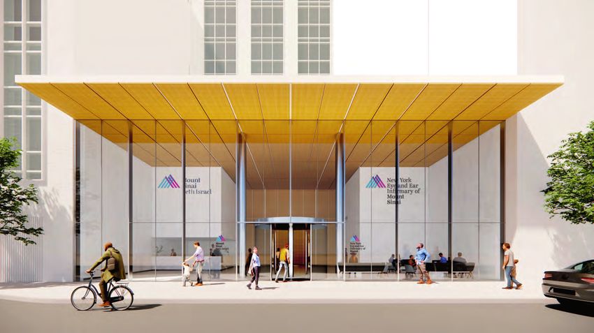
MESSAGE                   FUTURE                 TRAINING PROGRAM                            RESEARCH                    PATIENT STORIES            CLINICAL PROGRAMS                             NYEE STATS            ACCOLADES AND RETIREES                                FACULTY NEWS

continued from page 7

The Jorge N. Buxton, MD, and Douglas F. Buxton, MD, Microsurgical Education Center*

                                                                                                                                                    Modernized Emergency Department at Mount Sinai Beth Israel, co-branded with NYEE, with dedicated exam rooms for Ophthalmology and ENT cases.*
and Vision Research Institute and                and Regenerative Biology, whose lab                 capabilities, and to use stem cell and
Site Chair of Ophthalmology, The                 is making considerable headway in                   viral vector technologies to enable the
Mount Sinai Hospital and Mount Sinai             unraveling the biology of the human                 long-sought-after reconstitution of            Jorge N. Buxton, MD, and Douglas F.               trove of experience over the past 200            relocating,” says Dr. Tsai. “But what
Queens. “That means working as we                eye to uncover mechanisms of cell                   retinal cells.                                 Buxton, MD, Microsurgical Education               years to reimagine how we will remain            has been constant is the collective
have in the past with pharmaceutical             regeneration. The Department of                                                                    Center, one of the most advanced                  in the forefront of patient care and             spirit and ethos, the commitment to
and industry partners on research                Ophthalmology is streamlining the work              Education is also central to NYEE’s            training labs in the country.                     innovation going forward—something               our mission to serve the community.
with clinical potential, while                   of its research faculty by organizing               blueprint for the future. “We’re not                                                             our predecessors have had to do in               That is the very core of NYEE:
expanding projects initiated by our              them into teams to tackle challenges                just continuing our training programs          As NYEE looks forward into its third              years past.”                                     our incredible faculty, voluntary
own clinician-scientists, and those              such as finding upstream factors                    for residents, fellows, and medical            century of growth, the institution’s                                                               physicians, staff, and trainees infused
supported by the NIH.”                           that contribute to complex diseases                 students, but actually growing and             leadership recognizes that fundamental            While NYEE’s present location at                 with the spirit of our founders to serve
                                                 through novel analytical epidemiology               improving them, knowing how essential          change will impact virtually every part of        East 14th Street and Second Avenue               the community, train ophthalmic
Underscoring the broad sweep of                  and big data approaches. Also, teams                they are to providing the highest-quality      it. “This kind of change can be unsettling        in Manhattan’s East Village has                  leaders of tomorrow, and move the
our research is the work of talented             are being assembled to discover ocular              clinical care to our patients,” says Paul A.   for some, but it’s also an incredible             been its home since 1856, the site is            field of ophthalmology forward with
scientists like Timothy A. Blenkinsop,           biomarkers for systemic disease                     Sidoti, MD, Deputy Chair for Education         opportunity to grow and improve on the            actually NYEE’s eighth location. “Over           breakthrough innovation. And we will
PhD, Associate Professor of                      with the help of artificial intelligence            and Site Chair of Ophthalmology, NYEE.         original mission of NYEE,” asserts Dr.            the years, the institution has had to            take that esprit de corps with us no
Ophthalmology, Cell, Developmental,              and NYEE’s sophisticated imaging                    The centerpiece of that effort is the          Tsai. “It’s a chance for us to draw on our        evolve and grow, and that often meant            matter where we are situated.”

                                                                           Ambulatory Eye Clinics: Reception area and exam room*                    The Shelley and Steven Einhorn Clinical Research Center: Waiting room and ophthalmic imaging center*

8                                                                                                                                                   *Preliminary renderings, subject to change.                                                                                                     9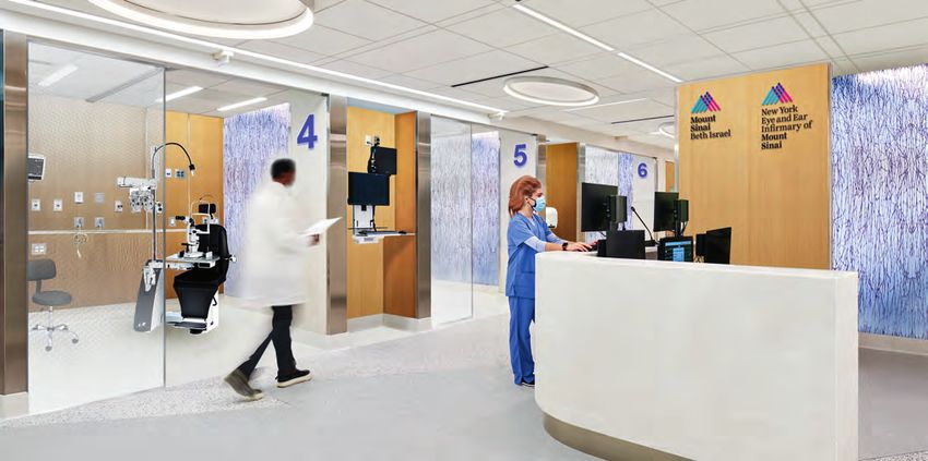
MESSAGE FUTURE TRAINING PROGRAM RESEARCH PATIENT STORIES CLINICAL PROGRAMS NYEE STATS ACCOLADES AND RETIREES FACULTY NEWS
Oldest Eye Hospital in America
Is Hitting a New Stride With Its
Training Program
As New York Eye and Ear Infirmary of Mount Sinai (NYEE) redraws its operational
blueprint to meet the demands of a pandemic-driven world, it remains more
committed than ever to expanding its educational program for ophthalmic residents
and fellows—one of the largest and most respected training initiatives in the country.
“Education is basic to our mission and exposes them to each of the them to faculty members to review
of providing outstanding clinical subspecialty clinics at NYEE, as and critique synchronously or at a
care, and for that reason, our well as the Jorge N. Buxton, MD, and later time. By the same token, the
program to train the next generation Douglas F. Buxton, MD, Microsurgical new capabilities will allow faculty to
of ophthalmologists will not only Education Center. videotape surgical procedures and
remain a top priority for New York techniques in the lab, then store them
Eye and Ear, it will continue to grow “We saw dramatic evidence of how in an online library for ready access
and improve in the years ahead,” these trainees were able to develop by residents as part of their surgical
says Paul A. Sidoti, MD, Deputy their skills in just three months here, training.
Chair for Education and Site Chair of prior to starting their first year of
Ophthalmology for NYEE. residency,” says Dr. Reddy. A number “The AV upgrades will allow us to
of trainees, he adds, took the Joint formally integrate lab activity into
There is no better marker of that Internship Program a step further the curriculum,” explains Douglas
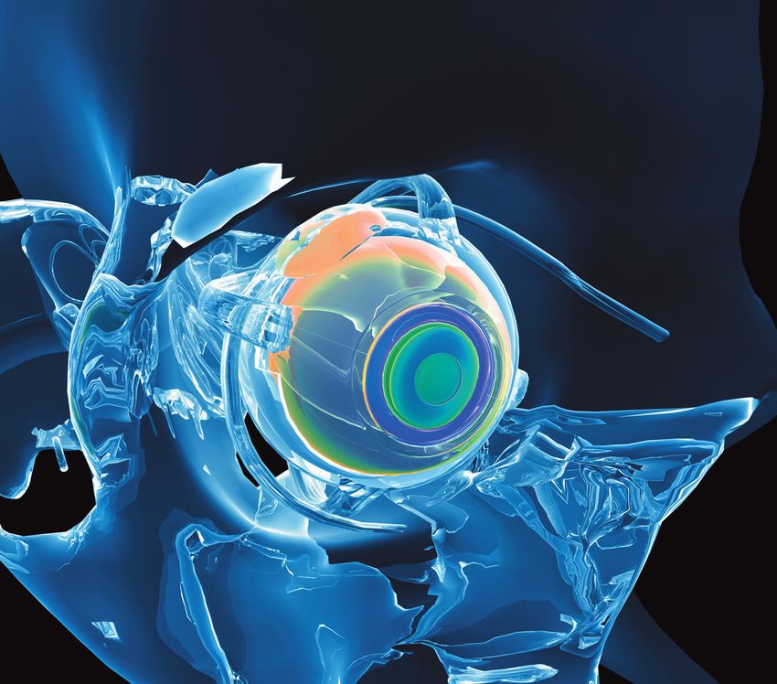
commitment than the novel Joint by using it as a platform to begin F. Buxton, MD, Clinical Professor of AUPO SCOR skills transfer course conducted in
June at NYEE. Top: Trainees developing advanced
Internship Program, which notched research projects. Ophthalmology at the Icahn School
phacoemulsification skills including stop and chop and
its first-year anniversary in July 2021. of Medicine at Mount Sinai, and son
phaco chop in the Buxton Microsurgical Center.
A special curriculum was developed Change is also critical to the future of of Jorge N. Buxton, MD, the first chief
Bottom: A dry-lab session with trainees practicing
for the 10 participating students, the Buxton Microsurgical Education of cornea service at NYEE and a advanced anterior segment skills.
whose one-year internship at Mount Center, where students learn and champion of modern microsurgery.
Sinai Beth Israel now includes nine practice in a wet lab setting the “The new ‘smart’ cameras are
months of general medicine and intricate microsurgical skills they’ll connected to the internet, which will
three months of ophthalmology soon bring to patients in the operating make it possible to stream real-time
training at NYEE. According to room. Under way are sophisticated lab activity to any location. And that
Harsha S. Reddy, MD, Ophthalmology audio-visual improvements to this creates the opportunity for worldwide
Residency Program Director and Site premier site. Specifically, high- educational classes from NYEE.”
Director for Oculoplastics, Orbital, resolution video cameras are being
and Reconstructive Surgery at NYEE installed at four of the wet lab’s 16 Technologically, the Buxton lab
and Mount Sinai Beth Israel, that microscope-equipped training is already among the leading
tailored curriculum teaches students stations, allowing residents to tape ophthalmic surgical training sites in
the basics of the ophthalmic exam their practice sessions and then relay the country. In 2006, a virtual reality
10 continued › 11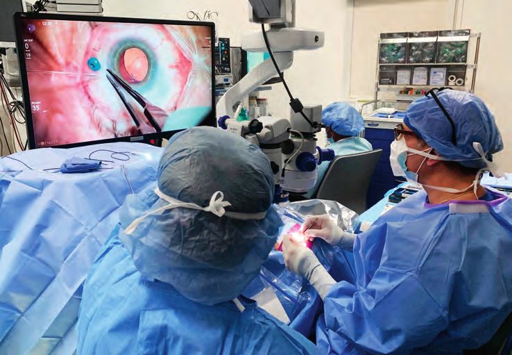
MESSAGE FUTURE TRAINING PROGRAM RESEARCH PATIENT STORIES CLINICAL PROGRAMS NYEE STATS ACCOLADES AND RETIREES FACULTY NEWS
continued from page 11
and techniques. The program,
known as SCOR (Surgical
Curriculum for Ophthalmology Recognizing
Residents), brought together
15 residents throughout the
A Legacy of
New York metropolitan area
for a combination of online and
Passion and
hands-on training prior to rolling Dedication to
out the first wet lab course to 74
third-year residents at a AUPO- Microsurgery
sponsored event in the fall of 2021.
“The small scale wet lab we held And Education
at NYEE generated very positive
feedback from trainees and faculty
alike, and that’s enabling us to
Left to right: Richard B. Rosen, MD,
further improve the curriculum James C. Tsai, MD, Amalia Buxton,
Left to right: Bernadette Maliwat, RN, voluntary attending John Flanagan, MD, and Michael Chua, MD,
to ensure the best possible Douglas F. Buxton, MD, and
resident class of 2021, performing cataract surgery
training experience for residents,” Julia Fallon, MD
says Steven Feldon, MD, MBA,
Executive Vice President of AUPO.
Since 2004, a newly renovated, fully Professor of Ophthalmology, who the virtual reality EyeSi Surgical
Another significant training-
equipped wet lab at NYEE known has taught at the Center for the past simulator and, most recently, the
related event has been NYEE’s
initially as the Jorge N. Buxton, MD, 25 years. Preceyes surgical robotic assistant
implementation of the Ngenuity®
Microsurgical Education Center has technology, have helped keep
3D Visualization System. This
bridged the gap between academic As the hub for hands-on training, the the Center on the cutting edge of
state-of-the-art technology
medicine and the operating room lab offers a range of courses that ophthalmic surgical training in the
provides surgeons with enhanced
for ophthalmologists-in-training. include corneal transplantation, United States.
magnification, detail, and depth
glaucoma filtering and tube implant
perception for visualizing the
When it came time to name the surgery, strabismus surgery, retina Ensuring that the lab remains
internal structures of the eye.
Center, Dr. Buxton was a natural surgery, and suturing and incision top-tier, the Jorge N. Buxton, MD,
Instead of peering through a
choice. In 1963, he was appointed techniques. The addition in 2006 of Microsurgical Education Foundation
microscope during surgery,
Surgeon-Director and the first was created by Dr. Buxton in 2010.
ophthalmologists put on special
Director of Cornea Service at NYEE. Under his executive directorship,
3D glasses and look at an 80-inch,
Later he was named Executive the Foundation has provided
high-definition video monitor that
Surgeon and Chair of the Medical funding over the years to promote
projects three-dimensional images
Board. Beyond titles, he had earned the Center’s core mission: offering
of the eye in real time. By allowing
a reputation as a renowned corneal ophthalmic microsurgical training in
Ronald Gentile, MD, a retina specialist, performing vitrectomy surgery using the Ngenuity trainees to view the fine detail
surgeon and a fierce champion of an environment thoroughly grounded
3D Visualization System. of what the surgeon sees in the
modern-day microsurgery. in experimentation, innovation, and
OR, the 3D Visualization System
EyeSi Surgical simulator was installed, special curricula created for retinal excellence.
constitutes a groundbreaking
“He was the original microsurgical
enabling ophthalmologists-in-training and glaucoma surgery. teaching tool at NYEE.
trainer at this hospital, and was Seventeen years later, the lab and
to experience simulated surgery
passionate about excelling in also the foundation are getting a new
for development of hand-eye-foot In acknowledgment of the “The Ngenuity system speaks to
procedures performed under 2004 grand opening of the Jorge N. Buxton, MD, name—the Jorge N. Buxton, MD, and
coordination before transitioning to stature NYEE’s training program our commitment to innovation and Microsurgical Education Center at NYEE.
magnification with delicate Douglas F. Buxton, MD, Microsurgical
the operating room. And recently, the has achieved, the Association staying in the forefront of surgical Left to right: Joseph Arrigo, MD, Douglas F.
microscopic instruments and Education Center—in recognition of
country’s first robotic assistant for of University Professors in technology,” says Dr. Sidoti. “And Buxton, MD, Arthur Tortorelli, Amalia Buxton,
materials,” says his son, Douglas F. the younger Dr. Buxton’s unwavering
ophthalmic microsurgery entered the Ophthalmology (AUPO) used the just as importantly, it speaks to our J. Robert Rosenthal, MD, Young Bin Choo, MD,
Buxton, MD, FACS, Surgeon Christopher Linstrom, MD, and Richard B. philanthropic support of NYEE
lab, where residents and fellows will specialty hospital’s campus as a continued emphasis on education
Director at NYEE and Clinical Rosen, MD residency and fellowship education.
soon have the unique opportunity to beta site in June 2021 for honing as the most powerful pathway to
train virtually on the device through advanced cataract surgery skills those goals.”
12 13
MESSAGE                    FUTURE   TRAINING PROGRAM   RESEARCH   PATIENT STORIES   		   CLINICAL PROGRAMS   NYEE STATS   ACCOLADES AND RETIREES   FACULTY NEWS

                                                                                                        For One Researcher,
                                                                                                        The Fight to Cure
                                                                                                        Blindness Is Personal

Timothy Blenkinsop, PhD,
in Central Park.

14                                                                                                                                                           15
MESSAGE FUTURE TRAINING PROGRAM RESEARCH PATIENT STORIES CLINICAL PROGRAMS NYEE STATS ACCOLADES AND RETIREES FACULTY NEWS
continued from page 15
Image illustrates transplantation of adult
retinal stem cell-derived retinal pigment epithelial
Timothy A. Blenkinsop, PhD,
needs no reminder of why A Promising cells (hRPESC-RPEs) on PET scaffolds
successfully engrafted underneath the macula
of a non-human primate model after 3 months.
reporting each morning
New Pathway
This is visualized live in vivo by combined fundus
photography and fluorescein angiography. This
to his science lab at the demonstrates the feasibility of using hRPESC-
RPE transplants to replace defective RPE as a
Icahn School of Medicine at
Mount Sinai in Manhattan Toward possible treatment for macular degeneration.
Image courtesy of the authors.
Retinal Cell
feels so effortless.
an additional rationale for initiating
clinical trials that transplant cadaver-
“Scientific inquiry, particularly in the
area of translational research, fills me
with at least the hope that my efforts
Replacement derived retinal pigment epithelium
cells into the eyes of humans.
may eventually contribute to improving
people’s lives,” says Dr. Blenkinsop, an
Associate Professor and investigator
Therapy “While we still do not know which
source will ultimately lead to
an effective cell replacement
who is focused on better understanding therapy in humans, all options
the biology of the human eye and its must continue to be investigated,”
mechanisms of regeneration. Just as Dr. Blenkinsop maintains. What
vigorous, though, is his appreciation for gives cells from cadaver human eyes
the awesomeness of science. Or, as a clear advantage, however, is their
he puts it, “Being the first to discover a abundant and diverse supply. More
new phenomenon always fills me with Macular degeneration often begins in the cells Blenkinsop, PhD, Associate cells remained stable, are safe, and than 100,000 eyes are donated
awe. For a brief moment, I alone hold a Professor of Ophthalmology, Cell, were able to recover and function annually to eye banks in the United
secret about human biology, which is
of the retinal pigment epithelium (RPE), as do Developmental, and Regenerative under the retina for at least three States alone, and each of those
then shared with others: this is a fleeting dozens of other RPE-related diseases that have Biology, Icahn School of Medicine at months without serious side effects, donations could be expanded 100-
feeling of elation I continue to chase.” Mount Sinai. “By taking advantage such as immune system attack or fold, thanks to the number of usable
triggered vision loss in upwards of 200 million of eye donations as an alternative to light sensitivity. Among the most cells for transplantation each could
Dr. Blenkinsop’s work is also personal. embryonic and induced pluripotent encouraging findings was that the yield. What’s more, these stored
His brother lost his eyesight 15 years ago
people globally. For that reason, researchers stem cells, cadaver-derived ocular transplanted RPE assumed in part the cells could be matched to individual
in a devastating automobile accident, have been scrambling to find the safest and most cells offer a distinct opportunity for normal function of the RPE, which is patients through blood and human
when Timothy was a PhD neuroscience donor compatibility with a virtually to support the photoreceptors. leukocyte antigen typing—in the
student at NYU. Soon afterward he effective way to rescue or even restore vision unlimited supply. Another special same manner that patients and
switched his studies from motor through RPE cell replacement therapy. attribute these cells have is they have The clinical goal of replacing donors are matched for heart or lung
coordination to vision, and translational lived a lifetime as RPE and therefore dysfunctional RPE with healthy cells transplantation—to minimize the
research became his new mantra. may be more mature and stable than is to prevent further retinal atrophy chance of immune rejection.
The Mount Sinai/New York Eye and The Mount Sinai/New York Eye The results were published in the other stem cell sources.” and vision loss, including blindness.
Ear (NYEE) Eye and Vision Research and Ear (NYEE) Eye and Vision February 2021 issue of the Stem If dormant photoreceptors are still “We’re still a few years away from
Institute (together with the Black Family Research Institute has now taken Cell Reports. For their study, the Icahn Mount present within the diseased area, human clinical trials,” Dr. Blenkinsop
Stem Cell Institute) has given him a this research an important step Sinai team and laboratory partners however, replacement technology notes, “but the results of our
generous stage to carry out that mission. further with the finding that retinal “Our findings suggest that human- with research centers in Singapore might also be able to actually restore latest research leave us greatly
“What I’m hopefully doing is laying the cells derived from the eyes of derived RPE stem cells are a safe collaborated to surgically implant, lost vision. To explore that potential, encouraged that we’ve created a
foundation for therapies that could one human cadavers are able to survive and highly promising source of cell underneath the primate’s macula, Dr. Blenkinsop plans to launch a study durable model that could potentially
day help people with severely impaired and partially regain their function replacement therapy for patients healthy RPE cells extracted from that draws on the same models of help countless numbers of patients
vision. People like my brother.” when transplanted into the with vision loss due to RPE donated human eyes. As they his recently completed investigation. by replacing lost or damaged
maculae of non-human primates. dysfunction,” says Timothy A. demonstrated for the first time, these He believes that work should provide RPE cells.”
16 17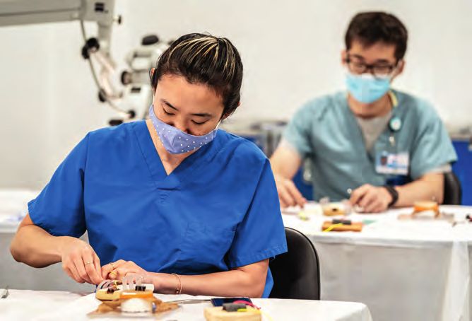
MESSAGE                   FUTURE                 TRAINING PROGRAM                             RESEARCH                     PATIENT STORIES          CLINICAL PROGRAMS                  NYEE STATS             ACCOLADES AND RETIREES                       FACULTY NEWS

Racing Against Time to
Discover a Gateway to COVID-19
Transmission
As face masks became commonplace in the wake of the pandemic, researchers
from the Mount Sinai/New York Eye and Ear (NYEE) Eye and Vision Research
Institute offered concrete evidence that another form of protection may be
necessary. The team showed that SARS-CoV-2, the virus that causes COVID-19,
can invade the body through not just the respiratory tract, but also the surface
cells of the eye, and that protective eyewear might be advisable, particularly when
people spend extended periods in areas with poor ventilation such as an airplane or
restaurant. The findings were published in the journal Cell Stem Cell (July 2021).

                                                                                                                                                    down. They found that SARS-CoV-2         The well-known spike protein of            community’s knowledge of the
                                                                                                                                                    suppresses the innate immune             SARS-CoV-2 tethers itself to ACE2,         biology of the virus, the Mount Sinai-
Limbal region of the cornea showing immunofluorescent staining of SARS-CoV-2 in green and the ACE2 receptor in red in the cornea of the human eye   system in the same manner as             while TMPRSS2 cleaves ACE2,                led research team advanced useful
                                                                                                                                                    found in cells of both the respiratory   enabling the virus to invade the host      suggestions on how it might be better
Timothy A. Blenkinsop, PhD, Associate             he says. “And through our research,                   in the lab a whole-eye model using          and intestinal tracts. SARS-CoV-2        cell and replicate. “We learned that       controlled. “Handwashing is important
Professor of Ophthalmology, Cell,                 we showed for the first time that viral               human pluripotent stem cells                hijacks the transcriptional machinery    ACE2 and TMPRSS2 are preferentially        because whenever we subconsciously
Developmental, and Regenerative                   infection is possible through the ocular              (hPSC). After exposing the hPSC-            suppressing cellular function to         expressed in the limbal cells of the       rub our eyes, we may be transferring
Biology, Icahn School of Medicine                 surface epithelium, including corneal,                derived model to SARS-CoV-2,                promote viral replication.               eye, so the necessary machinery for        the virus,” Dr. Blenkinsop points out.
at Mount Sinai, was one of the lead               limbal, and scleral cells. In fact, the               they again found that the putative                                                   SARS-CoV-2 entry is already in place,”     “And if we’re going to be in the same
investigators. “When patients infected            limbal region between the cornea and                  limbal cells were preferentially            Another question they asked was          explains Dr. Blenkinsop. “The bottom       indoor space with the same recycled
with the virus started complaining                sclera had by far the highest SARS-                   infected. They performed single-            why limbal cells were preferentially     line from our research is that the eye     air for more than a half-hour, I would
about eye problems, we were curious               CoV-2 viral titers.”                                  cell RNA sequencing to gain more            infected. Do limbal cells exhibit        is a potential vector for infection from   recommend eyewear to be on the safe
to know if SARS-CoV-2 was present                                                                       insight into the biology of the             the machinery for infection, which       SARS-CoV-2.”                               side. Given the Delta variant is more
in the ocular postmortem tissue of                To determine the susceptibility of                    infection, namely, which pathways           includes the receptor ACE2 and the                                                  transmissible, vigilance is needed more
individuals positive for SARS-CoV-2,”             the human eye, scientists developed                   are activated and which are shut            associated host protease TMPRSS2?        In addition to broadening the scientific   now than ever before.”

18                                                                                                                                                                                                                                                                           19
MESSAGE FUTURE TRAINING PROGRAM RESEARCH PATIENT STORIES CLINICAL PROGRAMS NYEE STATS ACCOLADES AND RETIREES FACULTY NEWS
An Innovative New Role for Fig. 2: Labeling of non-perfused and
OCT Angiography in the Battle re-perfused capillaries. Averaged
OCT angiography images of a sickle
cell HbSC disease patient with non-
Against Sickle Cell Retinopathy
perfused segments around the fovea
(top row) and enlarged inter-capillary
spaces in the temporal retina (bottom
row) for the baseline (A & D) and 1-hour
follow-up (B & E) imaging sessions.
Intermittently filled capillary segments
are labeled as non-perfusion (red)
when they are filled at baseline but not
Among the debilitating effects of sickle cell disease, which often strikes in the prime at 1-hour follow-up (C & F). Non-filled
segments at baseline which re-appear
of life, is sickle cell retinopathy, a condition that may go undetected until it has caused at 1-hour follow-up are considered as
permanent damage to the eye. That picture has turned considerably brighter, re-perfusion (cyan) (C & F).
however, with the development by researchers at New York Eye and Ear Infirmary
of Mount Sinai (NYEE) of an innovative new technique for sequential imaging of
retinal blood flow. In sickle cell patients, this technique can reveal how the disease is
progressing, as well as the effectiveness of treatment regimens such as hydroxyurea.
Their work was reported in a study published online in the May 2021 issue of
to a “sickled” shape when stressed. disease burden and the current physicians to gauge the impending
Biomedical Optics Express. These misshapen cells clump and activity of the sickle cell disease for danger of the sickling condition
jam the blood flow in capillaries, which the particular patient. until the patient experiences vision
become inflamed and occluded. loss,” observes Dr. Chui. “That’s why
“For the first time we’ve shown how Progressive regions of blocked “We learned from our study that OCT angiography could be a game
Fig. 1: OCT Angiography OCT (optical coherence tomography) circulation can lead to loss of vision in patients with no active sickle cell changer, leading to earlier diagnosis of
scan sequence angiography can be used to evaluate the retina, which may ultimately result retinopathy show minimal intermittent retinal issues and potentially avoiding
demonstrating the immediate status of sickle cell in blindness. fluctuation in capillary blood flow,” irreversible blindness and vascular
intermittent capillary filling explains co-author Richard Rosen, MD, complications in other parts of the
disease using dynamic retinal imaging
(frames 4 of 10). This is a
that depicts microscopic changes Currently, ophthalmologists rely on Chief of Retina Service, Distinguished body.”
sickle cell HbSS disease
in blood flow in the smallest blood static images from single scans of the Professor of Ophthalmology at the
patient on hydroxyurea
vessels,” says Toco Chui, PhD, Director retina, a practice that fails to capture Icahn School of Medicine at Mount Researchers speculate that the
treatment. Yellow arrows
point to two capillary of the David E. Marrus Adaptive Optics the dynamic nature of the disease Sinai, and Vice Chair and Director of technology could have implications
segments bordering the Imaging Laboratory at NYEE, and senior process. OCT angiography provides Ophthalmology Research at NYEE. “In that stretch well beyond sickle cell
foveal avascular zone of the author of the study. “This approach access to the most delicate capillaries patients with more frequent temporary disease. “Microcirculation in the
macula which demonstrate allows us to noninvasively monitor the and the ability to measure and map capillary blockage there is probably a capillaries of the retina provides a
intermittent capillary retinal microcirculation over time and microscopic vaso-occlusive events higher risk of permanent closure, which unique window on what is happening
perfusion during the 10 assess a sickle cell patient’s condition across short- and long-term intervals. can lead to significant vision loss.” in other parts of the body, like the brain
scan sequence. The time before or after initiation of therapy.” In their analysis of 27 patients, NYEE and major organs,” says Dr. Rosen. “For
of image acquisition is seen researchers imaged each subject This novel use of OCT angiography, that reason, OCT angiography could
in the upper left corner of 10 times in a row over a 10-minute a relatively new technology that provide a strong platform for detecting
Sickle cell disease is an inherited red
each OCT-A scan.
blood cell disorder that afflicts about span. An hour later, they repeated the NYEE has been in the vanguard of a variety of circulation threats in a non-
10 percent of African Americans. A procedure to determine which blood developing, could be an important tool invasive way.”
mutation in the hemoglobin gene vessels were repeatedly opening in the armamentarium of clinicians who
causes the protein to fold abnormally, and closing. From these studies they take care of patients with sickle cell
resulting in red blood cells that distort were able to characterize the level of disease. “It’s often impossible for these
20 21MESSAGE FUTURE TRAINING PROGRAM RESEARCH PATIENT STORIES CLINICAL PROGRAMS NYEE STATS ACCOLADES AND RETIREES FACULTY NEWS
A Novel Gene Therapy
glaucoma models that mimicked the
pathophysiology of human disease
with both high and normal intraocular
pressure. The team learned that
Approach to Help Prevent CaMKII regulated the survival of
retinal ganglion cells across many
of these pathologies, and that in the
Retinal Vision Loss small animal excitotoxicity model,
insults to the cell bodies or their
axons which form the optic nerve
led to inactivation of CaMKII and its
downstream signaling target CREB
(cAMP response element binding
protein).
Over the past decade, Bo Chen, PhD, has increasingly drawn national attention for his
breakthrough work in gene therapy to save and even restore the sight of people with “Intriguingly, we found that
reactivation of CaMKII and CREB
degenerative retinal disease. Dr. Chen, Associate Professor of Ophthalmology, and provided robust protection for retinal
ganglion cells,” notes Dr. Chen, “and
Neuroscience, and Director of the Ocular Stem Cell Program at the Icahn School of that CaMKII-mediated protection
Medicine at Mount Sinai, has now taken his lab’s research an intriguing step further slowed down the disease progression
in both glaucoma models.”
with a promising gene therapy approach to protecting and revitalizing retinal ganglion
Making that reactivation possible
cells compromised by severe injury to the optic nerve from diseases like glaucoma. was a gene therapy approach
deployed by the researchers to
introduce a more active type of
In a paper in the journal Cell (August CaMKII into the original retinal
2021), the investigator reported how Graphical Abstract: CaMKII-CREB signaling plays a major role in the maintenance of retinal ganglion cells to boost their activity.
the reactivation of a key enzyme ganglion cells (RGCs), and reactivation of CaMKII activity via AAV gene therapy in injury and
This modified version of CaMKII—
disease models in mice protects RGCs and preserves visual function and visually guided behavior.
known as CaMKII and its downstream with a mutated amino acid—was
signaling in retinal ganglion cells transferred to the targeted cells
through gene therapy provided through an adeno-associated
robust protection of retinal ganglion glaucoma, the leading cause of light that enters the eye into a viral vector.
cells and preserved vision in multiple irreversible visual impairment signal transmitted to the brain—do
diseases and injury mouse models. worldwide. Glaucoma affects an not regenerate. For that reason, “Our research showed that
“We uncovered evidence for the first estimated 76 million people, some neuroprotective strategies designed CaMKII could indeed be a valuable
time that CaMKII is a key regulator of of whom will progress to blindness to preserve the cell bodies (which therapeutic target to save retinal
the survival of retinal ganglion cells despite aggressive treatment to contain the DNA of the retinal ganglion ganglion cells and preserve vision in
in both normal and diseased retinas, reduce their intraocular pressure. cells) and their axons could be critical treating potentially blinding diseases
and could be a desirable therapeutic As Dr. Chen puts it, “The need for to preventing further vision loss. like glaucoma,” says Dr. Chen, winner
target for vision preservation in neuroprotective strategies to save of the Pew Scholars in the Biomedical
conditions that damage the axons vulnerable retinal ganglion cells has Dr. Chen and his team investigated Sciences Award given to young
and somas of retinal ganglion cells,” never been greater.” whether CaMKII could play such a investigators showing outstanding
says Dr. Chen, who moved his lab therapeutic role. They tested the promise. “The fact that manipulation
three years ago from Yale School of As ophthalmologists are well aware, enzyme across a wide range of of CaMKII would involve a one-time
Medicine to Mount Sinai/New York the biggest hurdle to restoring vision injury and disease animal models, transfer of a single gene only adds
Eye and Ear (NYEE) Eye and Vision loss from glaucoma and other retinal including optic nerve damage, to its vast potential to treat serious
Research Institute. diseases and injuries is the fact that excitotoxicity (where nerve cells are retinal conditions in humans.”
axons—the long nerve fibers that destroyed by the overactivation of
Dr. Chen’s work could prove allow retinal ganglion cells to process glutamate receptors that result in
particularly consequential for Dr. Chen and lab assistant in his laboratory visual information by converting damage to the cell structure), and two
22 23MESSAGE FUTURE TRAINING PROGRAM RESEARCH PATIENT STORIES CLINICAL PROGRAMS NYEE STATS ACCOLADES AND RETIREES FACULTY NEWS
Going Deeper Into the
Microworld of Ocular Disease
Retinal imaging has taken another bold leap at New York Eye and Ear Infirmary
of Mount Sinai (NYEE) with the development of non-confocal adaptive optics
processing that allows for visualization of the eye’s microvasculature down to
capillaries, the blood cells that move through them, and the aneurysms that develop
in diabetes and hypertension. The new enhancement to adaptive optics scanning
light ophthalmoscopy (AOSLO) increases the contrast and resolution of the already
state-of-the-art imaging platform, promising to provide ophthalmologists with Figure 1: A red blood cell rouleau (aggregation of red blood cells) in a patient with sickle cell disease imaged using non-confocal AOSLO. Yellow
unprecedented opportunities to diagnose and treat ocular disease. arrows indicate the appearance of red blood cell rouleau at Time 1 and Time 2 and disappearance at Time 3.
“There are only a handful of retinal conjugate plane to include Ophthalmology at Stanford University.
systems like this in the world with only the direct backscattered “We hope to take advantage of this
the ability to show features like the light, eliminating multiply scattered approach to explore cellular level
outpouching or herniation of blood (out-of-focus) light. Non-confocal regions that have been impossible for
vessels that are typical of diabetic AOSLO, on the other hand, harvests us to reach in the past, including retinal
retinopathy, and the intermittent the multiply scattered light to ganglion cells—which could be critical
flow of red blood cells through facilitate non-invasive visualization for more sensitive glaucoma detection
capillaries in patients affected of otherwise transparent retinal and monitoring—and vitreous
with sickle cell retinopathy (Figure vascular structures. macrophages, which are known to play
1),” says Richard B. Rosen, MD, an important role in vitreous-macular
Belinda Bingham Pierce & Gerald “Quad-detection non-confocal interface diseases (Figure 2).”
G. Pierce, MD, Distinguished Chair AOSLO allows us to see blood flow,
of Ophthalmology, Vice Chair blood cells, and other transparent Dr. Rosen believes that vitreous
and Director of Ophthalmology tissues and cells in the retina with detachments—a very common
Figure 2: Two macrophages imaged at A1) baseline and A2) after 90 minutes in a healthy subject using non-confocal AOSLO. Arrows indicate shape-
Research at NYEE and Chief of enhanced contrast,” explains Toco condition that affects older adults
shifting of the cell body over time. B) The chromo-temporal map shows the cell translocations over 2 hours. Both macrophages remain relatively
Retina Service for the Mount Sinai Chui, PhD, Associate Professor at where the gel-like vitreous shrinks,
stationary while their processes extend and retract in various directions over 2 hours. The background is composed of the framework of collagen
Health System. “Non-confocal the Icahn School of Medicine and and pulls away from the surface of the protein strands that compose the vitreous gel.
adaptive optics allows us to see Director of the David E. Marrus retina—are another important target
microscopic structures and Adaptive Optics Imaging Laboratory for non-confocal AOSLO imaging.
cellular-level movement with a and Computational Imaging at NYEE. “We hope it will enable us to study how
resolution and clarity never before these cells above the retinal surface approaches continue to be refined in advanced hardware development
possible.” Dr. Chui created the image are involved in abnormal vitreous the adaptive optics laboratory of that began 25 years ago,” says
processing software that is now detachment, which can lead to macular Dr. Chui, the potential applications Dr. Rosen. “It’s the next big step in
Traditional confocal AOSLO being used with AOSLO hardware puckering and holes, frequently for researchers and clinicians alike being able to explore the microworld
improved in vivo imaging of the installed at NYEE as part of a requiring surgery to repair.” are only starting to come into focus. of patients.”
retinal microvasculature by research collaboration with Alfredo “In many ways, non-confocal imaging
placing a spatial pinhole at the Dubra, PhD, Associate Professor of As these new image processing fulfills the promise of much of the
24 25MESSAGE FUTURE TRAINING PROGRAM RESEARCH PATIENT STORIES CLINICAL PROGRAMS NYEE STATS ACCOLADES AND RETIREES FACULTY NEWS
Coffee Alert for Those at Reassurance for Patients With
High Risk of Glaucoma Glaucoma Drainage Implants
Think hard about that third or fourth cup of coffee if glaucoma runs in your family. As ophthalmologists are well aware, increasing numbers of patients undergoing
cataract surgery have in place glaucoma drainage implants—shunts that bypass the
That’s the message from a new study robust data on genetics, caffeine eye’s natural drain and lower intraocular pressure (IOP). More of a medical mystery is
by Icahn School of Medicine at Mount consumption, and intraocular the short- and long-term impact of cataract surgery on the IOP of glaucoma patients
Sinai researchers that demonstrated pressure of participants. The results
the interaction of diet and genetics confirmed a prior glaucoma study with these devices. Is the procedure safe, or does it pose risks for patients whose
in glaucoma, the leading cause of reporting that greater caffeine intake
blindness in people over 60. The was associated with open-angle
vision has already been compromised?
paper, published in the June 2021 glaucoma in people with a family
issue of Ophthalmology, found that history of the disease.
individuals with the strongest genetic New findings by researchers at New Icahn School of Medicine at Mount (in 6 percent), and choroidal effusion
predisposition to elevated intraocular “Our study suggests that many genes York Eye and Ear Infirmary of Mount Sinai. “These findings should help (in 4 percent).
pressure (IOP) had an increased risk may interact with caffeine to increase Sinai (NYEE) should put the minds reduce concerns that postoperative
of glaucoma if they also had a high or decrease intraocular pressure, of both surgeons and patients at inflammation and cytokines “These surgeries tend to be more
caffeine intake. though only a handful appear to be ease. In the largest study of its type, released from cataract surgery complicated than normal cataract
statistically significant,” explains published in Ophthalmology Glaucoma could potentially stimulate scarring procedures, particularly given the
“Caffeine consumption by itself is Dr. Pasquale. “Further research is (November 2020), the team reported and thereby increase intraocular higher incidence of mature cataracts
not a robust risk factor for primary needed to determine which specific that the use of phacoemulsification pressure.” in this patient population, and the
open-angle glaucoma,” says Louis genes and their components are most in patients with glaucoma drainage requirement for pupil expansion,”
R. Pasquale, MD, Deputy Chair for important, and could thus potentially implants resulted in a reduction in IOP As for the potential mechanisms explains Dr. Wong. “That’s why
Research for the Department of serve as biomarkers to indicate which in the first week following surgery, behind the temporary IOP reduction, physicians need to carefully
Ophthalmology at Icahn Mount Sinai individuals are predisposed to higher and remained unchanged relative to Dr. Wong suggests that the irrigation consider the heightened risks. We
and Director of the Mount Sinai/New IOP and glaucoma.” the preoperative level over the next force during phacoemulsification recommend checking the central
York Eye and Ear (NYEE) Eye and two years. could produce micro-ruptures in corneal thickness and the corneal
Vision Research Institute, and lead Once those genes are confirmed, the capsule of the drainage device endothelial cell density prior to
author of the paper. “But if someone they might eventually open the door to “Our study reinforces the fact that reservoir, thus facilitating drainage of phacoemulsification when a drainage
has a strong family history of the widespread genetic testing designed cataract surgery can be done safely the eye’s aqueous humor. implant is in place. If precautionary
disease and they drink a lot of coffee, category for high eye pressure who to inform members of the public and prove beneficial to glaucoma steps like these are taken, there’s no
they’d be well advised to consider consumed three cups of coffee daily who are at the greatest risk of high patients with drainage implants since Still, cataract surgery in the company reason why patients can’t be assured
cutting back to two cups a day.” were nearly four times more likely to intraocular pressure and glaucoma. it can lower their intraocular pressure of glaucoma drainage implants is not excellent outcomes from their
have glaucoma than those who did Dr. Pasquale suggests that kind of on a transient basis, and reduce the without risks. Dr. Wong and his team surgery.”
While researchers have long known not consume caffeine and were at knowledge and awareness could number of glaucoma medications found a number of postoperative
that variants in multiple genes are more the lowest genetic risk quartile for help set the stage for powerful new needed during that period,” says complications, including cystoid
common in people with glaucoma, elevated eye pressure. approaches to battling glaucoma. lead author Sze Wong, MD, Assistant macular edema (in 10 percent of
the study is the first to pinpoint a “Genes are not destiny,” he asserts, Professor of Ophthalmology at the cases), corneal decompensation
large panel of genes that may interact For their research, the team led by “and finding environmental and dietary
to magnify the risk of glaucoma in Dr. Pasquale drew upon roughly strategies to mitigate the impact of
high-caffeine consumers. Specifically, 121,000 members of the UK Biobank. genetics could be a highly effective
investigators found that people in the This cohort proved to be an invaluable way to reduce the risk and burden of
top 25 percent of the genetic risk score resource inasmuch as it contained glaucoma.”
26 27MESSAGE FUTURE TRAINING PROGRAM RESEARCH PATIENT STORIES CLINICAL PROGRAMS NYEE STATS ACCOLADES AND RETIREES FACULTY NEWS
Cataract Surgery
In the Tightest of Spaces
Kira Manusis, MD, Co-Director of Cataract Surgery Services at New York Eye and
Ear Infirmary of Mount Sinai (NYEE), knew the patient referred to her in April 2021 for
cataract evaluation and surgery would be a challenge. But she didn’t appreciate the
full dimensions until she posted the case on a global chat room designed to advise
clinicians on difficult cases. Thousands of anterior segment specialists are in that
chat room, but only one responded.
The case involved a 55-year-old Not surprisingly, other ophthalmologists
woman whose vision had been around the city who had been
greatly impaired over the past approached by the patient’s family were
two years by hypermature (white) loath to take the case on. “It’s definitely
cataracts in both eyes. The real a feather in Dr. Manusis’s cap that she Caption
difficulty, though, wasn’t the cataracts. proceeded with this operation because
It was the patient’s corneas, rendered I don’t think too many surgeons could
so tiny by a hereditary condition that have managed it,” says the patient’s
standard phacoemulsification and brother, Dean Liambas. It was Mr. Paula Liambas with her brother Dean near their home in Queens
intraocular lens implantation in such Liambas who sought help for his sister,
a crowded surgical space without Paula, after her reduced vision caused to the intraocular lens that fixate the modifications to the surgery, including to the surgeon’s office to have the
damaging surrounding tissue would Top: An unusually small surgical area as illustrated her to repeatedly bump into objects lens in the eye—to size them to the tiny creating a scleral tunnel for the incision patch removed from the eye. “She
pose a huge technical hurdle. In by the surgical tools, with the cornea measuring around the house and fail to visually capsular bag. And the third approach, and use of a cystotome-assisted immediately smiled and clapped
addition to the microcornea and iris 6.0 x 7.5 mm in diameter. Bottom: Paula Liambas recognize people she knew well. “At considered a last resort, would be to capsulorhexis. her hands because she was seeing
coloboma in each eye, the case was after cataract surgery. one point we were crossing the street not insert the intraocular lens, leaving 100 percent better than before,”
further complicated by the fact that and she didn’t see the curb, causing her the patient to a future with aphakic To the pleasant surprise of Dr. Manusis remembers her brother, who was by
the patient's condition prevented to trip and fall,” he recalls. “I knew then glasses. and the fellow assisting her, the her side throughout the pre- and post-
usual imaging and eye measurements, something had to be done.” anatomy of the patient’s lens was found surgical periods.
prior to cataract surgery, from being Once the patient was under to be normal, enabling them to safely
performed. Prior to the day of surgery, Dr. Manusis’s anesthesia, Dr. Manusis was able to proceed with the preferred surgical The procedure on the second eye was
game plan was to perform cataract perform more precise measurements plan: implanting a normal, though also well tolerated by the patient, giving
“We had no idea what the patient’s extraction in each eye (one month apart) of the corneal diameter. While average slightly more flexible, acrylic intraocular her 20/150 vision in the left eye and
anatomy was going to look like under general anesthesia, with three corneas measure 10.5 x 11.5 mm lens into the capsular bag. And while 20/200 in the right eye one month after
once we got into the eye,” admits possible surgical pathways. The first— vertically and horizontally respectively, the extremely tight workspace and both surgeries. That change has since
Dr. Manusis, Assistant Professor of and most preferable—would be to place Paula’s corneas were determined reduced visibility due to the miniature- translated into a much-improved quality
Ophthalmology at Icahn School of the intraocular lens into the capsular to be 6.0 x 7.5 mm. The axial length size cornea proved taxing for the of life for Ms. Liambas and her brother.
Medicine at Mount Sinai. “It was one was so small that there were very few bag if the anatomy was found to be of the eye was noted to be 21.7 mm surgeons, the 40-minute procedure “She’s a lot more independent now,”
of the most difficult cases I’ve ever comparable human case studies; Dr. normal, with adequate zonular support. with keratometry readings of 48 x 51 went well. Mr. Liambas explains. “Before, I’d say,
handled because of all the unknowns Manusis even consulted books on The second possibility, in the event the diopters, which would lead to a readily ‘Paula, hold onto my arm so I can guide
and the cramped space in which veterinary ophthalmic surgery to help capsular bag was too small, would be to available intraocular lens power. How well was evident the next you.’ Now she lets go of my arm and
we had to operate.” The anatomy guide her course of treatment. cut the haptics—the flanges attached This allowed her to make several day when Ms. Liambas returned says, ‘I got it.’”
28 29You can also read