Optical Imaging: Which Modalities Should Every Endoscopist Know and Love? - Vani Konda, MD
←
→
Page content transcription
If your browser does not render page correctly, please read the page content below
Optical Imaging:
     Which Modalities Should
Every Endoscopist Know and Love?
                     Vani Konda, MD
  Baylor Scott and White Center for Esophageal Diseases
      Baylor University Medical Center, Dallas, Texas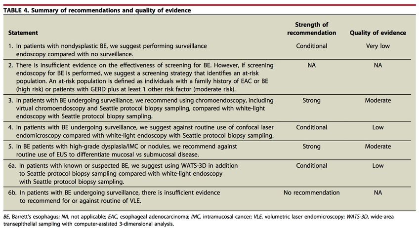
Disclosures
Current
• Exact Sciences (Advisory Board)
• Lucid (Research)
• Cernostics (Consulting)
• Medtronic (Consulting)
• Ambu (Consulting)

Past
• Olympus (Research)
• Pentax (Research)
• Mauna Kea Technologies (Speaking, Consulting)
Histology in Barrett’s Esophagus

                                   V. Konda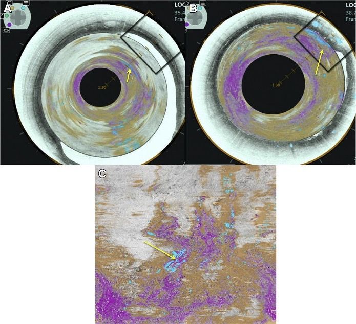
Standard Surveillance for Detection of Dysplasia
                • Endoscopic documentation                     •   Seattle Protocol
                    •   Longer segments are at higher risk          • Visible lesions
                        of neoplasia than short segments.           • Multiple levels for occult disease
                    •   Suboptimal documentation of                      • random 4QB q1-2 cm
                        length and biopsy protocol
                                                                         • q1 cm if history of dysplasia
                • Visible lesions                                   • Limitations
                    • Higher risk for harboring neoplasia                • Sampling error
                    • Can be subtle and challenging to                   • Lag time in diagnosis
                      detect                                             • Poor adherence with protocol is
                    • Variable detection of lesions                        associated with increased risk of
                         •   Community vs expert (60% v 87%)               missed neoplasia.
                                                                         • Longer segments are associated
                                                                           with poor adherence

                   Reid BJ et al. Am J Gastroenterol. 2000          Scholvinck et al. Endoscopy 2017
                   Peters et al. Dis Eso 2008                       Boys et al J Gastrointest Surg 2020
                   Wani et al. GIE 2019                             Curvers et al Eur J Gastro Hep 2008
High Quality Endoscopic Assessment: “5 L’s”
5L’s        Assessment                             Tools & Tips
Landmarks   Endoscopic Landmarks
            - Diaphragmatic Impression
            - Top of Gastric folds
            - Squamocolumnar Junction
Length      Length of Barrett’s segment            Prague Classification (C and M)
            Length and Extent of esophagitis       Los Angeles grading system
Look        Take time to inspect and evaluate      High resolution endoscope
            for subtle lesions                     Distal attachment cap
                                                   Chromoendoscopy and virtual chromoendoscopy
                                                   Inspection Technique
                                                   Recognition of Subtle Lesions
Lesions     Identify, Document, and Target         Paris Classification
            Visible lesions which have high risk   Tissue acquisition with EMR or targeted biopsies
            of harboring neoplasia
Levels      Assess multiple levels for occult      Seattle Protocol
            dysplasia                              Additional Tissue Acquisition techniques
Foundation for Better Detection

  Tools
  • High resolution endoscopy
  • Soft distal attachment cap
  • Enhanced endoscopic imaging

  Techniques
  • Suction, Irrigation & Mucolytics
  • Insufflation and Deflation
  • Tip deflection
  • Retroflexion

  Recognition
  • Inspect
    • Longer Inspection time is associated with higher rates of detection
      • Suspicious lesions (p=0.0001)
      • HGD/EAC (p=0.001)
      • >1 min / centimeter BIT
    • Train to recognize subtle, flat lesions

                                           Gupta et al. GIE 2012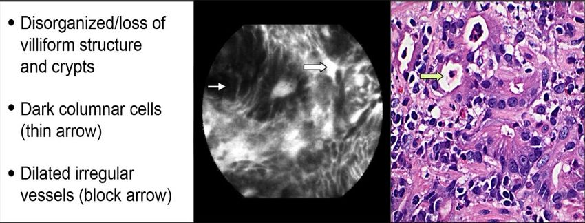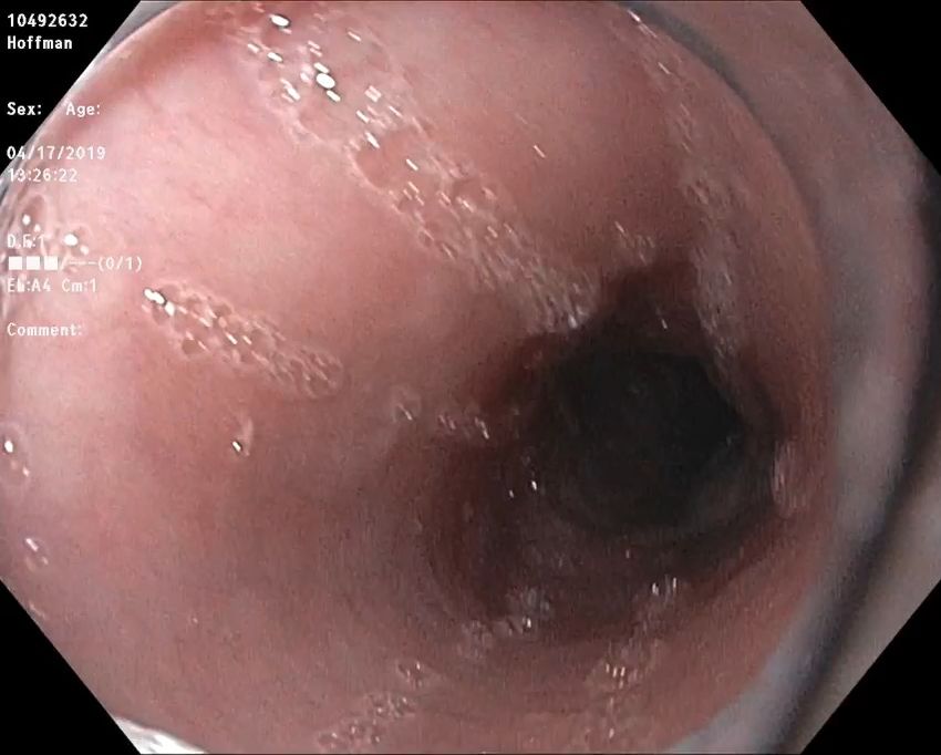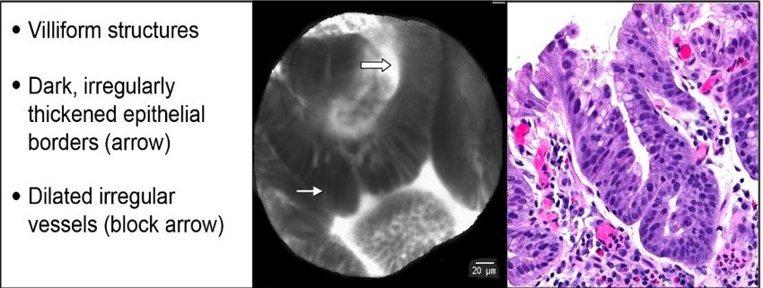
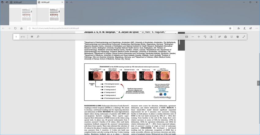
Recognition Training Module

                                       200

           Bergman et al Gastro 2019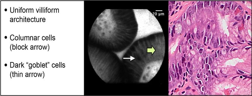
Diagnosis by EMR
• Endoscopic mucosal resection (EMR) should be
  done to stage any visible lesion in the setting of
  dysplasia.
• EMR more accurate than biopsies for assessing
  neoplasia in BE.
    • 1/3 – 1/2 of DX by biopsy up-staged or down-
      staged p/ EMR                                                  Downstaged,
                                                                       23.9%
    • Higher IOA among pathologists with EMR than
      with biopsy                                                                   Same, 49.5%
• EUS not recommended for early T staging
    • Meta-analysis (n=895, 11 studies) 75%                         Upstaged,
      accuracy                                                       26.6%
    • Overstaging and understaging of early T
      lesions
    • Appropriate for N staging
                  Konda et al. CGH 2014; Chennat, Konda et al. AJG 2009;
                  Thota et al. DDW 2014; Wani et al. CGH 2010; Wani et al. DDS
                  2013; Qumesya et al Dig Liver Dis 2018; Qumseya et al. GIE 2019
Digital Chromoendoscopy
• Narrow band imaging (NBI)
• Filtered Blue Light
• Enhances Mucosal pattern and Vascular Pattern
• Most studied
Sensitivity NPV Specificity
PIVI Threshold 90% 98% 80%
Overall 94.2% 97.5% 94.4%
Performance
Thosani et al. GIE 2016BING Criteria: Consensus Development
Regular Irregular
Mucosal
Vascular
Sharma et al. Gastro 2016NBI with Near Focus * Confidence in NBI with near focus 92% versus 74.1% p
Acetic Acid (AA)
• Acetic acid
  • Chromoendosocopy
  • Contrast agent
  • Enhances mucosal pit pattern
    temporarily with a whitish effect                        ASGE PIVI Meta-analysis (4 studies)

• Proposed Portmouth Criteria                                     Sensitivity        NPV           Specificity
  • Focal loss of acetowhitening &
    surface patterns                           PIVI Threshold     90%                98%           80%
  • Endoscopists performance
    improved using criteria to aid in          Overall            97%                99%           85%
    recognition                                Performance
     •   Sensitivity 98.1% (from 79.3%)
     •   NPV 97.4% (from 80.2%)

                                      Thosani et al. GIE 2016; Chedgy et al. Endoscopy 2017 ;
                                                      Kandiah et al. Gut 2018
Chromoendoscopy and Virtual Chromoendoscopy
In patients with BE undergoing surveillance, we recommend
using chromoendoscopy, including virtual chromoendoscopy and
Seattle protocol biopsy sampling, compared with white-light
endoscopy with Seattle protocol biopsy sampling.
206
Qumseya et al. GIE 2019Artificial Intelligence : Computer aided detection1
• ARGOS project
• 494,364 still images
• accuracy of 92% for detection dysplasia
• sensitivity of 95% & specificity of 85%
• CAD with convolutional neural networks
• Still images
• accuracy 93.7%
• sensitivity 95.6% & specificity 91.8%
• AUC 0.94
• 30 pre-recorded video clips
• per-lesion sensitivity of 95%
• per-patient negative predictive value of 100%
de Groof AJ et al. Gastro 2020Hashimoto R et al. GIE 2020
Samarasena J, Konda V et al. DDW 2021Confocal Laser Endomicroscopy
Technology System Depth Resolution Span
Endoscope Confocal Laser Scope & 0 – 250 0.7 micron 550 microns
based CLE Endomicroscopy processor microns
(eCLE) (CLE)
(not available)
Probe Based Confocal Laser Probe 65 microns 1 micron 240 microns
CLE Endomicroscopy Video
(pCLE) (CLE)
(commercially
available)Confocal Laser Endomicroscopy
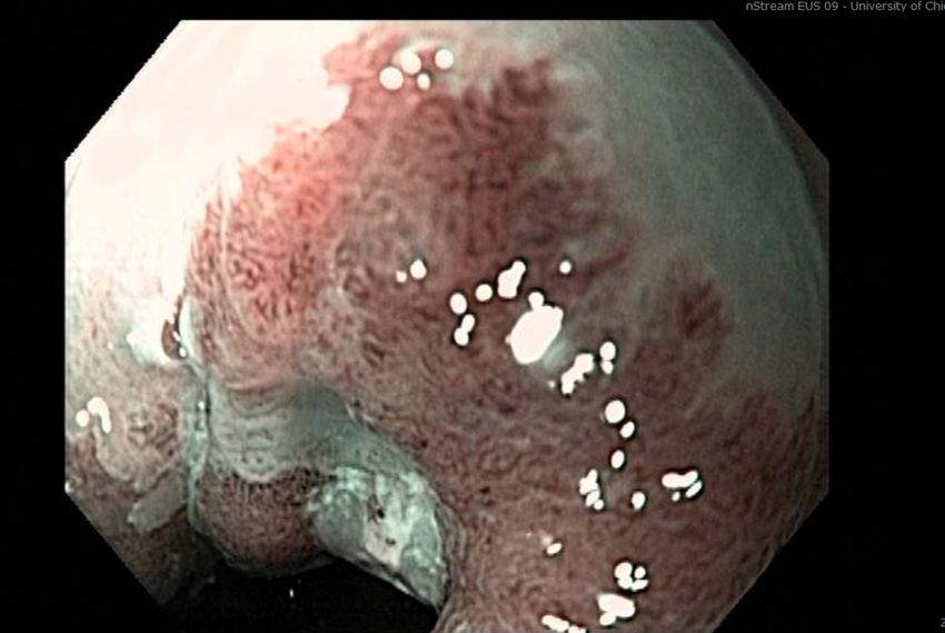
Normal Squamous Epithelium Non-dysplastic Barrett’s Esophagus
High-grade Dysplasia Adenocarcinoma
Wallace et al. Endoscopy 2011
Gaddam et al. AJG 2011CLE and PIVI Thresholds
Sensitivity NPV Specificity
PIVI Threshold 90% 98% 80%
Overall 90.4% 96.2% 89.9%
Performance
• Not recommended for widespread use in general surveillance
• Role in referral centers with high cases of dysplasia & expertise in CLE
Thosani et al. GIE 2016
ASGE Technology Committee GIE 2016Volumetric Laser Endomicroscopy
• Optical coherence tomography based
technology
• Laser probe in balloon catheter
• Enables micro architectural imaging down to 7
micron resolution and 3 mm deep
• Offers a cross sectional span of 6 cm
• Histologic correlation feasible with laser
marking
• Not currently commercially available
211OCT-SI Amsterdam
VLE
VLE- DA
Sensitivity 70% • Score of 8
Specificity 60% • Sensitivity
Accuracy 67% 83%
• Specificity
Sensitivity 86% 71%
Specificity 88%
Evans JA et al CGH 2006 Accuracy 87% Swager et al. GIE 2017
Leggett et al. GIE 2015OCT & VLE performance
OCT Studies Sensitivity Specificity
HGD/IMC Patients
OCT Off line 4 studies 89% 91%
Per lesion N=170
OCT Real time 3 studies 79% 94%
Per lesion n= 138
VLE Studies Sensitivity Specificity
HGD/IMC Patients
VLE 5 studies 85% 73%
Per lesion N=309
VLE Real time 3 studies 100% 55% 2
1
Per patient n= 35 3
Rodriguez et al. Endoscopy Int Open 2019Volumetric Laser Endomicroscopy
• Multicenter registry (1000 patients, known or suspected BE)
• VLE guided tissue acquisition in 71% of cases and treatment in 54%
• Multicenter study, 10 experts, Web based module with VLE videos of Region of Interest
• High confidence
• Accuracy 88%
• Sensitivity 83%
• Specificity 90%
• Fair agreement (kappa = 0.29)
• Multicenter study, 12 experts, Web based module with VLE videos of Full Scans
• High confidence : Correct neoplastic diagnosis (81 %) & Lesion location (73 %)
• Fair agreement (kappa 0.28)
• Computer aided detection algorithm developed and tested
• CAD : Accuracy 85%, Sensitivity 91%, and Specificity 82%
• VLE expert : 77%, 70%, 81% respectively
Smith et al Dis Esophagus 2019 Trindade et al. Gastro 2019
Struyvenberg et al Dis Esophagus 2020 Gora et al. GIE 2018
Struyvenberg et al Endoscopy 2021 Dong CGH 2021
214
Struyvenberg et al GIE 2020Tools you should know and love
Tools you should know and love
•Summary
• Remember the 5 L’s: Landmarks, Length, Look, Lesions, and Levels for a high-quality
  endoscopic assessment.
• Look carefully with tools like HRE, NBI, and consider a soft cap. Use good inspection
  technique, recognize subtle lesions, and spend adequate time.
• Advanced endoscopic imaging may improve detection and localization of neoplasia.
  These modalities may be more accessible with the help of computer aided
  detection.
• Visible lesions in the setting of dysplasia should be diagnosed with endoscopic
  mucosal resection which can have both a role in diagnosis and therapy.

                                                                                          2
                                                                                          1
                                                                                          7
You can also read