Oral Lesions in the Bit Area in Finnish Trotters After a Race: Lesion Evaluation, Scoring, and Occurrence
←
→
Page content transcription
If your browser does not render page correctly, please read the page content below
ORIGINAL RESEARCH
                                                                                                                                                   published: 12 July 2019
                                                                                                                                            doi: 10.3389/fvets.2019.00206

                                             Oral Lesions in the Bit Area in Finnish
                                             Trotters After a Race: Lesion
                                             Evaluation, Scoring, and Occurrence
                                             Kati Tuomola 1*, Nina Mäki-Kihniä 2 , Minna Kujala-Wirth 3 , Anna Mykkänen 4 and
                                             Anna Valros 1
                                             1
                                              Department of Production Animal Medicine, Research Centre for Animal Welfare, University of Helsinki, Helsinki, Finland,
                                             2
                                              Independent Researcher, Pori, Finland, 3 Department of Production Animal Medicine, Faculty of Veterinary Medicine,
                                             University of Helsinki, Helsinki, Finland, 4 Department of Equine and Small Animal Medicine, Faculty of Veterinary Medicine,
                                             University of Helsinki, Helsinki, Finland

                                             Oral lesions in the bit area are common in horses, but not comprehensively studied in
                                             harness racing horses. This study describes the type and occurrence of oral soft tissue
                                             lesions in the area affected by the bit, hereafter called the bit area, in trotters after a race.
                             Edited by:      Based on our results, we suggest a system for scoring lesions according to size, type
                       Jessica Gimpel,
        Pontifical Catholic University of    (bruise or wound), age, and depth (superficial or deep). The data was collected during a
                             Chile, Chile    welfare program for trotters, conducted by The Finnish Trotting and Breeding Association
                      Reviewed by:           (Suomen Hippos ry). The rostral part of the mouth of 261 horses (151 Standardbreds,
           Tamara Alejandra Tadich,
                                             78 Finnhorses, and 32 ponies) was examined after a race in a systematic manner, using
          Universidad de Chile, Chile
                Lesley Ann Hawson,           a bright light source without sedation or a mouth gag. The lip commissures (outside and
    Harness Racing Victoria, Australia       inside), bars of the mandible, buccal area near the second upper premolar teeth, tongue,
                   *Correspondence:          and hard palate were visually examined; bars of the mandible were also palpated. Points
                          Kati Tuomola
                kati.tuomola@helsinki.fi     were assigned to every lesion and then added together, such that each horse got an
                                             acute lesion score. Based on the score, the horses were divided into four groups (A—D)
                    Specialty section:       as follows: Group A, no lesions; B, mild lesions; C, moderate lesions; D, severe lesions.
         This article was submitted to
         Animal Behavior and Welfare,
                                             Of all the horses examined, 84% (219/261) had acute lesions in the bit area. In total, 21%
                a section of the journal     (55/261) had mild lesions, 43% (113/261) had moderate lesions, and 20% (51/261) had
        Frontiers in Veterinary Science
                                             severe lesions. Visible bleeding outside the mouth was observed in 2% (6/261) of the
         Received: 27 February 2019
                                             horses. Further, 5% of the horses (13/261) had blood on the bit when it was removed
           Accepted: 11 June 2019
            Published: 12 July 2019          from the mouth, even though no blood was visible outside the mouth. In conclusion, soft
                              Citation:      tissue lesions in the bit area were common in the Finnish trotters examined. Moreover,
            Tuomola K, Mäki-Kihniä N,        the absence of blood outside the mouth does not rule out serious injuries inside the
     Kujala-Wirth M, Mykkänen A and
Valros A (2019) Oral Lesions in the Bit
                                             mouth. The scoring system presented can be used for evaluating the severity of oral
 Area in Finnish Trotters After a Race:      lesions in different equestrian disciplines and populations to allow for comparable data
      Lesion Evaluation, Scoring, and
                                             across studies.
   Occurrence. Front. Vet. Sci. 6:206.
       doi: 10.3389/fvets.2019.00206         Keywords: animal welfare, bit, harness racing, horse, oral lesion, trotter

Frontiers in Veterinary Science | www.frontiersin.org                                1                                                   July 2019 | Volume 6 | Article 206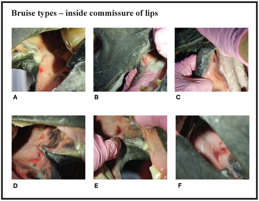
Tuomola et al.                                                                                                     Oral Mucosal Lesions in Trotters

INTRODUCTION                                                               and age (old or acute injury) of oral lesions in the bit area in a
                                                                           sample of trotters racing in Finland.
Bit-related oral lesions cause pain, and are a commonly reported
welfare problem for horses (1–4). Odelros and Wattle (5)
                                                                           MATERIALS AND METHODS
reported acute soft tissue injuries in the rostral mouth in 88%
(127/144) of Standardbred trotters examined in Sweden. Previous            Horses
studies have also reported oral lesions in other equestrian                The Finnish Trotting and Breeding Association (Suomen
disciplines, but dissimilar scoring systems make comparisons               Hippos ry) is responsible for licensing trainers, drivers, and
between studies challenging. In a study of Icelandic horses at             officials, as well as creating and monitoring the racing rules.
competitions in Iceland, Björnsdottir et al. (1) reported 36%              Since this study was part of the association’s welfare program
of horses with mild (up to 1 cm) lesions, and 8% with lesions              for trotters, the oral examination performed after the race was
that were more severe (over 1 cm) before the competition.                  compulsory. The horses (n = 261) were privately owned trotters
Of these 424 horses, 77 were re-examined after the race.                   that participated in 10 separate harness racing events (115 races)
Bit-related lesions were found in 60%. Notably, if the horse               at four race tracks in Western Finland (Pori, Tampere, Forssa,
had more than one lesion, only the most severe one was                     and Turku). Standardbred trotters (n = 151), Finnhorses (n =
included in their data. In a study of polo and racehorses (n               78), and ponies (n = 32) ranging in age from 3 to 15 years old
= 100) in England, Mata et al. (4) used a grading system                   were included in the study. Six of the Finnhorses participated to
from 0 to 5 to evaluate lip commissure and bar injuries, and               the monté race and all other horses participated into ordinary
another grading system to evaluate tongue injuries. In Denmark,            harness races.
Uldahl and Clayton (6) examined 3,143 horses from various                      Initially, the horses were randomly selected from the starting
disciplines after a competition performance (show jumping,                 lists, however practical constraints were taken into account in
dressage, eventing, and endurance). In total, 9.2% of the horses           order to maximize the number of horses examined in the limited
had lesions or visible bleeding from the mouth. However, only              time after finishing the race performance. The horses evaluated
the corners of the mouth were examined, not the oral cavity.               in previous races were excluded so that none was evaluated more
Lacerations of the skin and mucosa, as well as the presence                than once. The horses which were resisting the examination were
of blood on the skin and mucosa were reported separately,                  excluded from the study. We later checked whether the horses
but the four outcomes were combined into a single category                 competed again within 2 weeks.
for analysis.
    Tell et al. (7) concluded that riding a horse with a bit and           Oral Examination
bridle can cause lesions to the oral cavity. However, they noted           The horses were examined 5–20 min after the race at their
that oral lesions were also present in broodmares that were not            outdoor harnessing booth in the warm–up area. The rostral
regularly ridden with a bit, although to a lesser extent compared          part of the oral cavity was evaluated by the first author of this
to those ridden with a bit. In this study, ulcers more than 0.5 cm         study, who is a veterinarian experienced in oral examination
in diameter were considered as large.                                      of horses. The examination, which was modified from the one
    There have been various methods for lesion examination.                used in Icelandic horses (1), was carried out without sedation
After competition, Icelandic horses, polo horses, and racehorses           or a mouth gag; the horse was without the bridle, and wearing
have been examined without sedation or a mouth gag. Sedation               only its own halter. During the examination, the veterinarian
was also not used when examining trotters in Sweden after                  wore disposable nitrile gloves and a Lumonite Navigator 3000
competition, however a mouth gag and a light source was used,              headlamp set at 420–1,300 lumens. The examination began with
along with flushing the mouth with water (1, 4, 5). Tell et                the examiner standing on the left side of the horse. The tongue
al. (7) performed the examination on sedated horses with a                 was externally guided to the left side, allowing evaluation of
mouth gag and a light source. This examination was not related             the buccal mucosa near the second upper premolar tooth (106)
to competitions.                                                           and the mucosa at the inside of the lip commissure, on the
    According to the Finnish racing guidelines, official race track        contralateral (right) side. The tongue and palate were examined
veterinarians should only examine the horses after a competition           visually. If a sharp hook in 106 or 206 teeth was noticed while
if they show bleeding from the mouth. However, there is no                 examining the buccal area near those teeth, it was recorded and
information about the oral health of horses that do not show               the trainer was informed, but otherwise we could not palpate and
bleeding from the mouth after a race.                                      examine sharp enamel points without mouth gag. Finally, the left
    The lesion grading systems used in earlier studies are all             bar area was palpated, and the left external commissure of the lips
unique to each study, and none of them account for the number,             (the outside skin area) was examined. The same procedure was
depth, and size of the lesions. We thus believe that a quick oral          repeated on the right side of the horse (Video 1 in Supplementary
examination, which can be performed in the field environment               Material). Video recordings of some of the typical lesions were
and a simple, practical, and objective scoring system that includes        taken with a digital camera (Panasonic DMC-GX7). An assistant
both the number and severity of the lesions is needed to allow for         recorded the findings of the oral examination and the bit type
more accurate comparisons among studies. Our aim was to create             on a data sheet, which was a modified version of a former Vet
such a scoring system, and apply it to determine the occurrence,           Form 2 from the International Federation of Icelandic Horse
location, type (bruise or wound), size, depth (superficial or deep),       Associations (Data Sheet 1 in Supplementary Material).

Frontiers in Veterinary Science | www.frontiersin.org                  2                                          July 2019 | Volume 6 | Article 206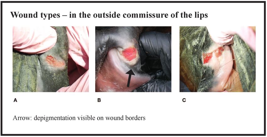
Tuomola et al.                                                                                                                               Oral Mucosal Lesions in Trotters

 FIGURE 1 | Bruise types and their points at the inside commissure of the lips. (A) 1 point, (B) 2 points, (C) 2 points, (D) 3 points, (E) 4 points, (F) 4 points.

Lesion Scoring                                                                            as an acute lesion. In some cases, a wound at the bars of the
Since many horses had several lesions, we established a score that                        mandible was accompanied by swelling (Figures 4B,C). We did
considered all lesions in each horse. Points were given for every                         not systematically record redness and swelling at the bars, but we
acute lesion that was detected, then added up to form a total                             noted both in some horses without bruises or wounds.
score for the horse. This acute lesion score combines the number,                             The horses were divided into four groups (A—D) according to
size, quality (bruise or wound), and depth (superficial or deep)                          their acute lesion score as follows: Group A (no acute lesions) =
of acute lesions. Old lesions were recorded separately. A bruise                          horses with 0 points; Group B (mild lesions) = horses with 1–2
(syn. contusion, hematoma) was determined as a discoloration of                           points; Group C (moderate lesions) = horses with 3–11 points
a superficially intact mucosa. Bruises were given points from 1 to                        (although this excluded horses with eight points for a single
4 according their size (maximum width) as follows: 1 cm but
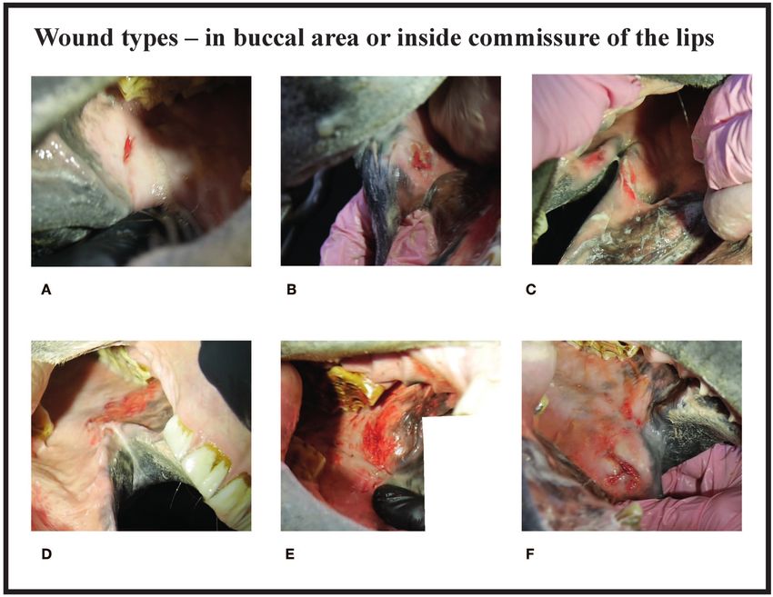
Tuomola et al.                                                                                                                            Oral Mucosal Lesions in Trotters

 FIGURE 2 | Wound types and their points in buccal area or inside commissure of the lips. (A) 4 points, (B) 6 points, (C) 6 points, (D) 8 points, (E) 10 points,
 (F) 10 points.

 FIGURE 3 | Wound types and their points in the outside commissure of the lips. (A) 6 points, (B) 6 points, (C) 8 points.

Frontiers in Veterinary Science | www.frontiersin.org                               4                                                    July 2019 | Volume 6 | Article 206
Tuomola et al.                                                                                                                             Oral Mucosal Lesions in Trotters

 FIGURE 4 | Wound types and their points at the bars of the mandible. (A) 4 points, (B) 6 points, (C) 8 points, (D) 8 points, (E) 8 points, (F) 8 points.

video recordings (Panasonic DMC-GX7; lens H-FS14140, 14–                                 were the most common bit type (75%, 195/261 horses), followed
140; settings: STD, MP4, 1,920 × 1,080 50 p 28 Mbps, recording                           by a straight bit type (18%, 48/261) and a double-jointed bit type
mode: M (movie), exposure: P, light metering method: multiple,                           (7%, 18/2019). The number of horses examined in this study
automatic focusing ON, optical zoom only) of the typical lesions                         represents 3.6% of all the trotters (n = 7,261) that competed in
were taken for lesion severity scale documentation purposes.                             2017 in Finland (10).
From the video files, a focused frame was saved as a JPG image file                         There were no acute lesions in 16% (42/261) of the horses
using a video editing software (Adobe Premiere Pro CC 2017).                             (Group A). We found that 21% (55/261) of the horses had
                                                                                         mild lesions (Group B), which meant they either had a 1 cm
Statistical Analysis                                                                     bruise or two bruises
Tuomola et al. Oral Mucosal Lesions in Trotters
FIGURE 5 | Scoring of oral lesions. Points for each acute lesion are added together to form the acute lesion score.
tissue (Figure 4D), and incision, a clean cut wound longer than
its width (Figure 2A). Deep wounds had a crater-like appearance
(Figures 4B–F) or extensive damage to the submucosal tissue
(Figures 2E,F).
Old Lesions
We found old lesions in 16% (41/261) of the horses, which were
characterized by depigmentation on the outside commissures of
the lips, scars, old wounds, or old bruises (Figure 10). Ten horses
(4%) had only old lesions and 31 horses (12%) had old lesions
together with acute lesions. The number of old lesions in any
single horse ranged from 0 to 3 (Figure 11).
Location of Acute Lesions
The most common place for a lesion was the inside commissure
of the lips, where 64% of all horses had lesions. The location
of lesions is presented in Figure 12. In some cases, the lesion
FIGURE 6 | Depigmentation in the outside commissure of the lips. Plain extended from the inside commissure of the lips to the buccal
depigmentation without scarring and thickening of the skin.
area (Figures 2D–F). The location of such lesions was recorded
as inside commissure of the lips.
appearances. The most common type was abrasion, a superficial Tongue and Hard Palate Lesions
injury to the mucosa (Figure 2D). Other types were laceration, a Nine horses had tongue lesions. Four had bitten their tongue
full thickness injury to the mucosa characterized by tearing of the (1.5%), three had bruises under the tongue (1.1%), and two
Frontiers in Veterinary Science | www.frontiersin.org 6 July 2019 | Volume 6 | Article 206Tuomola et al. Oral Mucosal Lesions in Trotters FIGURE 7 | Number of acute lesions in all horses. FIGURE 8 | The horses (n = 261) were divided in four different groups (A—D) according to their acute lesion score. FIGURE 9 | Acute lesion scores in all horses. had bruises at the sides of the tongue (0.8%). Only one the acute lesion score in this study because these lesions horse (0.4%) had a small lesion in the hard palate. Tongue were located so that they were not probably related to and hard palate lesions (n = 10) were not included in the bit. Frontiers in Veterinary Science | www.frontiersin.org 7 July 2019 | Volume 6 | Article 206
Tuomola et al.                                                                                                     Oral Mucosal Lesions in Trotters

 FIGURE 10 | Type of the lesions in horses.

                                                                         tip of its tongue, which caused the bleeding. The three horses that
                                                                         got highest points (22, 27, and 36) did not have blood anywhere.

                                                                         Timing of the Next Race Performance
                                                                         In total, 20% of the horses (51/261) had severe lesions (Group
                                                                         D). Of those, 65% (33/51) competed again within 2 weeks,
                                                                         13 competed within 1 week, and two competed again the
                                                                         following day.

                                                                         DISCUSSION
                                                                         We found a high occurrence of oral lesions in Finnish trotters
 FIGURE 11 | Number of old lesions.                                      examined. Over half of the horses (56%) had more than one acute
                                                                         lesion in the bit area. In fact, many had three to four lesions, and
                                                                         in some cases, as many as five or six. This scoring system enables
                                                                         an accurate evaluation of overall damage to the oral structures in
Teeth                                                                    the bitting area. In this respect, our method differs from previous
Ten horses (3.8%) had a sharp beak in 106 or 206 teeth and in five       studies on Icelandic horses, where the score was based only on
of the horses the beak may have caused worsening of the lesion           the most severe lesion detected (1).
(Figure 2E). In the five other horses the lesion was not adjacent            The horses were divided into four groups on the basis of their
to the beak.                                                             acute lesion score. Group A included horses with depigmentation
                                                                         of the corners of the lips, and/or an old scar or bruise, but without
Bleeding                                                                 acute lesions considered to cause acute pain. Group B included
Blood was visible outside the mouth in 2% (6/261) of the horses.         horses with mild lesions that might potentially cause discomfort
One of these horses had bitten its tongue. In 5% (14/261) of             or pain, but would likely heal quickly, and therefore have minor
the horses, blood was visible on the bit once removed from the           significance for the welfare of the animal. In contrast, Group D
mouth, but blood was not visible on the outside of the mouth.            included horses with either multiple lesions or large and deep
Blood was visible on the wound in the mouth in 5% (13/261) of            lesions that likely cause considerable pain and heal slowly. The
the horses, but not on the bit nor outside the mouth. Two of the         cut-off value for Group D was selected to include the horses
three horses excluded from the study had blood on the bit. For           with the most severe and potentially most painful damage. Group
those horses with blood on the outside of the mouth, the mean            C included moderately affected horses. However, some horses
acute lesion score was 15.2 (min 10, max 21, SD 4.4), and for            with potentially painful injuries fell into Group C, such as the
those with blood on the bit, the mean score was 12.7 (min 6, max         horse in Figure 3B. In the study of Icelandic horses, a lesion was
26, SD 5.8). Among all the horses, the mean lesion score was 5.6         graded as severe if it was an ulcer larger than 1 cm in diameter,
(min 0, max 36, SD 5.6). Of the 14 horses with blood on the bit,         with inflammation and/or soreness of the mucosa, or prominent
10 had severe lesions (Group D), and four had moderate lesions           thickening of the bars (1). In our study, some of the horses
(Group C). Of the six horses with blood outside the mouth, five          with superficial wounds larger than 1 cm fell into the moderate
had severe lesions (Group D) and one had moderate lesions                group (Group C), but if the horse had a deep 1 cm wound or
(Group C). The horse with the moderate lesions had bitten the            multiple moderate lesions it fell into the severe group (Group D).

Frontiers in Veterinary Science | www.frontiersin.org                8                                           July 2019 | Volume 6 | Article 206
Tuomola et al. Oral Mucosal Lesions in Trotters
FIGURE 12 | Location of acute lesions in all horses.
Thus, in our study, both Groups C and D include horses with We found that 26% of the trotters had lesions in the bars of
potential welfare problems, but the most severe cases are reflected the mandible. This is comparable to the 31% of Icelandic horses
in Group D. that had lesions after a competition (1). On the other hand, in
Our examination method is not a substitute for a full oral the study by Tell et al. (7) of riding horses and broodmares, no
or dental examination where sedation, mouth gag, and mirror horses had ulceration at the bars (n = 113). We found that it
are essential. Nevertheless, this quick and practical technique was important to examine and palpate carefully the area near the
allows information to be acquired regarding the rostral part of second lower premolar (306, 406), since we noticed that lesions
the oral cavity that is potentially affected by the bit. During in that area were hard to detect. We did not observe bone spurs
the examination, the horses experienced a slight and brief or swellings of the bone at the bars of the mandible, but the
inconvenience while their tongue was being held. In the study possibility of these lesion types should be kept in mind when
by Odelros and Wattle (5), the mouth was similarly evaluated examining the rostral part of the oral cavity (4, 11). In the study
without sedation, but with a mouth gag. They flushed the mouth by Mata et al. (4), polo ponies (n = 50) and racehorses (n = 50)
with tap water before the examination, which can improve had 28 and 30 bone spurs, respectively, in the mandible bars. No
visibility if the horse has a lot of mucus or food in the mouth. other lesions in the bars were mentioned. In future studies, lesion
In our study, 84% of the horses had acute lesions in the bit score points could be given for obvious swelling of the mucosa or
area after the race performance. This result is similar to a study the bone on the mandible bars.
of Swedish trotters, which reported lesions in 88% of horses (5). In our study, 26% of the horses had acute lesions in the
In Icelandic horses, 60% had lesions after a competition in 2012. buccal area near the maxillary 06 teeth, which is more than
A follow-up study reported 33% in 2014, and 43% in 2016 (1, 2). in broodmares (5%), but less than regularly ridden horses in
In our study, 20% of the horses had severe lesions, which is more Sweden (56%) (7). Some of the buccal lesions and lesions
than in Icelandic horses (8%). However, direct comparison of the extending from the inner lip commissure to the buccal area
results is difficult due to the different grading systems (1). may be related to sharp enamel points of 106 and 206 teeth,
Of the trotters examined in our study, 6% had acute lesions if present (Figures 2A,E). A driver pulling at the reins may
in the outside commissures of the lips. This result is similar cause the mucous membrane to glide over these teeth with
to the 9.2% of riding horses reported in a Danish study (6). increased pressure. However, the majority of the lesions in our
Broodmares (n = 20) not using a bit did not have lesions in the lip study were not near these potentially sharp enamel points, and
commissures (7), indicating that this type of lesion is likely due to therefore likely to be related to the bit rather than pressure
the use of bits. In the study by Mata et al. (4), polo ponies (n = 50), from sharp enamel points. Doherty et al. (12) have studied
and racehorses (n = 50) had 15 and 53 commissure ulcerations, noseband tightness in other equestrian sports and they found
respectively. The racehorses had a significantly higher prevalence that only 7% of the horses had a noseband in the two fingers
of commissure damage and a higher severity grading when classification, which is the general recommendation. It is possible
compared to the polo ponies. Assuming that a horse can have a that sometimes noseband or other trotters’ equipment’s might
maximum of two commissure ulcers (one per side), the number press mucosal membranes against the teeth and contribute to
of racehorses affected by this type of lesions would have been at lesions, but it has not been studied.
least 25%, which is much higher than in our study. We cannot, The oral mucosa consists of stratified squamous epithelium
however, directly compare our results to the Mata et al. (4) study (mucosal epithelium) and an underlying connective tissue, called
because different grading systems were used and because the the lamina propria (13). Since the mouth is the gateway to
prevalence of lesion per horse was not reported and the result the alimentary and respiratory tract, the oral mucosa is densely
may contain both inside and outside commissure lesions. innervated in order to monitor all entering substances. Free nerve
Frontiers in Veterinary Science | www.frontiersin.org 9 July 2019 | Volume 6 | Article 206Tuomola et al. Oral Mucosal Lesions in Trotters
endings are found in the mucosal epithelium and lamina propria. for careful evaluation of when the horse can be deemed fit for
The sensation of pain is initiated by a noxious stimulus, such as a competition again.
mechanical force causing tissue damage (13, 14). It is thus likely The presence of a veterinarian in harness racing events in
that lesions in the oral mucosa cause pain to the horse. Pain acts Finland is regulated by the Animal Welfare Act (26). According
as an important protective warning system to minimize tissue to the regulations in place during the preparation of this
damage (14–16). The horse is a flight animal, and its reaction to manuscript (2019), the veterinarian may remove a horse from
noxious stimuli is to escape the source (17). After experiencing a the race or order an oral examination and a health certificate
painful event, the horse can try to alter its behavior by learning examination before it is allowed to compete again, if it is noticed
to avoid potentially painful stimuli (16). Many trainers were that the equipment has damaged the horse (27). However, as we
surprised to learn that their horse had severe lesions. Signs have shown in this study, the absence of blood on the outside
of pain in horses are not always well-recognized, even though of the mouth does not rule out severe lesions inside the mouth.
pain affects the horses’ behavior and facial expressions (18– Moreover, it is often suggested that bleeding from the mouth is
21). When the signs are frequently witnessed, such as head due to the horse biting its tongue. However, we found that four
tossing, mouth opening or tongue lolling, people might begin horses had bitten their tongue and only one horse bled from
to regard such abnormal behavior as normal (20). If tissue the tongue.
damage is not prevented, the injured tissue causes inflammatory One explanation for the high occurrence of lesions in harness
pain. In this state, sensitivity is increased such that stimuli that racing may be the nature of the competition. Typically, 12−16
would not normally cause pain will cause it. If not treated, horses run together for a distance of 1,600−2,600 m. The horses
inflammatory pain can cause allodynia (reduced threshold to are highly aroused, and the drivers control the horses via reins
pain) or hyperalgesia (increased response to pain) (14, 15). In and bits. Since it is not desirable for the horses to fatigue during
this study, we did not evaluate soreness or pain to palpation, warm-up or early in the race, drivers might hold the reins with
since it would have been difficult to evaluate on horses with a greater tension. The horses that have been trained to respond
high sympathetic tone after a race performance. Interestingly, to stronger aids may have more oral injuries than horses given
the two horses with the highest acute lesion scores (27 and 36) lighter aids (4, 28). We did not study the amount of force applied
were extremely difficult to examine. The horse that received to the reins, but it is recommended in future studies.
27 points appeared to have “electric shocks” when its muzzle One limitation of our study is that the horses were not
was touched. Two of the three horses that were too difficult examined before the race, since we did not want to disturb the
to examine and were excluded from the study, had blood on competitors. Björnsdottir et al. (1) examined 77 horses before
the bit. We suggest that these difficulties during examination and after the competition. Of these, 43% horses had lesions
and extraordinary behavior were related to oral pain. Cook and already before, and 60% after the competition. Specifically in
Kibler (3) compared the behavior of 66 horses with and without a the bar region, however, there was a clear increase (8–31%) of
bit. The study was based on a questionnaire to riders, who had lesions after the competition (1). Based on the acute clinical
switched from a bitted to a bit-free bridle. From the answers, appearance of the lesions in our study, many were likely to have
69 pain signals were evaluated, and they noticed a 43−100% been acquired during the racing event, either during the warm-
reduction in pain signals in 65 horses when ridden without up or the actual race. Alternatively, the racing event may have
the bit. Minimizing injuries and pain by rapid diagnosis and worsened pre-existing lesions.
treatment are a part of the Five Domains of animal welfare
(22, 23). Even slight discomfort can cause the horse to focus on
the pain rather than on performance (16, 24). CONCLUSIONS
Persistence of the inflammatory response delays wound
In conclusion, soft tissue lesions in the bit area were a common
healing (13). Foreign material in the wound, such as dirt, debris,
finding after a race performance in Finnish trotters examined.
and sutures can cause an intense inflammatory reaction that
Lesions are easily left unnoticed, since they are inside the mouth
interferes with normal wound healing. A bit can be considered
and usually do not bleed. Importantly, while blood on the bit is
as a “foreign material” in the mouth, potentially preventing
a strong indication that the horse has severe lesions inside the
wound healing (25). On the other hand, profuse blood supply and
mouth, the absence of blood on the bit and especially outside the
the moist environment in the mouth enhance wound healing,
mouth does not rule out severe injuries inside the mouth. The
compared to skin. The time required to replace all the cells in
scoring system described here is practical, fast, and well-tolerated
the epithelium has been estimated to be 52−75 days in the skin,
by the horses, and can be used to evaluate the severity of lesions
41−57 days in the gingiva, and 25 days in the buccal mucosa
at the race track. This study paves the way for future work in oral
(13, 25). Healing of the lesions on the skin on the external
health of trotters.
lip commissures may therefore take more time. Collagen is
deposited rapidly in the wound within 5–20 days, thus increasing
tissue tensile strength, although as many as 150 days may DATA AVAILABILITY
be required to regain normal tissue strength (13, 25). In our
study, 33 horses with severe lesions competed again within 2 The datasets for this manuscript are not publicly
weeks, and it is thus not likely that their lesions were healed available because the data was collected during a
completely before the next race. We thus suggest that when welfare program for trotters, conducted by The Finnish
lesions, especially severe ones, are recorded, there is a need Trotting and Breeding Association (Suomen Hippos ry).
Frontiers in Veterinary Science | www.frontiersin.org 10 July 2019 | Volume 6 | Article 206Tuomola et al. Oral Mucosal Lesions in Trotters
Requests to access the datasets should be directed manuscript. MK-W, AM, and AV contributed to interpreting the
to kati.tuomola@helsinki.fi. results and preparation of the manuscript. All authors read and
approved the final manuscript.
ETHICS STATEMENT
FUNDING
The study did not include procedures to animals of a
type that requires formal approval from an animal ethics The study was partly funded by Suomen Hippos ry.
committee. The study was, however, considered ethically
acceptable by the University of Helsinki Viikki Campus ACKNOWLEDGMENTS
Research Ethics Committee (Statement 8/2018). The study
information was published as an announcement in the The authors would like to thank Suomen Hippos ry and Katja
national newspaper for trainers (Hevosurheilu) and on Hautala and Reija Junkkari for making this study possible. We
the internet page of The Finnish Trotting and Breeding would also like to thank the horse owners and trainers for their
Association (www.hippos.fi) prior to the study. Anonymity interest and positive attitude toward the study, Pirkko Valmari for
of the trainers and drivers was maintained. During the the advice on the scoring system, Mirjami Miettinen for advice
examination, the horses experienced a slight inconvenience on the oral examination form, Jarno Mäenpää, and Riitta-Mari
when the tongue was held, which lasted no more than 1– Tulamo for their assistance.
2 min. In general, the horses tolerated the examination well.
The examination was ceased and the horse was excluded SUPPLEMENTARY MATERIAL
from the study if it was difficult to examine (three out of
264 horses). The Supplementary Material for this article can be found
online at: https://www.frontiersin.org/articles/10.3389/fvets.
AUTHOR CONTRIBUTIONS 2019.00206/full#supplementary-material
Video 1 | Oral examination. An educational video on how the horse owners and
KT contributed to the study design, performed the oral trainers could examine the rostral part of the mouth for lesion prevention or
examinations, data collection and analysis, and preparation of early detection.
the manuscript. NM-K recorded all findings, contributed to data Data Sheet 1 | Oral examination form. A modified version of a former Vet Form 2
collection and analysis, video recordings, and preparation of the from the International Federation of Icelandic Horse Associations.
REFERENCES 10. The National Equine Competence Assocation of Finland. Hevostalous lukuina
(The Horse Industry by The Numbers). Ypäjä: Hippolis (2017). Available online
1. Björnsdóttir S, Frey R, Kristjansson T, Lundström T. Bit-related at: http://www.hippolis.fi/UserFiles/hippolis/File/Hevostalouslukuina2017_
lesions in Icelandic competition horses. Acta Vet Scand. (2014) 56:40. lopullinen.pdf
doi: 10.1186/s13028-014-0040-8 11. Cook WR. Damage by the bit to the equine interdental space
2. Björnsdóttir S, Frey R, Kristjansson T, Lundström T. Welfare indicator for and second lower premolar. Equine Vet Educ. (2011) 23:355–60.
competition horses. Bit-related lesions. In: Poster Presentation, Nordic Equine doi: 10.1111/j.2042-3292.2010.00167.x
Veterinary Congress. Norway (2018). 12. Doherty O, Casey V, McGreevy P, Arkins S. Noseband use in Equestrian
3. Cook WR, Kibler M. Behavioural assessment of pain in 66 horses, with sports–an International study. PLoS ONE. (2017) 12:e0169060.
and without a bit. Equine Vet Educ. (2018) in press. doi: 10.1111/eve. doi: 10.1371/journal.pone.0169060
12916 13. Nanci A, Wazen R. Repair and regeneration of oral tissues. In: Nanci A, editor.
4. Mata F, Johnson C, Bisho C. A cross-sectional epidemiological Ten Cates’s Oral Histology. Development, Stucture and Function. St. Louis, MO:
study of prevalence and severity of bit-induced oral Elsevier; Mosby Inc. (2013). p. 278–340.
trauma in polo ponies and race horses. J Aplied Anim 14. Woolf CJ. Pain:moving from symptom control toward mechanism-
Welf Sci. (2015) 18:259–68. doi: 10.1080/10888705.2015.10 specific pharmacologic management. Ann Intern Med. (2004) 140:441–451.
04407 doi: 10.7326/0003-4819-140-8-200404200-00010
5. Odelros E, Wattle O. Influence of racing on oral health in Standardbred 15. Muir WW. Anaesthesia and pain management in horses. Equine Vet Educ.
trotters. In: Poster Presentation, Nordic Equine Veterinary Congress. (1998) 10:335–40. doi: 10.1111/j.2042-3292.1998.tb00905.x
Norway (2018). 16. Sneddon LU, Elwood RW, Adamo SA, Leach MC. Defining
6. Uldahl M, Clayton H. Lesions associated with the use of bits, nosebands, spurs and assessing animal pain. Anim Behav. (2014) 97:201–12.
and whips in Danish competition horses. Equine Vet Educ. (2018) 51:154–62. doi: 10.1016/j.anbehav.2014.09.007
doi: 10.1111/evj.12827 17. Taylor PM, Pascoe PJ, Mama KR. Diagnosing and treating pain in the horse:
7. Tell A, Egenvall A, Lundström T, Wattle O. The prevalence of oral where are we today? Vet Clin North Am Equine Pract. (2002) 18:1–19.
ulceration in Swedish horses when ridden with bit and bridle and doi: 10.1016/S0749-0739(02)00009-3
when unridden. Vet J. (2008) 178:405–10. doi: 10.1016/j.tvjl.2008. 18. Pehkonen J, Karma L, Raekallio M. Behavioral signs associated with equine
09.020 periapical infection in cheek teeth. J Equine Vet Sci. (2019) 77:144–50.
8. Knottenbelt DC. Iatrogenic and idiopathic disorders. In: Pascoe’s doi: 10.1016/J.JEVS.2019.03.005
Principles and Practice of Equine Dermatology. London: Saunders Elsevier 19. Gleerup K, Forkman B, Lindegaard C, H Andersen P. An equine pain face. Vet
(2009). p. 335–338. Anaesth Analg. (2014) 42:103–14. doi: 10.1111/vaa.12212
9. Scott DW, Miller WH. Pigmentary Abnormalities. In: Equine 20. Lesimple C, Hausberger M. How accurate are we at assessing others’ well-
Dermatology. St. Louis, MO: Saunders, Elsevier Science (2003). being? The example of welfare assessment in horses. Front Psychol. (2014)
p. 591–5. 5:21. doi: 10.3389/fpsyg.2014.00021
Frontiers in Veterinary Science | www.frontiersin.org 11 July 2019 | Volume 6 | Article 206Tuomola et al. Oral Mucosal Lesions in Trotters
21. Dalla Costa E, Minero M, Lebelt D, Stucke D, Canali E, Leach Conflict of Interest Statement: The authors declare that this study received
MC. Development of the Horse Grimace Scale (HGS) as a pain funding from Suomen Hippos ry. The data was collected during a welfare program
assessment tool in horses undergoing routine castration. PLoS ONE. for trotters, conducted by Suomen Hippos ry. The funder informed the trainers
(2014) 9:doi: 10.1371/journal.pone.0092281 of the study on their website and in their newspaper. The funder approved the
22. Mellor DJ. Moving beyond the “Five Freedoms” by Updating the “Five proposed data collection method but had no further role in the study design,
Provisions” and Introducing Aligned “Animal Welfare Aims.” Animals. collection, analysis or interpretation of the data or preparation of the manuscript.
(2016) 6:59. doi: 10.3390/ani6100059 The decision to submit the report for publication is made by the authors, and
23. Mellor DJ. Operational details of the five domains model and its key approved by the funder. KT works as a veterinarian in races at Porin Ravit Oy,
applications to the assessment and management of animal welfare. Anim. which is one of the tracks, where horses were examined, but she was not on duty
(2017) 7:60. doi: 10.3390/ani7080060 during the research period. The authors declare that the research was conducted in
24. Scoggins RD. Bits, Bitting and Dentistry. In: Proceedings of the Annual the absence of any commercial or financial relationships that could be construed
Convention of the AAEP. San Diego, CA (2001). p. 138–151. as a potential conflict of interest.
25. Fossum TW, Hedlund CS, Hulse DA, Johnson AL, Howard HB, Willard MD,
Caroll GL. Surgery of the Integumentary System. In: Small Animal Surgery. Copyright © 2019 Tuomola, Mäki-Kihniä, Kujala-Wirth, Mykkänen and
Mosby Inc (2002). p. 136. Valros. This is an open-access article distributed under the terms of the
26. Animal Welfare Act 247/1996 16 §. Finland (1996). Creative Commons Attribution License (CC BY). The use, distribution or
27. The Finnish trotting and breeding association. Ravikilpailusäännöt (Rules of reproduction in other forums is permitted, provided the original author(s)
Racing) 2019 47§. Finland (2019). and the copyright owner(s) are credited and that the original publication in
28. Clayton H, Singleton WH, Lanovaz J, Cloud GL. Measurement of rein tension this journal is cited, in accordance with accepted academic practice. No use,
during horseback riding using strain gage transducers. Exp Tech. (2003) distribution or reproduction is permitted which does not comply with these
27:34–6. doi: 10.1111/j.1747-1567.2003.tb00112.x terms.
Frontiers in Veterinary Science | www.frontiersin.org 12 July 2019 | Volume 6 | Article 206You can also read