Pediatric Tibial Shaft Fractures - Kyle Schweser MD University of Missouri Core Curriculum V5 - Orthopaedic Trauma Association
←
→
Page content transcription
If your browser does not render page correctly, please read the page content below
Pediatric Tibial Shaft
     Fractures
       Kyle Schweser MD
      University of Missouri

                               Core Curriculum V5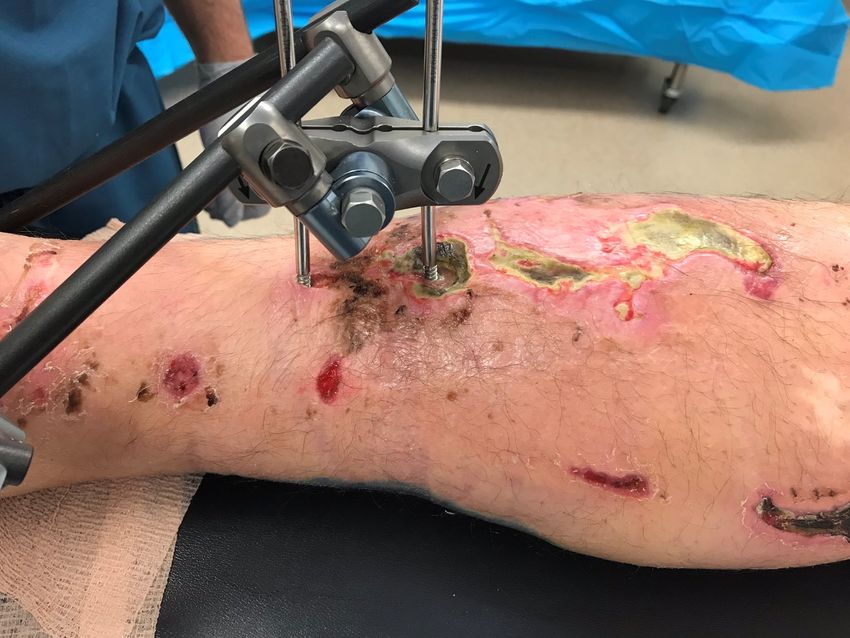
Disclaimer
• All clinical and radiographic images provided are used with
  permission of Kyle Schweser, MD, Sumit Gupta, MD, Chris Souder,
  MD, unless otherwise specified.

                                                             Core Curriculum V5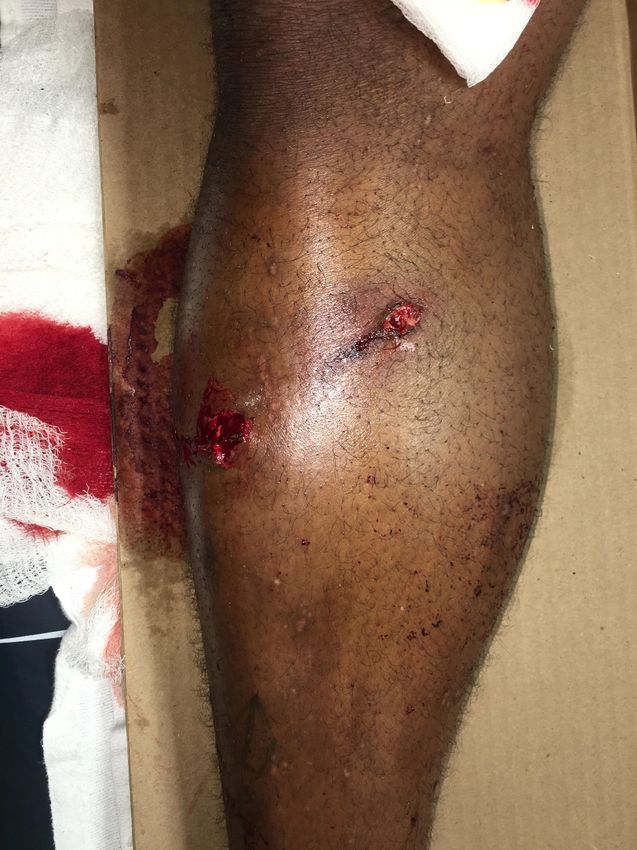
Objectives
• Discuss important initial evaluation of tibial shaft fractures
• Create an algorithm for treatment of tibial shaft fractures
• Identify specific fracture types and complications

• This lecture also contains a Compartment Syndrome Supplement at
  end of presentation

                                                                   Core Curriculum V5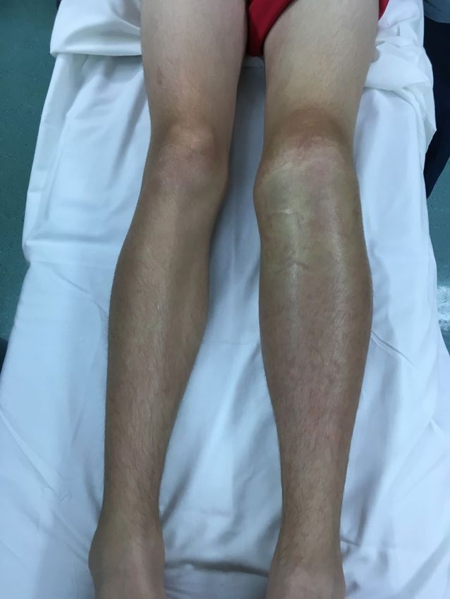
Growth of the Tibia/Fibula
• Growth from two physes
   •   Proximal tibia: 6mm/yr
   •   Distal tibia: 5mm/yr
   •   Proximal fibula: 6.5mm/yr
   •   Distal fibula: 4.5 mm/yr
• Proximal tibial physis closes posterior to anterior
• Tibial tubercle closes proximal to distal
• Distal tibial physis closes central to medial then lateral
               Waters, P. M., Skaggs, D. L., Flynn, J. M., & Rockwood, C. A. (2020). Chapter 5: Physeal Injuries and Growth
               Disturbances. In Rockwood and Wilkins' fractures in children (pp. 930-966). Philadelphia, PA: Wolters Kluwer.

                                                                                                                               Core Curriculum V5
Incidence
• 15% of all long bone injuries in children
   • 39% in the diaphysis
   • Third most common (femur>humerus>tibia)
• Average age: 8
• Boys > Girls
• 26% of abused children
   • Second most common fracture in abuse
      • 1st is humerus
   • Most commonly occurs in the proximal metaphysis in abuse
             *Raducha, Jeremy E. MD1; Swarup, Ishaan MD2; Schachne, Jonathan M. BA2; Cruz, Aristides I. Jr. MD, MBA3;
             Fabricant, Peter D. MD, MPH2 Tibial Shaft Fractures in Children and Adolescents, JBJS Reviews: February 2019 -
             Volume 7 - Issue 2 - p e4
                                                                                                                              Core Curriculum V5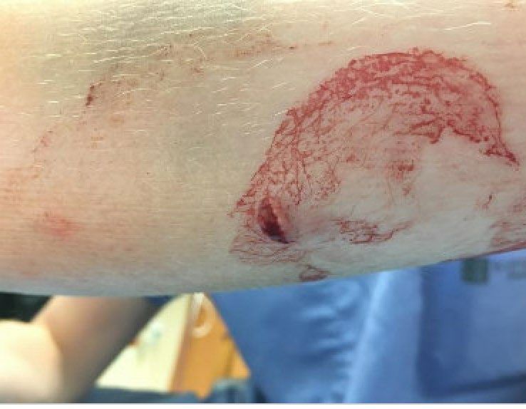
Mechanism and Characteristics
• Younger children
   • Torsional mechanism
• Older children
   • Sports/motor vehicle trauma
• Short oblique/transverse is most common pattern
• 2/3 have no fibula fx
   • Risk of varus malalignment
• 1/3 have an associated fibula fx
   • Risk of valgus malalignment
• Malalignment risk regardless of operative vs
  conservative management
   • Up to 40% malunion risk even in operative patients
            *Raducha, Jeremy E. MD1; Swarup, Ishaan MD2; Schachne, Jonathan M. BA2; Cruz, Aristides I. Jr. MD, MBA3;
            Fabricant, Peter D. MD, MPH2 Tibial Shaft Fractures in Children and Adolescents, JBJS Reviews: February 2019 -
            Volume 7 - Issue 2 - p e4                                                                                        Core Curriculum V5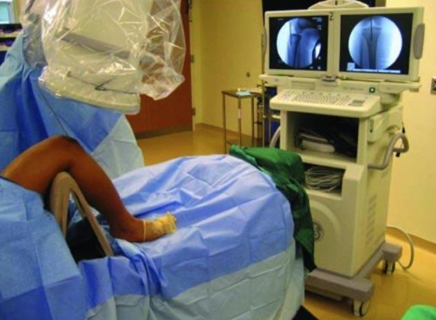
Evaluation
• History
   • Mechanism important in pediatric fractures
   • History of multiple fractures should raise concerns
• Physical Exam
   • Examine entire body
   • Don’t miss an open fracture
• Imaging
   • Image entire fractured bone
   • Image joint above and below
   • Skeletal survey if abuse suspected

                                                           Core Curriculum V5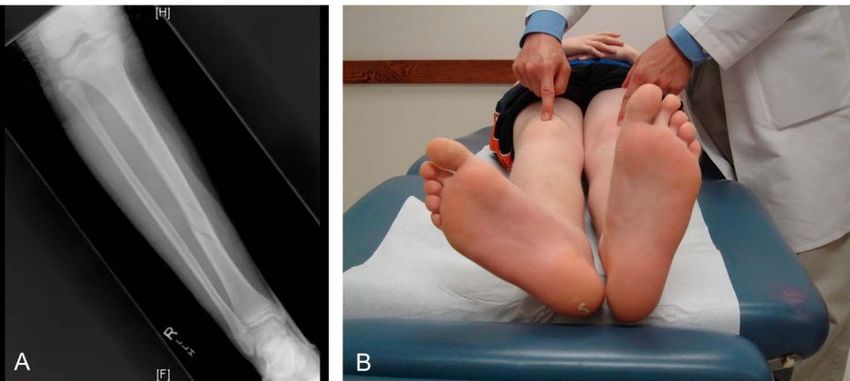
History
• History
   • Same for adults
      • Mechanism, quality/quantity of pain, location of fracture, etc
   • Different from adults
      • Need to rule out abuse

• Most pediatric tibia fractures present after a known
  trauma
   • NAT should always be considered in the young children
      • Especially non-ambulatory
   • Most NAT injuries are metaphyseal

                                                                         Core Curriculum V5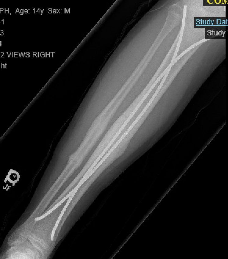
Exam
• Evaluation of the whole body
   • Bruising/skin lesions common in abuse
   • Areas of tenderness/deformity
       • Especially in those who can’t communicate
• Anteromedial tibia is subcutaneous
   • Any laceration should raise concern for an open fracture
• Perform a good neurovascular exam
   • Doppler pulses if not palpable
• Children do not malinger
   • Refusal to bear weight is a sign something is wrong
   • Limping, but an ability to walk, does not eliminate fracture
       • Toddler’s fracture

                                                                    Core Curriculum V5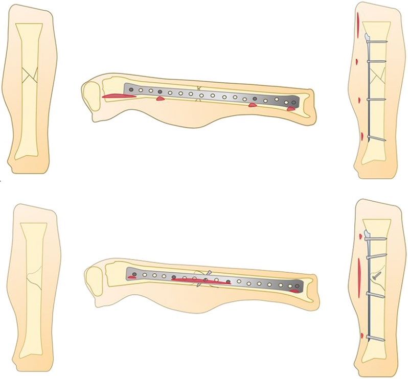
Compartment Syndrome
• Incidence
   • Up to 11% of pediatric tibia fractures
   • 40% of pediatric compartment syndrome due to tibia fxs
   • >80% associated with high energy
• The 6 P’s for older children can still apply
   • Pain out of proportion, paresthesias, pulselessness
   • Pallor, poikilothermia, and paresis
• Typical findings unreliable in younger children
   • Use the three A’s
       • Analgesic requirement increasing
       • Anxiety
       • Agitation
                    *Hak DJ. Acute Compartment Syndrome in Children. 2019 Sep 3. In: Mauffrey C, Hak DJ,
                    Martin III MP, editors. Compartment Syndrome: A Guide to Diagnosis and Management.

                                                                                                           Core Curriculum V5
                    Cham (CH): Springer; 2019. Chapter 13
Imaging Injury film
• All tibia fractures should include
• 2 view full length tibia films
• Ankle and knee joints must be included
• Can forgo in young children if the tibia film captures the foot and
distal femur
• If initial x-rays negative, but high clinical suspicion 3w f/u XR
• Protect, repeat x-rays in 1-2 weeks
• If abuse is suspected
• Skeletal survey
• Advanced imaging often not necessary
• Distal third fractures may require CT to evaluate joint/physeal
involvement Periosteal reaction now visible
Core Curriculum V5Fracture Patterns
• Fracture pattern associated with mechanism
• Low energyrotational
• Spiral fractures of the tibia
• Fibula fracture either
• At a different level
• Not present
• Direct impact (sports, nonaccidental)
• Transverse fracture
• High energy
• Short oblique or butterfly fragment
• Comminuted (axial load)
• Fibula fracture close to the same level
Low energy torsion-no High Energy
fibula fracture Core Curriculum V5Classification
• No current classification system
• Describe location (proximal, midshaft, distal)
• Describe pattern (oblique, transverse, segmental, etc)
• Describe mechanism and age
Core Curriculum V5Treatment
3m f/u (5y/o)
• Algorithm based on multiple factors
• Patient factors
• Fracture characteristics
• The majority can be treated closed
• Most heal quickly
• Toddlers: 3-4 weeks
• Juvenile: 6-12 weeks
• Nonunions are rare
• Usually occur in the setting of infection, open fractures, bone loss
• Certain treatment methods can increase nonunion rates (i.e. external fixators)
Core Curriculum V5Closed Treatment
• Most uncomplicated tibial shaft fractures can be
casted
• Cost-effective
• Goal is acceptable alignment
• Acceptable initial alignment controversial
• Based on age
• Older patients accept less deformity
• Secondary to limited remodeling potential
• Intact periosteal hinge (especially low energy)
facilitates reduction
• As age increases more consideration for surgery
*Hogue GD, Wilkins KE, Kim IS. Management of Pediatric Tibial Shaft Fractures. J Am Acad
Orthop Surg. 2019 Oct 15;27(20):769-778
Core Curriculum V5Closed Treatment
Acceptable initial alignment for closed treatment*
Younger than 8 Older than 8
• Coronal • Coronal
• Varus: 10° • Varus: 5°
• Valgus: 5° • Valgus: 5°
• Sagittal • Sagittal
• Apex anterior: 10° • Apex anterior: 5°
• Apex posterior: 5° • Apex posterior: 0°
• Shortening: 10 mm • Shortening: 5 mm
• Rotation: 5° • Rotation: 5°
• Cortical Overlap > 50% • Cortical Overlap > 50%
Waters, P. M., Skaggs, D. L., Flynn, J. M., & Rockwood, C. A. (2020). Chapter 25: Fractures of the shaft of
the tibia and fibula. In Rockwood and Wilkins' fractures in children (pp. 930-966). Philadelphia, PA:
Wolters Kluwer.
Core Curriculum V5Closed Treatment
• Remodeling is limited and not reliable in the tibia
• Capacity is greatest in the plane of joint motion
• Apex anterior easily accommodated with knee flexion for
ambulation
• Apex posterior requires knee hyper-extension
• Initial slight varus > valgus alignment potentially ok with intact
fibula because of a tethering effect
• With an intact fibula, the overgrowth in healing occurs only
on the tibia, causing a valgus force as the tibial growth is
temporarily increased compared to the fibula
• However, must be monitored as an intact fibula is a risk factor
for eventual varus malalignment
10 y/o M with 10month f/u
• Rotation poorly corrected with remodeling
Core Curriculum V5Closed Treatment
• Casting principles
• Long leg (above knee) cast
• Can provide rotational control if necessary
• Can prevent weight bearing if knee is bent
• If applying the cast in stages (for young children
who can’t cooperate)
• Apply cast for a short leg, creating your mold, allowing
to set
• Then apply long leg component with knee flexed 20-
40 degrees
Core Curriculum V5Associated fibula fx
Closed Treatment Intact fibula
• Tibia fracture with associated fibula fracture
• Apply a varus mold
• Valgus collapse is common
• Tibia fracture with intact fibula
• Apply a valgus mold
• Varus collapse is common60%
• Foot plantarflexion can prevent apex
posterior angulation
Waters, P. M., Skaggs, D. L., Flynn, J. M., & Rockwood, C. A. (2020).
Chapter 5: Physeal Injuries and Growth Disturbances. In Rockwood
and Wilkins' fractures in children (pp. 930-966). Philadelphia, PA:
Wolters Kluwer.
Core Curriculum V5Closed Treatment
• Bivalve cast in patients with swelling concerns
• Consider overnight inpatient monitoring in those requiring reduction
• Weekly radiographs are taken until bridging callus is seen
• Cast wedging in clinic can be utilized to correct interval displacement
• Can be done within first 3 weeks from fracture
Core Curriculum V5Closed Treatment: Wedging
• Technique
• An opening is made in the patients cast
• Typically done at the apex of deformity
• Opening wedges can have cast material,
or premade wedges, inserted in the gap
• Once corrected, the cast is overwrapped
with new cast material
• Care must be taken to protect soft tissue
Proprietary cast wedges
*Hogue GD, Wilkins KE, Kim IS. Management of Pediatric Tibial Shaft Fractures. J Am Acad
Orthop Surg. 2019 Oct 15;27(20):769-778
Core Curriculum V5Closed Treatment
• Tibial diaphyseal fractures
• Long leg cast for 4-6 weeks
• Transition to patellar-tendon bearing
cast vs short leg cast vs removable for
further protected weightbearing
• No immobilization necessary after
sufficient healing noted
*Hogue GD, Wilkins KE, Kim IS. Management of Pediatric Tibial Shaft Fractures. J Am Acad
Orthop Surg. 2019 Oct 15;27(20):769-778
*Raducha, Jeremy E. MD1; Swarup, Ishaan MD2; Schachne, Jonathan M. BA2; Cruz, Aristides I.
Jr. MD, MBA3; Fabricant, Peter D. MD, MPH2 Tibial Shaft Fractures in Children and Adolescents,
JBJS Reviews: February 2019 - Volume 7 - Issue 2 - p e4 Core Curriculum V5Closed Treatment
• Cast controversies
• WB vs NWB
• Low-energy tibia fractures can weightbear as tolerated-
but with increased risk of displacement
• Kids tend to self regulate, especially the younger they
are
• Toddler’s fractures are typically stable enough to allow
WB *Silva, Mauricio, et al. "A comparison of two approaches for the closed treatment of low-energy tibial
fractures in children." JBJS 94.20 (2012): 1853-1860.
*Joshua W.B. Klatt, MD; Alan K. Stotts, MD; John T. Smith, MD Isolated Pediatric Tibial Shaft Fractures Do
Not Need to be Treated in Above-Knee Cast, Fri., 10/15/10 Knee, Tibia & Pediatrics, Paper #61, OTA-2010
*Bauer, Jennifer M. MD, MS*; Lovejoy, Steven A. MD† Toddler’s Fractures: Time to Weight-bear With Regard
to Immobilization Type and Radiographic Monitoring, Journal of Pediatric Orthopaedics: July 2019 - Volume
39 - Issue 6 - p 314-317 Core Curriculum V5Open Fractures
• Treat the same as adults initially
• Early antibiotic administration
• Type 1 and 2First generation cephalosporins
• Type 3Add aminoglycoside
• Soil contaminationAdd penicillin
• Fresh water contaminationAdd fluroquinolone
• Confirm tetanus status and update if needed
Core Curriculum V5Open Fractures
• Timing to debridement
• Controversial
• Consider all factors
• Emergent
• Compartment syndrome
• Neurovascular injury
• Significant contamination
• Urgent
• Moderate contamination
• Skin at risk
• Irreducible fracture
• Within 24 hrs
• Clean wounds
• Reduced fractures
• Stable patients
Core Curriculum V5Surgical Treatment
• Indications for surgery
• Open fractures
• Compartment syndrome
• Soft tissue defect not conducive to casting
• Multiple extremity fractures
• Unacceptable alignment after closed reduction
• Segmental fractures
• Neurovascular injuries
Core Curriculum V5Surgical Treatment
• Majority of surgical patients tend to be
• Older (> 10 years)
• Both tibia and fibula fractured (length
instability issues)
• High degree of initial displacement
Core Curriculum V5Surgical Treatment
• Surgical options
• Pinning and casting
• External fixators
• Uniplane
• Multiplane
• Intramedullary nails
• Rigid
• Flexible
• Plate fixation
• While most can be treated closed, surgical
fixation rates are increasing
Core Curriculum V5Surgical Treatment
• Pinning and Casting
• Typically utilized with unstable patterns and compromised soft
tissue
• Type 1 open, transverse fracture
• Type 2 and 3 open fractures typically require more rigid
stabilization than wires provide
• Can be used in both open and closed fractures
• When possible, pins should be placed away from compromised
tissue
• Pins should avoid tendons/sheaths and neurovascular structures
• Cast placed over pins
• Pins removed at 4 weeks
• Additional casting vs boot depending on healing
Core Curriculum V5Surgical Treatment
• External fixation
• Indications similar to adults
• High energy fractures
• Soft tissue not amenable to fixation or casting
• Length unstable fractures
• Can be used for definitive, or temporary treatment
• Unique complications compared to other operative
techniques
• Pin site infections
• Slower union rates
• Requires procedure for removal
Core Curriculum V5Surgical Treatment
• Uniplane external fixators
• Typically used in an acute traumatic setting
• Converted to other forms of treatment when able
• Can convert to casting once soft tissue stabilize
• Can be used definitively
• Prolonged NWB period
• Older patients (>12 y/o) have increased risk of
• loss of reduction
• Delayed healing
• Younger patients have increased risk of leg length discrepancy
• Secondary to local hyperemia or growth factor release stimulating
physeal growth
*Hogue GD, Wilkins KE, Kim IS. Management of Pediatric Tibial Shaft
Fractures. J Am Acad Orthop Surg. 2019 Oct 15;27(20):769-778 Core Curriculum V5Surgical Treatment
• Multiplane external fixators
• Typically reserved for
• Significant bone loss
• Significant soft tissue injury
• Allows flap placement/management
• Late presentation/malunion with large deformity
• Benefits
• More stable than uniplane
• Allows for immediate WB
• Ability to adjust alignment (especially multi-directional correction)
• Constructs are bulky and expensive
Core Curriculum V5Surgical Treatment
• External Fixator
• Pin placement should be outside of the
zone of injury
• Zone of injury includes the soft tissue
injury, not just bony injury
Picture of an adult patient, but demonstrates the
consequences of pin placement w/in zone of injury
Core Curriculum V5Surgical Treatment
• External Fixator
• Ok to span the knee, or ankle, if necessary
• Plan pin/bar/strut placement to allow for
treatment/coverage of soft-tissues
• Circular fixation allows multiple smooth
tensioned wires in short segments and
small bones
• Can combine with AFO splint to prevent
equinus contracture
• Pins should not be placed closer than 1cm
to the physis
Core Curriculum V5Surgical Treatment
• Flexible nails
• Can be utilized in both open or closed fractures
• Similar rates of infection in open fractures when compared
to closed
• Fractures need to be length stable
• Ideal is a transverse, diaphyseal fracture
Core Curriculum V5Surgical Treatment
• May require immobilization after insertion
• Shorter than with casting alone
• Indications:
• Unacceptable alignment after CRC
• Polytrauma
• Floating knee injuries
• Extensive soft-tissue injury
• Associated compartment syndrome
• Spastic patients
Core Curriculum V5Surgical Treatment
• Flexible nails
• Benefits
• Avoids the physes
• Soft tissue friendly
• Shorter immobilization time compared to
casting
• Earlier WB when compared to casting
• Require additional procedure for removal
• 9-12 months post op
• Implant can irritate the skin
• Union 8-20 weeks
Core Curriculum V5Surgical Treatment
• Flexible nails
• The nails should be contoured prior to insertion
• Entry site is distal to the proximal physis in the
metaphysis
• Equal sized nails are used
• Stability is imparted through:
• Three-point fixation in the canal (similar to three
point mold on a cast)
• Fracture reduction/bone contact
• Nails should cross proximally and distally in the canal
only
• Do NOT twist nails
• The apex of the curves should be at the same level in
the mid-diaphyseal region
Core Curriculum V5Surgical Treatment
• Flexible nails
• Do not twist the nails
• Loses three point fixation
• Nails should be same size
• Larger nail exerts more force
• Potential deformity
Deformity secondary Twisted nails
to larger medial nail
Core Curriculum V5Surgical Treatment
• Flexible nails
• Complication rates of 15-20%
• Rates increase with age of patient
• In delayed in union in age > 14years
Gordon JE, Gregush RV, Schoenecker PL, Dobbs MB, Luhmann SJ. Complications after titanium elastic
nailing of pediatric tibial fractures. J Pediatr Orthop. 2007 Jun;27(4):442-6. doi:
10.1097/01.bpb.0000271333.66019.5c. PMID: 17513967. Core Curriculum V5Surgical Treatment
• Flexible nails
• 20% compartment syndrome rate
• Higher rates in those >50kg
• Increased forces needed for reduction
• Increased rates with comminuted fractures
• Tend to be higher energy fractures
• Initially short, then brought out to length with nail
• Loss of periosteal support
• Higher rate in those presenting with a neurologic injury
• More soft tissue energy
• Higher energy mechanisms
Pandya NK, Edmonds EW, Mubarak SJ. The incidence of compartment syndrome after flexible nailing of
pediatric tibial shaft fractures. J Child Orthop. 2011 Dec;5(6):439-47. doi: 10.1007/s11832-011-0374-y.
Epub 2011 Nov 1. PMID: 23205145; PMCID: PMC3221761. Core Curriculum V58 year old male, closed midshaft tibia fracture Immediate post operative images
Core Curriculum V510 months post op Post op removal
Core Curriculum V5Surgical Treatment
• Rigid IMNs
• Indicated in those with narrow or closed proximal
tibial physis/apophysis
• Nearing skeletal maturity
• Very limited data regarding their use in skeletally
immature
• Nail principles are the same as in adults
• Stability imparted by a “press fit” of the nail within the
canal augmented by locking
• If considering, and proximal physis is open
• Obtain a bone age assessment
• Tanner and Whitehouse method for calculation
• Ensure that IMN spans to proximal physis
• Theoretical decreased chance of physeal bar formation
Core Curriculum V5Surgical Treatment
• Rigid nails
• Benefits
• Allows for immediate weight bearing (load sharing)
• High level of stability
• Soft tissue friendly
• No need for immobilization
• No patient weight limit
• Anterior knee pain
• Can cause physeal arrest and growth disturbances
• Court-Brownt et al. examined 36 patients with open physes
(age 13-16)
• No evidence of shortening or growth arrest
*Court-Brown CM, Byrnes T, McLaughlin G. Intramedullary nailing of tibial diaphyseal
fractures in adolescents with open physes. Injury. 2003 Oct;34(10):781-5.
Core Curriculum V5Surgical Treatment
• Rigid nailing
• No study comparing supra- vs infra-
patellar starting sites in adolescents
• Both safe in existing adult literature
• Ensure proper start site on a true AP
and lateral of the knee
• Do not cross physis with proximal
locking screws
• Reaming is similar for adults, and
intramedullary press fit is desired
• Ensure guide wire does not cross distal
physis
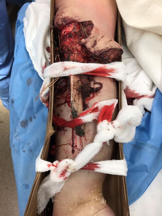
Core Curriculum V515 year old male, MVC, Type 3a open Post debridement/External fixation
Core Curriculum V5Hand X-ray to determine bone age Intraoperative fluoro image of start site
Core Curriculum V5Immediate Post op 12 months post op
Core Curriculum V5Surgical Treatment
• Plate osteosynthesis
• Difficult to give true indications
• When done properly, is appropriate for all ages/fracture
types
• Can be done in both open, and closed, fractures
• Open fractures should have minimal contamination and
allow for soft tissue coverage
• Should be done in a soft tissue friendly way
• Open fractures can expose the fracture for you, allowing
for easy access for fixation
• Percutaneous bridge plating possible for comminuted
fractures
• Acts as an internal “external” fixator
Murphy M, et al. Minimally Invasive Medial Plate
• Simple fractures should undergo rigid fixation Osteosynthesis of High-Energy Pediatric Tibia
Fractures. J Orthop Trauma. 2020;34(8): e272-e281
Core Curriculum V5Surgical Treatment
• Plate osteosynthesis
• Benefits
• Allows early mobilization
• Earlier weight bearing than cast or external fixator
• Lower cost
• Can remain extra-physeal
• Can plan to remove screws that bridge the physis if
required to adequate fixation
• Medially based plates can irritate soft tissues
• Requires larger incisions than other methods
• Can compromise blood supply if done improperly
Core Curriculum V5Surgical Treatment
• Plate osteosynthesis
• Bridge plating
• Small incision are made both proximally and distally
on the tibia
• Length and alignment are held manually, or with an
external fixator
• A submuscular path is created, followed by plate
insertion
• Several screws are then placed proximal and distal to
the fracture
• Needs to have a long working length in comminuted
fractures
Murphy M, et al. Minimally Invasive Medial Plate
Osteosynthesis of High-Energy Pediatric Tibia
Fractures. J Orthop Trauma. 2020;34(8): e272-
e281
Core Curriculum V5Surgical Treatment
• Plate osteosynthesis
• Compression plating
• Direct fracture reduction (open fractures,
small incisions) is necessary
• Oblique fracture: lag
screw/neutralization plate
• Transverse fracture: compression plate
• Alternative: percutaneous lag screw with
percutaneous neutralization plate
insertion
Murphy M, et al. Minimally Invasive Medial Plate
Osteosynthesis of High-Energy Pediatric Tibia
Fractures. J Orthop Trauma. 2020;34(8): e272-
e281
Core Curriculum V58 year old high energy, length unstable fracture 4 months post op after percutaneous bridge plating
Core Curriculum V5Specific Fractures
• Toddler’s fracture
• Proximal tibial metaphyseal fracture
• Distal tibial metaphyseal fractures
• Bicycle spoke injuries
• Pathologic fractures
Core Curriculum V5Toddler’s Fracture
• Childhood accidental spiral tibial fracture
(CAST fracture)
• Young, ambulatory child
• 9 months - 3 years old
• Low energy twisting mechanism
• Tripping
• Falling
• Spiral or oblique tibia fracture
• Fibula frequently intact
Core Curriculum V5Toddler’s Fracture
• Presents with limping or “toe touch” standing
• Above knee vs below knee cast vs walker boot
• Evidence supports below knee for 3-4 weeks
• Newer evidence suggests walker boot is sufficient
• Inherently stable fractures (thick periosteum)
• Child will self-regulate weight bearing
• Rapid healing
• ~4w
Core Curriculum V5Proximal Tibial Metaphyseal Fracture
• Common area of injury
• Most commonly seen in 3-6 year old
• Low energy mechanism
• Jumping on a trampoline—hyperextension
• Going down a slide and leg gets caught—valgus
• Typically involves tibial tubercle physis
Core Curriculum V5Proximal Tibial Metaphyseal Fracture
• Treated closed with above knee cast
• Varus mold may help prevent deformity
• If unable to get closed reduction, soft
tissue may be interposed
• CRPP required for fractures with
unacceptable reduction in the cast
Core Curriculum V5Proximal Tibial Metaphyseal Fracture
• Cozens phenomenon
• Transient valgus overgrowth
deformity of tibia
• Remodeling typically occurs over
12-24 months
• Surgery rarely required
• Lateral temporary
hemiepiphysiodesis if spontaneous
correction does not occur
Waters, P. M., Skaggs, D. L., Flynn, J. M., & Rockwood, C. A. (2020). Chapter 5:
Physeal Injuries and Growth Disturbances. In Rockwood and Wilkins' fractures
in children (pp. 930-966). Philadelphia, PA: Wolters Kluwer
Core Curriculum V5Distal Tibial Metaphyseal Fracture
• Often a greenstick injury
• Anterior cortex impacted
• Posterior cortex displaced
• Aim of treatment is prevention of recurvatum
deformity
• Closed treatment
• Long leg cast
• Foot in plantarflexion (helps reduce posterior
displacement)
• Can combine with percutaneous pinning
• Open treatment
• ORIF with plating for significant instability/loss of
reduction
Core Curriculum V510 year old male with a type 2 open fracture. Pins removed at 6 weeks, 1 year follow up shown
Core Curriculum V5Bicycle Spoke Injuries
• This is a soft tissue injury with an underlying fracture
• Occurs when a child’s foot becomes trapped in bike wheel
• Tibia often a spiral fracture
• Management is concerning soft tissue of the foot
• Should be admitted and observed for 24-72 hrs
• Well padded splint with elevation initially
• Observe the skin and treat accordingly
• Tibial management
• Cast after soft tissue improvement
• External fixator appropriate for severe soft tissue injuries
Core Curriculum V5Pathologic fractures
• Common tibial pathology
• Fibrous dysplasia
• Typically Heals with conservative therapy
• Nonossifying fibroma
• Heals with conservative therapy
• Aneurysmal bone cyst
• Requires surgery for both the cyst and fracture
• Unicameral bone cysts
• Heals with conservative therapy
• Surgery to resolve persistent cyst and prevent refracture
• Malignant lesions
• Nonoperative first during tumor workup
• Definitive management developed in conjunction with tumor therapy
Core Curriculum V5Complications
• Compartment syndrome
• Angular deformity
• Rotational deformity
• Does not remodel
• Growth arrest
• Infection
Lovejoy SA, Mehlman CT. The Community Orthopaedic Surgeon Taking Trauma
Call: Pediatric Tibia Fracture Pearls and Pitfalls. J Orthop Trauma. 2017;31
Supple 6: S22-S26
Core Curriculum V5Angular Deformity
• Correction overtime after union based on age
• Girls 1-8, Boys 1-10: moderate correction after union
• Girls 9-12, Boys 11-12: about half of the angulation
corrected over time
• Over 13: about a quarter of the angulated corrected
• Single plane deformity corrected better than
multiplanar
• Varus corrects more than valgus
• Most correction occurs in first 18 months after
injury Waters, P. M., Skaggs, D. L., Flynn, J. M., &
Rockwood, C. A. (2020). Chapter 5: Physeal Injuries
and Growth Disturbances. In Rockwood and
Wilkins' fractures in children (pp. 930-966).
Philadelphia, PA: Wolters Kluwer
Core Curriculum V5Angular Deformity
• Unacceptable malunion is not well documented
• Long term data regarding outcomes is not available
• Guided growth can be used for metaphyseal
deformities in skeletally immature
• Osteotomy is mainstay form of malunion corrective
surgery
Core Curriculum V5Leg-Length Discrepency
• Shortening at fracture site
• Growth arrest
• Proximal/distal tibial fractures
• Fracture extension into the physis
• Treatment violates the physis
• Tibial overgrowth
• Fracture healing stimulates the physis
• General age cut off: 10 years
• Younger—overgrowth
• Older—arrest
• Treatment depends on size of discrepancy and age of patient
Core Curriculum V5Infection
• Less common than in adult population
• More common in open injuries
• 2-10% occurrence
• Higher Gustilo-Anderson typehigher the risk
• Treatment options depend on situation
• Must control infection
• Debride to healthy, bleeding tissue
• Treatment mirrors adults in terms of source control,
staging, antibiotics
• Abx coated nails in those approaching skeletal maturity
*Cruz AI Jr, Raducha JE, Swarup I, Schachne JM, Fabricant PD. Evidence-based
update on the surgical treatment of pediatric tibial shaft fractures. Curr Opin
Core Curriculum V5
Pediatr. 2019 Feb;31(1):92-10214 year old with type 3a open fracture after an
MVC, status post external fixation/debridement
Core Curriculum V52 months post op: periosteal reaction, increased Treated with Rigid IMN fracture gap, draining wound Core Curriculum V5
Treated with debridement, hardware removal,
antibiotic nail insertion, and IV antibiotics

                                    Core Curriculum V5
Treated with further debridement, antibiotic spacer placement, and
rigid intramedullary nailing
Core Curriculum V5Bone grafted with autograft and graft substitute utilizing a pseudomembrane
induced staged osteosynthesis (Masquelet technique)
*Masquelet AC. Muscle reconstruction in reconstructive surgery: soft tissue repair and long
bone reconstruction. Langenbecks Arch Surg. 2003 Oct;388(5):344-6.
Core Curriculum V518 months s/p bone grafting
Core Curriculum V5Summary
• Most pediatric tibial shaft fractures can be treated conservatively
• Casting is cost effecting
• However, surgical management is increasing in frequency
• Allows earlier weight bearing/mobilization
• NAT and compartment syndrome should not be missed
• Treatment is typically dictated by age, fracture type, location, and soft
tissue
• Remodeling occurs less in the tibia than the femur
• Remodeling occurs less in older patients
Core Curriculum V5References
• Waters, P. M., Skaggs, D. L., Flynn, J. M., & Rockwood, C. A. (2020). Chapter 5:
Physeal Injuries and Growth Disturbances. In Rockwood and Wilkins' fractures in
children (pp. 930-966). Philadelphia, PA: Wolters Kluwer.
• Raducha, Jeremy E. MD1; Swarup, Ishaan MD2; Schachne, Jonathan M. BA2; Cruz,
Aristides I. Jr. MD, MBA3; Fabricant, Peter D. MD, MPH2 Tibial Shaft Fractures in
Children and Adolescents, JBJS Reviews: February 2019 - Volume 7 - Issue 2 - p e4
• Hak DJ. Acute Compartment Syndrome in Children. 2019 Sep 3. In: Mauffrey C,
Hak DJ, Martin III MP, editors. Compartment Syndrome: A Guide to Diagnosis and
Management. Cham (CH): Springer; 2019. Chapter 13
• Hogue GD, Wilkins KE, Kim IS. Management of Pediatric Tibial Shaft Fractures. J
Am Acad Orthop Surg. 2019 Oct 15;27(20):769-778
• Joshua W.B. Klatt, MD; Alan K. Stotts, MD; John T. Smith, MD Isolated Pediatric
Tibial Shaft Fractures Do Not Need to be Treated in Above-Knee Cast, Fri.,
10/15/10 Knee, Tibia & Pediatrics, Paper #61, OTA-2010
Core Curriculum V5References
• Bauer, Jennifer M. MD, MS*; Lovejoy, Steven A. MD† Toddler’s Fractures: Time to Weight-bear
With Regard to Immobilization Type and Radiographic Monitoring, Journal of Pediatric
Orthopaedics: July 2019 - Volume 39 - Issue 6 - p 314-317
• Andreacchio A, Alberghina F, Marengo L, Canavese F. Pediatric tibia and femur fractures in
patients weighing more than 50 kg (110 lb): mini-review on current treatment options and
outcome. Musculoskelet Surg. 2019 Apr;103(1):23-30.
• Court-Brown CM, Byrnes T, McLaughlin G. Intramedullary nailing of tibial diaphyseal fractures in
adolescents with open physes. Injury. 2003 Oct;34(10):781-5.
• Cruz AI Jr, Raducha JE, Swarup I, Schachne JM, Fabricant PD. Evidence-based update on the
surgical treatment of pediatric tibial shaft fractures. Curr Opin Pediatr. 2019 Feb;31(1):92-102
• Masquelet AC. Muscle reconstruction in reconstructive surgery: soft tissue repair and long bone
reconstruction. Langenbecks Arch Surg. 2003 Oct;388(5):344-6.
• Bible JE, McClure DJ, Mir HR. Analysis of single-incision versus dual-incision fasciotomy for tibial
fractures with acute compartment syndrome. J Orthop Trauma. 2013 Nov;27(11):607-11. doi:
10.1097/BOT.0b013e318291f284. PMID: 23515126.
Core Curriculum V5Compartment Syndrome
Supplement
Core Curriculum V5Compartment Syndrome
• Diagnosis is clinical most commonly
• When to use invasive (pressure) monitoring
• Obtunded or preverbal patients
• Unclear clinical picture with high suspicion
• Atypical presentation (e.g. atraumatic, DVT, etc.)
• Exertional compartment syndrome
• Ways to monitor invasively
• Transducer-tipped catheters
• Arterial line set up
Collinge C, Kuper M. Comparison of Three Methods for
Measuring Intracompartmental Pressure in Injured Limbs of
Trauma Patients. J Orthop Trauma. 2010;24(6):364-8.
Core Curriculum V5Compartment Syndrome
• Diagnosing with pressure monitoring
• Less clear in children
• Children have higher resting compartment pressures
• Calculate Delta P (△P)
• Subtract compartment pressure from Diastolic blood pressure (DBP)
• ΔP=DBP–compartment pressure
• If ΔP ≤ 20 mm Hg (30 mm Hg in adults)
• Diagnostic for compartment syndrome
• Can subtract from mean arterial pressure as well
• If within 30 mm Hg of MAP, then diagnostic
• Blood pressure should be taken prior to anesthesia, if possible
Livingston, Kristin S. MD; Glotzbecker, Michael P. MD; Shore, Benjamin J. MD, MPH,
FRCSC Pediatric Acute Compartment Syndrome, Journal of the American Academy
of Orthopaedic Surgeons: May 2017 - Volume 25 - Issue 5 - p 358-364 doi:
10.5435/JAAOS-D-15-00655
Core Curriculum V5Compartment Syndrome
• Measure within 5 cm of the fracture
• Where to take the Pressure
• Lateral compartment
• Just anterior to the posterior border of the fibula
• Must be taken in the peroneal muscle belly
• Anterior compartment
• 1 cm lateral to anterior tibia
• May be less depending on size of patient
• Deep posterior compartment
• Immediately posterior to the posteromedial border of the tibia
• Superficial posterior compartment
• Two finger breadths posterior to posteromedial border of the tibia
• Alternative: middle of medial calf
Core Curriculum V5Compartment Syndrome
• Tips on pressure monitoring
• Zero the system just prior to needle insertion
• Zero the system with the needle at the angle its being
inserted
• Insert needle perpendicular to skin
• Insert until a “pop” is felt to ensure entry into
compartment
• Monitor pressure within 5 cm of the fracture (if
present)
• Strongly consider sedation for children
• Document BP prior to induction
Core Curriculum V5Compartment Syndrome
• Arterial line monitoring
equipment
• 18-gauge needle
• High-pressure tubing
• Pressure transducer
• Associated equipment
• Adjustable transducer stand
• Pressure monitor
• 10—20mL syringe
• Three-way stopcock
• Sterile saline with pressure bag
Core Curriculum V5Compartment Syndrome
• Arterial line technique
• Set up the arterial line transducer, tubing, pressure bag as normal
• Adjust transducer to be at the same height as the leg
• Attach stopcock to transducer tubing
• Opposite to the tubing, insert needle (can have its own tubing)
• On the remaining opening, attach syringe
• Flush the system with saline from the bag
• Level the needle at site of compartment measuring
• Zero the monitor
• Insert the needle without changing position of hand
• Close system off to transducer and inject 1cc saline
• Open system to transducer and monitor pressure
*Steps can vary depending on system. Alternate description in video: https://otaonline.org/video-
library/45036/procedures-and-techniques/multimedia/16731429/acute-compartment-syndrome-of-the-lower-
extremity Core Curriculum V5Compartment Syndrome
• Requires emergent surgical intervention
• In the setting of delayed (> 24 hrs) presentation
• No change in treatment for acute presentation
• Children have improved recovery rates compared to adults in this setting
• Limit initial muscle debridement, even if ischemic, as recovery is likely
Lin, James S.a; Samora, Julie Balcha,,b Pediatric acute compartment syndrome: a
systematic review and meta-analysis, Journal of Pediatric Orthopaedics B: January 2020 -
Volume 29 - Issue 1 - p 90-96 doi: 10.1097/BPB.0000000000000593
Core Curriculum V5Compartment Syndrome
• Dual vs Single incision fasciotomy
• Limited evidence in pediatric literature
• Adult literature shows no difference
• Primary closure
• Infection
• Need for coverage
• No difference, as long as complete release of
all compartments
Neal M, et al. The Efficacy of a Single-Incision Versus Two-
Incision Four-Compartment Fasciotomy of the Leg J Orthop
Trauma. 2016;30(5):e164-8
Bible JE, McClure DJ, Mir HR. Analysis of single-incision versus dual-incision fasciotomy
for tibial fractures with acute compartment syndrome. J Orthop Trauma. 2013
Nov;27(11):607-11. doi: 10.1097/BOT.0b013e318291f284. PMID: 23515126.
Core Curriculum V5Compartment Syndrome
• Dual incision
• Anterolateral incision
• Incision along length of fibula
• Length varies in pediatric population
• Avoid proximal neck and distal subcutaneous fibula
• Find intermuscular septum
• Incise 1 cm on either side of septum for anterior and lateral compartment release
• Protect superficial peroneal nerve at distal incision
• Posteromedial
• Incision posterior to posterior border of tibia
• Distance from posterior border of tibia varies in pediatric population
• Incise fascia overlying superficial posterior compartment
• Find Soleal bridge attached to tibia and release for deep posterior
• Video for technique: https://otaonline.org/video-library/45036/procedures-and-
techniques/multimedia/16776673/dual-incision-release
Core Curriculum V5Compartment Syndrome
• Single Incision
• Same as anterolateral incision in dual incision technique
• First find intermuscular septum
• Incise 1 cm on either side for anterior and lateral compartment
• Continue further posterior to lateral compartment
• Release fascia over superficial posterior compartment
• Deep posterior is found
• In the interval between superficial posterior and lateral compartments
• Follow the interosseous membrane along back of fibula
• Release the deep compartment off this membrane
• Images for technique on slides 22-27
• Video for technique: Single Incision Four Compartment Parafibular Fasciotomy |
Procedures & Techniques | OTA Online Trauma Access
Core Curriculum V5Core Curriculum V5
Core Curriculum V5
Superficial         Anterior compartment
peroneal n.      Lateral compartment
              Superficial posterior
              compartment

              Superficial peroneal n.

                          Core Curriculum V5
Lateral compartment
Fibula
Superficial posterior
compartment
Lateral compartment
Fibula
Flexor Hallicus Longus m.
Superficial posterior
compartment
Core Curriculum V5You can also read