PERINATAL NEUROPATHOLOGY: A PRACTICAL APPROACH - Society for Pediatric Pathology
←
→
Page content transcription
If your browser does not render page correctly, please read the page content below

PERINATAL
       NEUROPATHOLOGY:
     A PRACTICAL APPROACH
                     John M. Lee MD PhD

                     Attending Pathologist and
         Ivan S. Ciric Chair of Neurosciences NorthShore
                     University Health System

                        Clinical Professor
                   Pritzker School of Medicine
                      University of Chicago

Disclosures
-Cornelli Consulting, Milan, Italy: Hold a joint patent for a
glycosaminoglycan for the treatment of Alzheimer’s disease
and other related neurological disorders, ie. anti-anxiety effects

-Editor-in-Chief, Journal of Neuropathology and Experimental
Neurology, American Association of Neuropathologists

-Member of the Medical and Science Advisory Board for
Ceremark Pharma LLC

                                                                     1
GROSS DESCRIPTION:
                   Autopsy Brain Cutting

Perinatal Brain Cutting
Gross description
The brain is carefully removed and fixed in formalin (with or without acetic acid) for a minimum of 7-10 days
prior to full gross examination. After fixation, the brain is [intact and shows two cerebral hemispheres / mostly
intact / partially fragmented / completely fragmented]. The external surface of the brain is [white and shiny/
dusky] and there [is / is not] anterior-posterior blunting. The leptomeninges [appear congested and/or show
possible subarachnoid hemorrhage].

[No/primary/secondary/tertiary] sulcation is present. The calcarine fissure [is / is not] present. The parieto-
occipital fissure [is / is not] present. [Add any other details about sulcation
pattern_______________________________________]. The corpus callosum [is/is not] present.

The [right / left] hemisphere is serially sectioned and shows [describe any lesions or findings such as,
intra/periventricular hemorrhage, parenchymal hemorrhage(s), white matter cystic change etc.
_________________________________________________________

[Other notes_________________________________________________________________
Sections:
B1 - brainstem and cerebellum
B2 - frontal lobe
B3 - temporal lobe with hippocampus
B4 - parietal lobe
B5 - occipital lobe
B6 - thalamus/subcortical nuclei
B7 -
B8 -

                                                                                                                    2
UNSW EMBRYOLOGY

UNSW EMBRYOLOGY

                  3
MICROSCOPIC DESCRIPTION:

                           4
Normal Cortical Lamination (Near Term)

                                         5
Neuronal Maturation

Pseudoverrucous Lamination (Normal up to 28 weeks)

                                                     6
Acute Perinatal Stress
 Subarachnoid Hemorrhages
Germinal Matrix Hemorrhages
Choroid Plexus Hemorrhages
 Hypoxic-Ischemic Changes

Acute Perinatal Stress
Subarachnoid Hemorrhages
Germinal Matrix Hemorrhages
Choroid Plexus Hemorrhages
 Hypoxic-Ischemic Changes

                              7
Obsteticalpathology.com

              Subarachnoid Hemorrhage

                                        8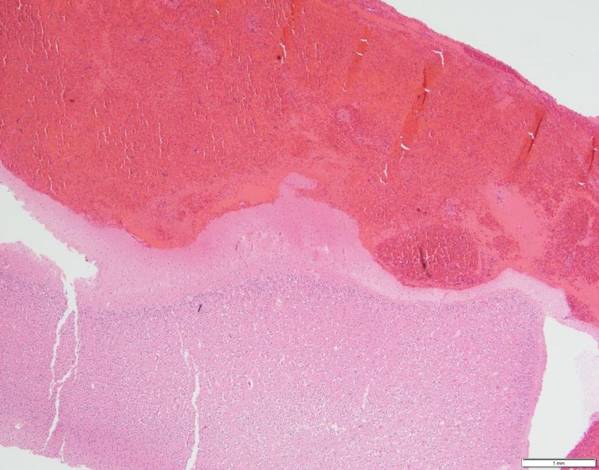
Subarachnoid Hemorrhage
                             Parenchymal Hemorrhages
   Obsteticalpathology.com

                                                       9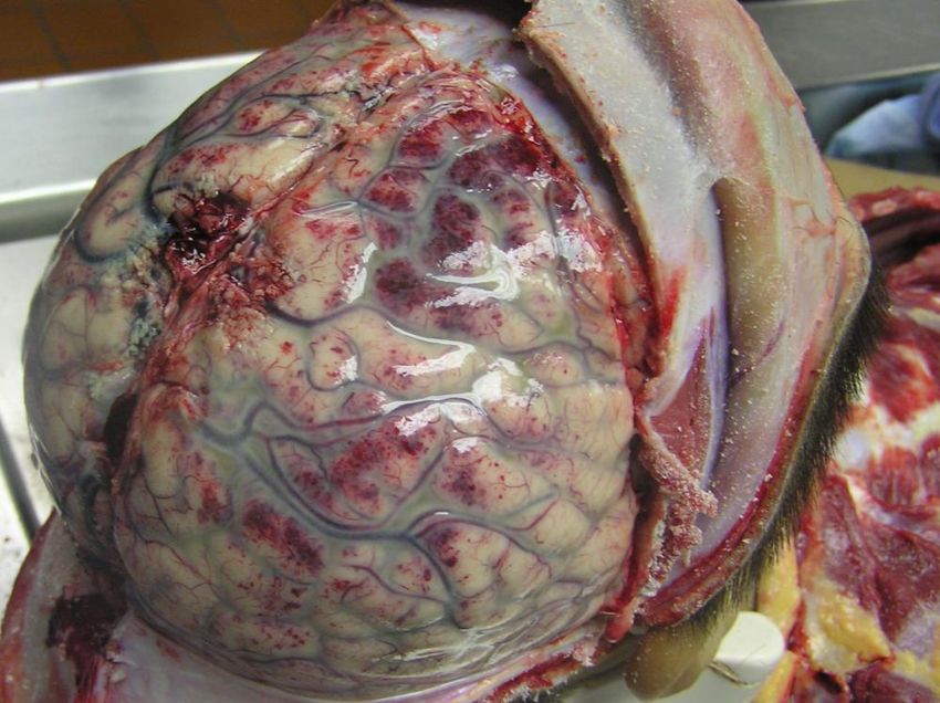
Acute Perinatal Stress
   Subarachnoid Hemorrhages
 Germinal Matrix Hemorrhages
  Choroid Plexus Hemorrhages
   Hypoxic-Ischemic Changes

GERMINAL MATRIX HEMORRHAGE

                               10
Germinal Matrix Hemorrhage
Obsteticalpathology.com
Acute Perinatal Stress
Subarachnoid Hemorrhages
Germinal Matrix Hemorrhages
Choroid Plexus Hemorrhages
Hypoxic-Ischemic Changes
Metabolic Encephalopathy
11Alzhiemer Type II Astrocytes Shrunken Eosinophilic Neurons
(Metabolic Encephalopathy) (Acute Anoxic-Ischemic Encephalopathy)
Kinney and Volpe (Volpe’s Neuropathology of Newborn 6th Ed)
Pons
Shrunken Eosinophilic Neurons
(Acute Anoxic-Ischemic Encephalopathy)
12Pons
Shrunken Eosinophilic Neurons
(Acute Anoxic-Ischemic Encephalopathy)
Thalamus
Shrunken Eosinophilic Neurons
(Acute Anoxic-Ischemic Encephalopathy)
13Prolonged Perinatal Stress
Chronic Hypoxic-Ischemic Changes
Periventricular Leukomalacia
Parenchymal Calcifications
Prolonged Perinatal Stress
Chronic Hypoxic-Ischemic Changes
Pontosubicular Necrosis
Periventricular Leukomalacia
Parenchymal Calcifications
14Slideserve.com
Nuclear Karyorrhexis
From Semanticscholar.org
15Prolonged Perinatal Stress
Chronic Hypoxic-Ischemic Changes
Periventricular Leukomalacia
Parenchymal Calcifications
Oushsc.edu Periventricular Leukomalacia
16Major Congenital Malformations
Agenesis of the Corpus Callosum
Down Syndrome
Holoprosencepaly
Anencephaly
Lissencephaly Spectrum
Polymicrogyria
Heterotopias
Agenesis of the Corpus Callosum
17Agenesis of the Corpus Callosum
Agenesis of the Corpus Callosum with Hydrocephalus
18Agenesis of the Corpus Callosum
•Complete and incomplete (partial) types
–Partial is usually only missing the
splenium
•Isolated (silent clinically or subtle) or seen in
association with other malformations (i.e.
holoprosencephaly)
•May be sporadic but typically associated with
syndromes: Aicardi, Andermann, Meckel
•Possible pathogenesis:
–Probst bundle of misdirected fibers
–Mechanical defect suggested by
hamartoma/ lipoma
A. Viaene Pediatric Neuropathology: Malformations AANP Teaching
Rounds, Feb 24, 2021
Holoprosencephaly
19Holoprosencephaly
Pediatric Neuropathology: A Text-
Atlas Figures 2.7-3 and 2.7-5
Holoprosencephaly
Berry, RS and Andrews SW, Images in
Forensic Pathology Acad Forensic Pathol (2014) 4: 258-260
20Holoprosencephaly Etiology
•Material diabetes mellitus
•Infections: Toxoplasmosis, syphilis, rubella
•Teratogens: Ethanol, retinoic acid, cholesterol
synthesis inhibitors
•Genetic factors:
–Cytogenetic abnormalities seen in 50% of cases
•Trisomy 13 most frequent
–Smith-Lemli-Opitz syndrome (DHCR7)
–Other Mutations
A. Viaene Pediatric Neuropathology: Malformations AANP Teaching
Rounds, Feb 24, 2021
Anencephaly
21Anencephaly
• In the United States the prevalence of
anencephaly was 9.4 per 100,000 live births
in 2001
• Between 1999 and 2004, there were 2,116
cases reported
– though this number greatly underestimates the
actual occurrence because many anencephalic
fetuses are spontaneously aborted or electively
terminated
Anencephaly
• Anencephaly is a severe defect which is
ultimately incompatible with life
– Infants born alive generally die within hours
with some surviving a few days or rarely a few
weeks
22Anencephaly
• Anencephaly is characterized by an open
defect in the calvaria (skullcap) and the
skin, causing the cranial neural tissue to be
exposed
• Neural tissue that is exposed to amniotic
fluid in utero is damaged
– Results in a fibrotic, vascular mass with scant
neural tissue, termed area cerebrovasculosa
Anencephaly
• Portions of the skull including the frontal,
parietal and occipital bones are absent
giving the characteristic appearance of
“bulging” eyes (note eye are formed)
• Underdevelopment of the pituitary gland
(posterior usually not anterior) with adrenal
gland hypoplasia
• Absent neck
23Anencephaly
• Cranial and facial abnormalities are also
seen in anencephaly
– Result of abnormal induction of neural crest
tissue
• The forebrain and variable amounts of the
upper brainstem are typically involved
Head 11.1 cm in circumference (50th percentile = 17.3 cm)
Approximate biparietal diameter 2.9 cm (50th percentile = 4.71 cm)
Inner canthal distance 1.0 cm (50th percentile = 1.25 cm)
24Absent skull bones and spinal rachischisis, with a portion
of red soft tissue located in the left parietal area (“area
cerebrovasulosa”).
CORTEX AND LEPTOMENINGES WITH
“AREA CEREBROVASULOSA”
25LEPTOMENINGES WITH “AREA CEREBROVASULOSA”
26Embryology
• Closure of the cranial neuropore occurs around
• 3-4 weeks after conception
• Lack of closure results in the absence of the
calvarium and the brain
• Neural tissue that is exposed to the in utero
environment is damaged resulting in a fibrotic,
vascular mass with scant neural tissue
remaining
– This is known as “area cerebrovasculosa”
Associated Abnormalities
• Other common abnormalities seen with
anencephaly are cardiac, pulmonary, renal
and skeletal
• Affected by anencephaly and rachischisis
– Renal defects are seen in 17%
– Cardiac defects are seen in 4%
27Lissencephaly/Agyria
• Smooth or unconvoluted: agyria or lissencephaly
• Reduced number and widened sulci: Pachygyria or
macrogyria
• Histology:
– Agyria with or without 4 cortical layers =
Lissencephaly type I (Miller-Dieker)
– Lissencephaly Type II (Cerebro-ocular dysplasia)
Appearance
• Small, misshapen skull, low brain weight
– Abnormal face
• Thickened cortical ribbon with reduced
white matter
• Ventricular dilation and nodular
heterotopias
• Miller-Dieker best known syndrome
28Agyria in Lissencephalay Type I Miller-Dieker
Lissencephalay Type I Miller-Dieker
29The cortical surface is
smooth and the ribbon
greatly thickened, while
the greatly reduced white
matter contains a large
heterotopia (yellow
arrow).
30Lissencephaly Type II (Cerebro-ocular dysplasia) in an 18-week-old fetus
Lissencephaly
Age matched control
31Polymicrogyria
• Hyperconvoluted cortical ribbon by miniature, thin
gyri
• Varying degrees of neurologic disability
• Heterogenous Etiology:
– Intrauterine ischemia
– Intrauterine infection
– X-linked Aicardi syndrome
– Metabolic diseases
• Pelizaeus-Merzbacher, Leigh’s syndrome, etc.
– Perioxisomal disorders
• Zellweger’s, ALD
32Polymicrogyria
Polymicrogyria
33Polymicrogyria
Neuronal Heterotopias
• Perioxisomal, mitochodrial or chromosomal
disorders
• Often associated with epilepsy, particularly
myoclonic seizures
• Fetal insults: maternal hyperthermia,
mercury poisoning, radiation
• Can be nodular or laminar (rarer)
34Subependymal Heterotopias
Neurons in white matter
35Major Perinatal Brain Infections
Bacterial Meningitis
Viral Meningitis
TORCH (Toxo, CMV and Herpes)
Routes of Entry
• Hematogenous
– Most common
• Usually arterial
• Can be retrograde through anastomotic venous circulation of the face
• Direct implantation
– Usually trauma
• Can be iatrogenic – lumbar puncture
• Local extension
– Sinus, oral, spinal osteomyelitis
• Via peripheral nervous system
– Viral
• Indirect damage to the brain
– Microbial toxins
– Secondary Inflammatory responses
– Immune-mediated (auto-immune antibodies)
36Meningitis
Inflammation of the Meninges
• Usually bacterial (Acute)
• Viral (Self-limiting)
• Fungal (Immune-compromised)
Common Bacterial Causes
• Neonatal:
– E. coli, group B strep.
• 3 months – 3 years:
– Pneumococcus (Streptococcus pneumonia)
– H. influenza –less common presently due to vaccination
• Young adults:
– Neiserria meningitis
• Adults:
– Pneumococcus
• Age extremes:
– L. monocytogenes, Pneumococcus
37Morphology
• Meninges are opaque and thickened
• Exudate
• Inflammation can extend into vessels causing
vasculitis, thrombosis, and hemorrhage
• Leptomeningeal fibrosis with subsequent
hydrocephalus may be long term sequelae,
particularly due to the polysaccharides of
Pneumococcus
Bacterial meningits
38Meningitis
Meningitis
39Encephalitis
Bacteria
40CSF Findings
ORGANISM FLUID CELLS PROTEIN GLUCOSE PRESSURE
QUALITY PRESENT Relative to
plasma levels
BACTERIA CLOUDY PMNs VERY LOW HIGH
HIGH
VIRUS CLEAR LYMPHS SLIGHTLY NORMAL
HIGH
TB LYMPHS MODERATE MILDLY
HIGH LOW
Viral Meningitis
• Usually self-limiting without complications
• Many possible agents
– Mumps
– Enteroviruses
– EBV
– Coxsackie
41Meningeal and scanty perivascular infiltrate of lymphocytes
Encephalitis
General Features of Viral Encephalitis
Using Poliomyelitis as a Case Study
• Lytic infection of motor neurons
(neurotropism) with encephalomyelitis
• Macroscopic lesions rare
42Polio: Microscopic
• Usually worse than clinically apparent
• Anterior horn, motor nuclei of pons and
medulla, reticular formation and cerebellar
nuclei (neurotropism)
• Lymphocytic cuffing
• Leptomeningeal inflammation
• Neuronophagia
Meningeal and scanty perivascular infiltrate of lymphocytes
43Microglial nodules (viral
encephalitis)
Herpes Simplex
44Herpes Zoster (chicken pox)
in the dorsal root ganglion
CMV
45Congenital CMV
(calcifications)
Cytomegalovirus
• Particularly important in fetal/neonate
population - TORCH
• Common opportunistic infection in AIDS,
affecting the CNS in 10-20% of cases
• Large intracytoplasmic and intranuclear
inclusions
46CSF Findings
ORGANISM FLUID CELLS PROTEIN GLUCOSE PRESSURE
QUALITY PRESENT
BACTERIA CLOUDY PMN VERY HIGH LOW HIGH
VIRUS CLEAR LYMPHS SLIGHTLY NORMAL
HIGH
TB LYMPHS MODERATE MILDLY
HIGH LOW
Fungal Diseases
• Can present as meningitis, encephalitis or brain
abscess (rare in perinatal cases; found in pediatric
transplant and hematopoietic malignancies)
• Cryptococcus is a common form of fungal
meningitis (diagnosed by India Ink stain of CSF)
• Aspergillus or Candida most common via
hematogenous route
• Zygomycetes (Mucor) usually direct spread from
sinuses
47Angioinvasive aspergillus
References and Acknowledgments
1. Ellison and Love: Neuropathology 2c 2004 Elsevier LTD and Elsevier
2005
1. Kinney and Volpe (Volpe’s Neuropathology of Newborn 6th Ed)
2. Pediatric Neuropathology: A Text-Atlas Figures 2.7-3 and 2.7-5
4. Berry, RS and Andrews SW, Images in
Forensic Pathology Acad Forensic Pathol (2014) 4: 258-260
5. A.Viaene Pediatric Neuropathology: Malformations AANP Teaching
Rounds, Feb 24, 2021
48You can also read