Posttraumatic clostridial necrotizing fasciitis of the head and neck with descending necrotizing mediastinitis treated by video-assisted thoracoscopic
←
→
Page content transcription
If your browser does not render page correctly, please read the page content below
Kovačić I et al. Posttraumatic clostridial necrotizing fasciitis of the head and neck ... – Med Jad 2021;51(1):89-95
 Professional paper                                                                                      ISSN 1848-817X
 Stručni članak                                                                                Coden: MEJAD6 51 (2021) 1

       Posttraumatic clostridial necrotizing fasciitis of the head and neck with
    descending necrotizing mediastinitis treated by video-assisted thoracoscopic
                            surgery – report of a case

       Posttraumatski klostridijski nekrotizirajući fascitis glave i vrata s descendentnim
     nekrotizirajućim medijastinitisom, liječen video-asistiranim torakoskopskim kirurškim
                                 pristupom – prikaz bolesnika

        Ivan Kovačić, Marijan Kovačić, Jakov Mihanović, Martina Šarec Ivelj, Anela Tolić,
                                          Ivan Bačić*

                                                         Summary
           Background: Necrotizing fasciitis of the head and neck with subsequent descending necrotizing
       mediastinitis is a highly lethal condition. The clostridial origin has a particularly aggressive course.
           Case presentation: Herein we present a case of a 22-year-old male with clostridial necrotizing fasciitis of
       the head and neck complicated with descending necrotizing mediastinitis after a traumatic scalp wound. Three
       days after having sutured the wound at the Emergency Department, the patient became septic with marked
       cellulitis of the head and neck soft tissue. Urgent surgical wound debridement of necrotic tissue along with
       transcervical drainage of the upper mediastinum was performed. The patient was ventilator dependent,
       receiving vasoactive support and empiric broad-spectrum antibiotic therapy. Left-sided pleural effusion and
       CT signs of infection descent into the middle and lower mediastinum mandated further surgical intervention.
       A video-assisted thoracoscopic surgical approach was used to drain and debride the affected mediastinal and
       pleural spaces leading to a gradual stabilization of the patient followed by being transferred to the Surgical
       Department. Microbiological analysis revealed Clostridium perfringens as an infective agent. Further
       recovery was uneventful and the patient was dismissed on postoperative day 24.
           Conclusion: Posttraumatic clostridial gas gangrene of the head and neck is a fulminant and life-threatening
       infection. It requires urgent clinical and radiological assessment. Treatment should be multidisciplinary based.
       The invasiveness and extent of surgery should be tailored for individual patients. Video-assisted thoracoscopic
       surgery technique is a safe and appealing approach to all mediastinal compartments. It offers less invasiveness,
       superior visibility and equal efficiency in terms of tissue debridement and mediastinal as well as pleural
       drainage.
           Key words: necrotizing fasciitis, mediastinitis, Clostridium perfringens, gas gangrene, video-assisted
       thoracoscopic surgery
                                                          Sažetak
           Uvod: Nekrotizirajući fascitis glave i vrata s posljedičnim descendentnim nekrotizirajućim
       medijastinitisom, potencijalno je smrtonosna bolest. Kada je uzrokovana klostridijom ima osobito agresivan
       tijek.
           Prikaz bolesnika: Prikazujemo 22-godišnjeg mladića s nekrotizirajućim fascitisom glave i vrata, s
       razvojem descendentnog nekrotizirajućeg medijastinitisa. Infekcija je nastala putem razderotine vlasišta
       zadobivene u prometnoj nesreći. Tri dana nakon primarnog zbrinjavanja rane u Objedinjenom hitnom
       bolničkom prijamu, bolesnik je postao septičan sa znacima flegmone glave i vrata. Podvrgnut je hitnom

   *
     Zadar General Hospital, Department of surgery, Zadar (Ivan Kovačić, MD; Jakov Mihanović, MD, Ivan Bačić,
PhD, MD), Department of otorhinolaryngology and head and neck surgery (Marijan Kovačić, MD), Department of radiology
(Anela Tolić, MD); Polyclinic „Bagatin“, Department of general, plastic, reconstructive and aesthetic surgery, Zagreb
(Martina Šarec Ivelj, MD)
   Correspondence address / Adresa za dopisivanje: Jakov Mihanović MD, General hospital Zadar, Bože Peričića 5,Zadar,
Croatia; Cell: +385 98 544 401. E-mail: mihanovic@gmail.com
   Primljeno/Received 2020-08-30; Ispravljeno/Revised 2020-12-16; Prihvaćeno/Accepted 2020-12-17

                                                                                                                          89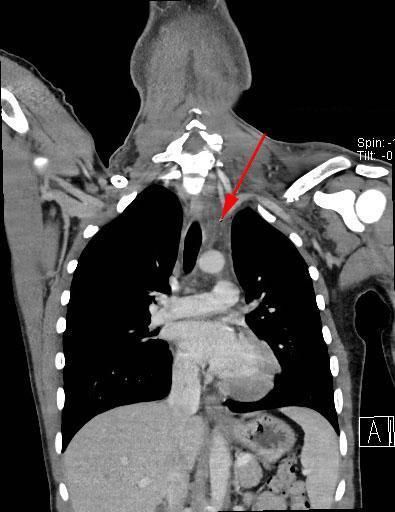
Kovačić I et al. Posttraumatic clostridial necrotizing fasciitis of the head and neck ... – Med Jad 2021;51(1):89-95

       debridmanu nekrotičnih areala i transcervikalnoj drenaži gornjeg medijastinuma. Za vrijeme boravka u
       Jedinici intenzivnog liječenja bio je ovisan o respiratoru, uz vazoaktivnu potporu i empirijsku antibiotsku
       terapiju širokoga spektra. Lijevostrani pleuralni izljev i CT znakovi širenja infekcije u donji medijastinum,
       bili su indikacija za daljnju kiruršku intervenciju. Video-asistirani torakoskopski kirurški pristup korišten je
       za drenažu i debridman inficiranog medijastinalnog i pleuralnog prostora, što je dovelo do postupne
       stabilizacije bolesnika koji je naposljetku premješten na kirurški odjel. Mikrobiološkom analizom obriska rane
       izoliran je Clostridium perfringens kao uzročnik infekcije. Daljnji oporavak je bio bez komplikacija i bolesnik
       je otpušten 24. dan od prijama.
           Zaključak: Posttraumatska klostridijska plinska gangrena glave i vrata, fulminantna je i potencijalno
       fatalna infekcija. Zahtijeva žurnu laboratorijsku i radiološku obradu. Liječenje zahtijeva multidisciplinarni
       pristup. Opseg kirurškoga zahvata treba pažljivo odmjeriti i prilagoditi pojedinom bolesniku. Video-asistirana
       torakoskopska kirurška tehnika pruža pouzdan i pogodan pristup svim medijastinalnim prostorima, uz
       minimalnu invazivnost, izvrsnu vidljivost i podjednaku učinkovitost.
           Ključne riječi: nekrotizirajući fascitis, medijastinitis, Clostridium perfringens, plinska gangrena, video-
       asistirana torakoskopska kirurgija
                                                                                               Med Jad 2021;51(1):89-95

                       Introduction                              dangerous type of infection readily leading into
                                                                 toxemia, multiorgan dysfunction and death. When the
    Necrotizing fasciitis (NF) is a progressive infection        infection starts in the head or neck, it spreads rapidly
of the fascial tissue with secondary spread to the               in the fascial planes descending into the loose
underlying soft tissue. It spreads rapidly, and tissue           mediastinal tissue aided with gravity and negative
loss is extensive and systemic toxemia striking. Blood           intrathoracic inspiratory pressure which makes a
supply to the affected areas is distribubed leading to           distinct clinical entity named descending necrotizing
ischemia and necrosis. It can occur in any part of the           mediastinitis (DNM).10-12 Patients with DNM are at
body. The most common sites are the extremities,                 risk of septic shock with a doubling mortality rate
abdominal wall and perineum. The head and neck are               compared to isolated NF of the neck.1,7,13 Pearse
affected in less than 5%.1-3 Regarding the causative             described descending necrotizing mediastinitis in
agent, NF is divided into several types:                         1938, most commonly having dental/odontogenic or
    Type I is the most common, with polymicrobial                oropharyngeal origin.14 The condition is not rare as
etiology, caused by both aerobic and anaerobic                   generally considered. Sarna and colleagues reported
species.                                                         mediastinal descent from the necrotizing infections of
    Type II is the monomicrobial, most commonly                  the neck in 40-45% of cases.10 Despite improved
caused by group A beta hemolytic streptococcus,                  medical therapy, the mortality is still high, with case
rarely Staphylococcus aureus.                                    series published in the last 3 years having mortality
    Type III and IV are caused by fish and amphibian             rates ranging from 18-33%.15,16 Herein we present a
pathogens (Vibrio species, Aeromonas hydrophila)                 rare case of posttraumatic clostridial gas gangrene
and fungi respectively being extremely rare.4,5                  affecting the head and neck with DNM successfully
    NF with the formation of gas collections within the          treated by a minimally invasive surgical approach.
affected tissue is a distinct illness historically known as
gas gangrene. There are two main subtypes; gas                                           Case report
gangrene caused by clostridia species and non-
clostridial gas gangrene. Clostridial gas gangrene has a             A healthy 22-years old male was brought to our
grimmer clinical course than the non-clostridial entity.6        hospital’s ER because of injuries sustained in a motor
Clostridium is a genus of anaerobic Gram-positive                vehicle accident. At arrival he was alert, hemo-
bacteria widely inhabiting soils and the intestinal tract        dynamically stable, apparently intoxicated with a large
of animals, including human large bowel and healthy              scalp laceration and clinically evident left clavicle
lower reproductive tract of females. Clostridium                 fracture. Cranial and cervical CT showed no signs of
becomes a pathogen in anaerobic environment                      injuries to the brain or spine. The wound was dirty,
typically in extensive, deep, soiled wounds and in               soiled with dirt and grass particles. It was appropriately
crush injuries.5-7 Clinical signs of the clostridial             debrided, copiously irrigated and loosely sutured.
infection develop when bacteria toxins and enzymes               Since his clavicle fracture needed surgery, he was
start to damage the tissue. Initially manifesting as             admitted to the surgical ward where the next day
cellulitis, it rapidly progresses deeper in a form of            subcutaneous emphysema of the scalp was noticed.
miozitis and finally mionecrosis.7-9 It is an extremely          The wound was explored in general anaesthesia,

90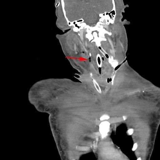
Kovačić I et al. Posttraumatic clostridial necrotizing fasciitis of the head and neck ... – Med Jad 2021;51(1):89-95

wound murky secretion was sampled for microbiology               was widened but without gas or liquid collections
and intravenous antibiotic therapy with co-amoxy                 (Picture 1). The patient was taken for urgent surgery in
clavulanic acid and clindamycin was started. During              general anaesthesia. Three separate incisions around
the next 48 hours, the patient became febrile,                   the wound through the scalp in bitemporal and
tachypneic, tachycardic, hypotensive and oliguric.               occipital directions were made. Extensive necrosis of
Further swelling, redness and tenderness of the scalp,           galeal fascia and periost from the frontal muscle to the
face and neck were noticed. Laboratory tests showed              occipital region was found. Also superficial and deep
an increase in the leukocyte count (13.4 to 19.1x10⁹/L),         temporal muscle fascia was necrotic as well as the
C-reactive protein level (from 135 to 159 mg/L) and              subcutaneous fatty tissue. The temporal muscle itself
creatinine level (from 65 to 152 μmol/L). Erythrocyte            had areas of necrosis. After extensive debridement,
count, platelets count, haemoglobin concentration and            several broad drains were placed along the incisions.
haematocrit were lowered as follows (2.5x10¹²/L,                 Superficial and deep neck fascial spaces were incised
110x109/L, 9.1 g/L, 27%). The patient was sent for a             and debrided. The infrahyoid muscles were only
CT scan of the head, neck and thorax which revealed              minorly affected with necrotic process. Anterior and
gas collections with tissue edema along deep and                 posterior mediastinum was drained through cervical
superficial facial and neck compartments. Mediastinum            incisions (Picture 2).

Picture 1 Head, neck and chest CT: (a) tissue edema along deep and superficial facial and neck compartments,
                        (b) widened mediastinum with fat tissue stranding (red arrows).
    Slika 1. CT glave, vrata i prsnog koša: (a) edem tkiva duž dubokih i površnih odjeljaka lica i vrata, (b)
                        prošireni medijastinum s nitima masnog tkiva (crvene strelice).

                                                                                                                       91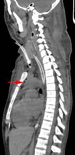
Kovačić I et al. Posttraumatic clostridial necrotizing fasciitis of the head and neck ... – Med Jad 2021;51(1):89-95
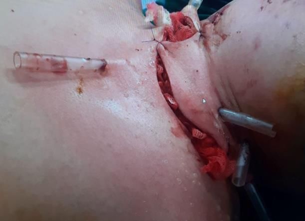
Picture 2 Surgical wound after neck incision and
transcervical upper mediastinal drainage
Slika 2. Kirurška rana nakon reza vrata i
transcervikalne drenaže gornjeg medijastinuma
Postoperatively, the patient required mechanical
ventilation and hemodynamic support in the Intensive
care unit (ICU). The wounds were regularly dressed
and drains irrigated. The microbiology report was
available at the time identifying Clostridium
perfringens needing a switch to targeted antibiotic
therapy (vancomycin, meropenem and clindamycin).
Despite intensive care treatment, the patient’s
condition did not improve. Skin redness and
subcutaneous emphysema crepitations were noticed in
both the pectoral and axillary regions. A follow-up CT
scan 24 hours after surgery revealed increasing pleural
effusion on the left side with air bubbles in lower Picture 3 Follow-up chest CT: (a) left sided pleural
mediastinum (Picture 3) effusion, (b) air in anterior mediastinum, (c) air in
posterior mediastinum (red arrows)
Slika 3. Kontrolni CT prsnog koša: (a) lijevostrani
pleuralni izljev, (b) zrak u prednjem medijastinumu,
(c) zrak u stražnjem medijastinumu (crvene strelice)
The bilateral temporal regions and neck contained the next several days, he was weaned off the ventilator
air bubbles despite incisions and drains (Picture 4). The which allowed his transfer to the surgical department
above described signs of descending mediastinitis on postoperative day 7. Further treatment goals
urged for a step-up surgical procedure. To secure the (regular wounds dressing, hyperalimentation and
airway, surgical tracheostomy was created. Temporal ambulatory independence) were achieved. His clavicle
regions and cervical incisions were re-explored and fracture healed spontaneously in a good position
drained, and several incisions were placed along the obviating the need for orthopedic surgery. The patient
bilateral pectoral and axillary regions. We proceeded was discharged 24 days after the initial injury.
with video-assisted thoracoscopic surgery (VATS)
through the left sided uniportal approach which served
well to evacuate the pleural effusion, incise and
debride mediastinal necrotic tissue and leave a thoracic
drain. A day after the surgery, the patient stabilized
hemodynamically, his renal function improved and
laboratory inflammation parameters decreased. Over
92Kovačić I et al. Posttraumatic clostridial necrotizing fasciitis of the head and neck ... – Med Jad 2021;51(1):89-95
Picture 4 Follow-up CT: (a, b, c) progression of air collections in the neck despite initial incisions (red arrows).
Slika 4. Kontrolni CT: (a, b, c) napredovanje sakupljanja zraka na vratu unatoč početnim urezima (crvene
strelice).
Discussion yet nonspecific early sign. The principles of diagnostic
and management of NF/DNM are the same regardless
Isolation of Clostridia from the tissue and gas of the underlying bacteria.17-20 The Laboratory Risk
collections seen on CT scans confirmed the diagnosis Indicator for Necrotizing Fasciitis is a scoring system
of clostridial gas gangrene and DNM. Typical CT of blood test findings devised by Wong and colleagues
signs of mediastinal infection are unenveloped fluid in 2004.21 Our patient had a confirmatory score of > 6.
collections and soft-tissue gas infiltration. The Treatment of NF should start immediately on the
widening of the mediastinal space is the most constant grounds of clinical suspicion coupled by CT findings.
93Kovačić I et al. Posttraumatic clostridial necrotizing fasciitis of the head and neck ... – Med Jad 2021;51(1):89-95
Surgical intervention should be prompt and aggressive. References
It is necessary to remove all of the dead and devitalized
tissue with drains left in the wound pockets and tunnels 1. Weiss A, Nelson P, Movahed R, Clarkson E, Dym H.
allowing intermittent or continuous irrigation. Cervical Necrotizing fasciitis: review of the literature and case
surgical intervention has standardized indications report. J Oral Maxillofac Surg 2011;69:2786-2794.
2. Ord R, Coletti D. Cervico-facial necrotizing fasciitis.
unlike mediastinal drainage which is still contro-
Oral Dis 2009;15:133-141.
versial.22 We performed initial mediastinal drainage 3. Wong CH, Wang YS. The diagnosis of necrotizing
trans-cervically but it was insufficient to halt the fasciitis. Curr Opin Infect Dis 2005;18:101-106.
descent of the infection to the lower mediastinum 4. Haywood CT, McGeer A, Low DE. Clinical
which was eventually reached by VATS. The first experience with 20 cases of group A streptococcus
successful VATS treatment of DNM was reported by necrotizing fasciitis and myonecrosis: 1995 to 1997.
Isowa and colleagues in 2004.23 VATS has a variety of Plast Reconstr Surg 1999;103:1567-1573.
uses in thoracic surgery having several important 5. Frank DN, Wysocki A, Specht-Glick DD, et al.
advantages compared to open thoracotomy. Besides Microbial diversity in chronic open wounds. Wound
being minimally invasive, it provides an excellent Repair Regen 2009;17:163-172.
6. Japanese Society of Chemotherapy Committee on
visualisation of the pleural cavity which is crucial for
guidelines for treatment of anaerobic infections;
efficient mediastinal debridement and prevention of Japanese Association for Anaerobic Infection
iatrogenic injuries. Cho and colleagues presented a Research. Chapter 2-5-2. Anaerobic infections
series of 17 patients with DNM treated minimally (individual fields): anaerobic infections of the head
invasively with efficiency that was comparable to a and neck. J Infect Chemother 2011;17 Suppl 1:67-71.
more extensive surgical approach.24 Our study 7. Panda NK, Simhadri S, Sridhara SR. Cervicofacial
contributes to the body of evidence opposing concerns necrotizing fasciitis: can we expect a favourable
on the efficacy of VATS to achieve ideal drainage and outcome? J Laryngol Otol 2004;118:771-777.
irrigation in severe cases. The role of tracheostomy in 8. Aldape MJ, Bryant AE, Stevens DL. Clostridium
the treatment of DNM is also debatable. Some authors sordellii infection: epidemiology, clinical findings,
and current perspectives on diagnosis and treatment.
avoid tracheostomy to prevent infection spreading
Clin Infect Dis 2006;43:1436-46.
while others praise its importance. Scheduled 9. Childers BJ, Potyondy LD, Nachreiner R, et al.
tracheostomy prevents compromising the airway Necrotizing fasciitis: a fourteen-year retrospective
and avoids a lot of trouble when performed study of 163 consecutive patients. Am Surg 2002;
emergently.16,18,25 Medical treatment includes main- 68:109-116.
taining an optimal nutritional status, targeted antibiotic 10. Sarna T, Sengupta T, Miloro M, Kolokythas A.
therapy and early physical rehabilitation. Hyperbaric Cervical necrotizing fasciitis with descending
oxygen therapy is a useful adjunct in any anaerobic mediastinitis: literature review and case report. J Oral
infection.13,26 We did not use it in our case because Maxillofac Surg 2012;70:1342-50.
once surgical therapy was addressed properly, 11. Singhal P, Kejriwal N, Lin Z, Tsutsui R, Ullal R.
Optimal surgical management of descending
recovery was swift and uneventful.
necrotizing mediastinitis: our experience and review
Posttraumatic clostridial gas gangrene of the head of literature. Heart Lung Circ 2008;17:124-128.
and neck is a fulminant and life-threatening infection. 12. Kruyt PM, Boonstra A, Fockens P, Reeders JW, van
It requires urgent clinical and radiological assessment, Lanschot JJ. Descending necrotizing mediastinitis
prompt surgical intervention and adequate antibiotic causing pleuroesophageal fistula. Successful treatment
therapy. In cases of mediastinal involvement, tailored by combined transcervical and pleural drainage. Chest
surgical drainage is indicated depending on the 1996;109:1404-07.
patient’s clinical condition, the extent of mediastinitis, 13. Sumi Y. Descending necrotizing mediastinitis: 5 years
and local expertise in minimally invasive surgery. The of published data in Japan. Acute Med Surg 2014;2:
best results are obtained with a multidisciplinary 1-12.
14. Pearse HE. Mediastinitis following cervical suppuration.
approach. VATS is a safe and appealing access to the
Ann Surg 1938;108:588-611.
middle and lower mediastinum. It offers far less 15. Wei D, Bi L, Zhu H, He J, Wang H. Less invasive
invasiveness, superior visibility and equal efficiency in management of deep neck infection and descending
terms of mediastinal and pleural debridement and necrotizing mediastinitis: A single-center retro-
drainage. spective study. Medicine (Baltimore) 2017;96:e6590.
16. Ma C, Zhou L, Zhao JZ, et al. Multidisciplinary
treatment of deep neck infection associated with
descending necrotizing mediastinitis: a single-centre
experience. J Int Med Res 2019;47:6027-40.
94Kovačić I et al. Posttraumatic clostridial necrotizing fasciitis of the head and neck ... – Med Jad 2021;51(1):89-95
17. Lancerotto L, Tocco I, Salmaso R, Vindigni V, 22. Abu-Omar Y, Kocher GJ, Bosco P, et al. European
Bassetto F. Necrotizing fasciitis: classification, Association for Cardio-Thoracic Surgery expert
diagnosis, and management. J Trauma Acute Care consensus statement on the prevention and
Surg 2012;72:560-566. management of mediastinitis. Eur J Cardiothorac Surg
18. Kovačić M, Kovačić I, Dželalija B. Nekroticni fascitis 2017;51:10-29.
vrata [Necrotizing fasciitis of the neck]. Acta Med 23. Isowa N, Yamada T, Kijima T, Hasegawa K, Chihara
Croatica 2013;67:53-59. K. Successful thoracoscopic debridement of
19. Lee JW, Immerman SB, Morris LGT. Techniques for descending necrotizing mediastinitis. Ann Thorac
early diagnosis and management of cervicofacial Surg 2004;77:1834-37.
necrotising fasciitis. J Laryngol Otol 2010;124:759- 24. Cho JS, Kim YD, I H, Lee SK, Jeong YJ. Treatment
764. of mediastinitis using video-assisted thoracoscopic
20. Lin C, Yeh FL, Lin JT, et al. Necrotizing fasciitis of surgery. Eur J Cardiothorac Surg 2008;34:520-524.
the head and neck: an analysis of 47 cases. Plast 25. Kovačić I, Kovačić M. Descendentni nekroticni
Reconstr Surg 2001;107:1684-93. medijastinitis iskustvo jednog centra [Descending
21. Wong CH, Khin LW, Heng KS, Tan KC, Low CO. necrotizing mediastinitis single center experience].
The LRINEC (Laboratory Risk Indicator for Lijec Vjesn 2014;136:186-191.
Necrotizing Fasciitis) score: a tool for distinguishing 26. Tilkorn DJ, Citak M, Fehmer T, et al. Characteristics
necrotizing fasciitis from other soft tissue infections. and differences in necrotizing fasciitis and gas forming
Crit Care Med 2004;32:1535-41. myonecrosis: a series of 36 patients. Scand J Surg
2012;101:51-55.
95You can also read