Relations of Brain Activity and Movement in Animal Hypnosis
←
→
Page content transcription
If your browser does not render page correctly, please read the page content below
ty from the areas deep within the brain were
Adapted Primary Literature for Teaching
                                                    large and rhythmic, averaging about 4/7 waves
                                                    per second. This so-called ―theta rhythm‖ is
                                                    prominent in rabbits that are awake and alert.
                                                    4. For those test sessions that did not terminate
   Relations of Brain Activity and                  quickly, the EEG signals eventually turned into
   Movement in Animal Hypnosis                      looking like those that occur during sleep. That
                                                    is, the cortex waves became larger and slower
                                                    and the theta rhythm disappeared to become
Original research report: Klemm, W. R. 1966.
                                                    more irregular and slower.
Electroencephalographic-behavioral dissocia-
                                                    5. On eight occasions in six rabbits, brief epi-
tions during animal hypnosis. Electroencepha-
                                                    sodes of epilepsy-like seizures occurred in the
lography and Clinical Neurophysiology. 21:
                                                    EEG during hypnosis, but there was no corres-
365-372.
                                                    ponding body movement. On other occasions,
                                                    such seizures were deliberately produced by
Adapting author: W. R. Klemm, Texas A&M
                                                    intravenous injection of seizure-producing
University
                                                    drugs (amphetamine, pentylenetetrazol, or di-
                                                    clonine). Hypnosis could abolish the seizure
Abstract                                            body movements, but EEG signs of seizures
1. Seventeen young, adult rabbits had recording     would persist.
electrodes implanted under anesthesia into var-     6. The "arousal" EEG, and especially the elec-
ious parts of the brain. After recovery from sur-   trical seizures, represented a conspicuous ex-
gery, the electrical activity (electroencephalo-    ample of EEG-behavioral dissociation. That is,
gram, EEG) from these electrodes was recorded       the usual correlations between behavioral state
before and after 76 test sessions in which the      and brain state did not occur.
experimenter produced hypnosis by turning the       7. The author concluded that this dissociation
rabbit on its back and holding all limbs and        state indicates that brain activity and behavior
head still for a few seconds.                       do not always correspond. Movement can be
2. Hypnosis appeared readily, because rabbits       disconnected from what is happening else-
are especially susceptible to the manipulation.     where in the brain. Perhaps the hypnosis mani-
The behavior of hypnotized rabbits was one of       pulations trigger activity in certain brain areas
immobility for a few minutes, after which the       that block movement without affecting level of
rabbit terminated the state on its own by right-    arousal and awareness.
ing itself. Immediately after inducing hypnosis,
a rabbit’s limbs extended and most muscles be-
came tense. But after a couple of minutes, some-    Introduction
times a more relaxed state followed in which        Most people have heard about ―hypnotized‖
muscle tone decreased and heart and respirato-      animals in which the animal is put into an ap-
ry rates slowed.                                    parent trance-like, immobile state. This state oc-
3. The brain activity was the same during the       curs rather easily in some species, such as the
first few minutes after hypnosis as it was before   opossum (recall the saying, ―playing possum‖),
hypnosis. The activity was typical of awake         birds, guinea pigs, and rabbits. The issue for the
rabbits. That is, EEG activity from the part of     author was whether hypnotized animals are re-
the brain controlling movements (―motor cor-        ally as asleep as they seem to be.‖Brain waves‖
tex‖) was small but very high frequency (that it,   (―electroencephalogram,‖ EEG for short) are re-
is showed many short-duration waves). Activi-       cordable from scalp electrodes by high-powered
     Page 1 of 5
amplifiers. What are actually recorded are the skull over a relatively ―silent‖ area over the
voltages from small electrical currents coming nose. Thus, amplifiers recorded the voltage dif-
from the brain. Voltages are very small, usually ference at each active electrode and the silent
less than 100 microvolts. electrode over the nose.1 Dental cement covered
Many prior studies had shown a clear asso- all electrodes and exposed scalp and held eve-
ciation with state of wakefulness and certain rything in place and sealed the area to prevent
patterns in the EEG. So, the author supposed infection. Electrodes were connected to a con-
that if hypnotized animals were really asleep, nector, also anchored in the cement, which
their EEG would look like it would during could be used for later connection to a cable
sleep. from the EEG machine during recording.
There is some electrical activity present at
the nose that comes from the underlying olfac-
tory bulb. However, the author in separate tests
recorded this activity and found it to be unique.
The size is small, compared to the signal that
was coming from electrodes in the brain.
Scientists call this ―reference recording.‖
Voltage difference between the signal at a brain
electrode is ―referenced‖ to the very small activ-
ity at the nose electrode. In some rabbits, this
standard ―reference‖ electrode arrangement
was not used. In these, electrode wires were
placed in pairs with exposed tips separated by
only 2mm. Thus, the activity detected came on-
ly from the immediate vicinity of the tip pair,
and there was no chance that the signal was
―contaminated‖ by olfactory bulb activity. In
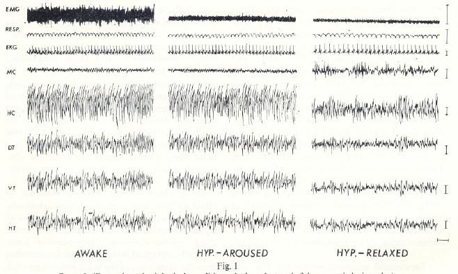
Top: rabbit immediately after hypnosis occurred. Bottom:
rabbit after several minutes later. Is the rabbit asleep or any case, the electrical activity was similar, but
awake? smaller.
Other electrodes were implanted to monitor
dorsal neck muscles, breathing movement, and
Methods the heart. Several rabbits had electrodes placed
Four days prior to hypnosis testing, seventeen
in leg muscles to monitor limb muscle tone.
young adult, anesthetized rabbits had elec-
After surgery, each rabbit received daily
trodes implanted into various parts of brain. doses of antibiotic and rest for three days. All
The electrodes were small wires insulated with animals were observed for at least 2 weeks after
varnish except at the tips so that the voltages
surgery, with no indication of infection or other
detected were coming mainly from the nerve
adverse influences of the surgery. After the ex-
cells nearest the exposed tips. Electrodes were periments, each animal was sacrificed with an
implanted with a micrometer-driven instrument overdose of anesthetic. The brain was bathed
(―stereotaxic‖ apparatus) that allowed precise
with formalin and examined grossly and micro-
three-dimensional placement. Electrodes were
scopically to confirm the exact location of im-
implanted into the hippocampus, thalamus, and planted electrodes.
hypothalamus. Another electrode was a stain-
less-steel screw in the skull overlying the motor
cortex. Another screw electrode was inserted in 1EEG amplifiers amplify the difference between voltage at
one electrode and another.
Page 2 of 5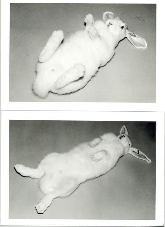
A lightweight cable connected the electrodes             small in size and of high frequency (many
to a traditional EEG machine used. Cable was                  waves per second)(Fig. 1). In the subcortical
suspended over the animal’s head with an elas-                areas with implanted wires, the activity was
tic band so as to permit relatively free move-                large and rhythmic (4-7 waves per second). This
ment. Amplifiers were set to record brain sig-                is typical ―theta‖ activity that is well-known in
nals between 1 and 75 Hz (roughly equivalent                  rabbits to indicate brain activation and
to ―waves per second‖). The data channels for                     This arousal activity was similar to that
muscle and respiration were set differently tor               known to occur in ―dream sleep.‖ A major dif-
optimal recording of those signals because they               ference, however is that in hypnosis the body
occur at a different frequency range.                         showed no signs of the twitches or rapid eye
     After a few minutes of recording from an                 movements that occur during dreaming. Also,
alert and behaving rabbit, hypnosis was in-                   there was some tone in neck muscles, which in
duced by gently turning the rabbit onto its back,             dream sleep totally disappears. During hypno-
while simultaneously using both hands to pre-                 sis in rabbits, the eyes remained open and fixed.
vent any limb movement and to hold the head                       In those trials where hypnosis lasted longer
still. The hypnosis terminated in a few minutes               than usual, brain and body activity were more
when the rabbit spontaneously righted itself.                 typically associated. That is, brain activity was
Inverting the rabbit in a V-shaped trough pro-                of the sleep type (cortex activity became larger
duced much longer periods of hypnosis. The                    and slower; subcortical activity lost the theta
reason is probably the extra contact and pres-                rhythm and became more irregular) and muscle
sure on the skin.                                             tone, and heart and respiratory rates decreased
     Recording sessions occurred each morning                 further (Fig. 1, right panel). Upon righting itself
over four-months. Testing occurred for a given                to terminate hypnosis, the activity patterns al-
rabbit at least four times at two-day intervals.              ways returned to the awake type.
During each test session, recording alternated
from before and during hypnosis.
     In some sessions, seizure-inducing drugs
were injected via ear vein to determine what
effect if any hypnosis would have on the brain
and behavioral effects of the drugs. As a con-
trol, random injections of sterile isotonic2 salt
solution was injected.

Results
Hypnosis was easy to induce in all rabbits. Im-
mediately after induction, limbs that had been                Fig. 1. Note that brain activity is about the same during
extended to resist the manipulation relaxed.                  the awake and hypnotized state (left and middle panel).
                                                              In long-duration hypnosis states, the activity could
Muscle recordings indicated that muscle tone                  change as seen on the right, indicating that the rabbit
decreased. Heart and respiratory rates de-                    might have fallen asleep. Top trace in all three panels is
creased slightly.                                             muscle activity, which decreases in hypnosis. Second and
   The EEG consisted of an ―arousal‖ pattern in               third traces reflect breathing movement and heart activi-
which the activity over the motor cortex was                  ty. Fourth trace is activity from the motor cortex. Other
                                                              traces are from electrodes implanted in the brain. Calibra-
                                                              tion marks on the right and at bottom are 100 μV and 1
2 Concentration that is osmotically neutral – that is, does   second. — note that these are essentially plots of voltage
not alter the osmotic balance between cells and extracellular versus time)
fluids.
      Page 3 of 5
On eight occasions, EEG signs of seizures (including his own) showing similar dissocia-
occurred during hypnosis, representing an even tions during hypnosis.
more dramatic indication that brain activity and The ability of hypnosis to stop seizures of
body activity need not correlate. This ―dissocia- movement but not in the brain itself had not
tion‖ could be repeated at will by injecting cer- been previously discovered. The seizures were
tain seizure-producing drugs (Fig. 2). similar to those reported by others with these
drugs in non-hypnotized animals. Control tests
showed these seizures were not electrical arti-
fact.
The author cited the similarity of these brain
seizures during immobility with those he had
seen in some anesthetized epileptic dogs.
The author also cited a recent paper in
which a variety of drugs could produce a dis-
sociation in the response of the brainstem’s
―arousal system‖ to sensory stimulation. The
EEG response could be fast or slow and the re-
sponse to single shocks could be abolition, in-
Fig. 2. Different types of brain seizures seen during hyp-
crease, decrease, or no change.
nosis, without drug (left panel) and with various seizure-
inducing drugs. Note the absence of corresponding mus- The author cited other research showing that
cle seizures (top trace). This is important because it shows dissociations could occur in other conditions.
that the seizure activity is not artifact that could have Large doses of atropine cause sleep-like brain
been generated by movement of body or cables during activity, yet the injected animal or human re-
recording. Neither animal nor cable moved during hyp-
mains awake. A certain anti-depressant drug
nosis, without or without seizure activity.
can do the same thing.
The opposite kind of dissociation can also
Control recordings using the paired wire re-
occur in which the brain activity is of the awake
cording revealed that these seizures were uni-
type, yet the animal or human seems to be
quely different and not artifact. Thus, the signal
sleeping. This occurs during dreaming, injection
must have originated in the recorded brain
of high doses of the tranquilizer, reserpine, or
areas (data not shown here). Such recording
simultaneous injection of the stimulant physos-
from olfactory bulb revealed low voltage, fast
tigmine and a tranquilizer, and in certain com-
activity (20-50/sec) that was not affected by
atose neurological conditions.
hypnosis nor the seizure-inducing drugs. Other
As speculation for the cause of the hypnosis
control tests included inverting the rabbit with-
dissociation, the author proposed that hypnosis
out achieving hypnosis. Such rabbits struggled
produces a ―release phenomenon.‖ The idea is
and muscle activity was greater than in the
that during hypnosis most brain activity is free
usual undisturbed awake state. The EEG was
to do what it will, while neural pathways that
unchanged from the awake type.
normally produce movement are inhibited.
There are neurons and clusters of neurons that
Discussion have inhibitory effects. The author in other
These results confirmed that EEG and behavior work had identified a couple of areas in brain
can be dissociated. An awake-type EEG can oc- (pons, midline thalamus) where electrical sti-
cur when the animal seems to be asleep. This mulation actually made hypnosis more pro-
can even include brain seizures in the absence found. Such stimulation presumably activated
of movement. The author cited other reports neurons that inhibit movement.
Page 4 of 5He concluded that this study shows the limits
of our understanding of how sensory input and
motor output interact.
###
Page 5 of 5You can also read