Report of Five Cases and Literature Review of Psoas Abscess
←
→
Page content transcription
If your browser does not render page correctly, please read the page content below
Hançerli et al. Report of Five Cases and Literature Review of Psoas Abscess

DOI: 10.4274/iksstd.2021.84756
İKSSTD 2021;13(3):252-7
                                                                                                          ORIGINAL INVESTIGATION / ÖZGÜN ARAŞTIRMA

Report of Five Cases and Literature Review of Psoas
Abscess
Beş Olgunun Sunumu ve Psoas Apsesinin Literatür Taraması
    Cafer Özgür Hançerli,                     Halil Büyükdoğan,                    Can Eren Ünlü,                Anıl Agar,            Cemil Ertürk
University of Health Sciences Turkey, Kanuni Sultan Süleyman Training and Research Hospital, Clinic of Orthopaedic and Traumatology, İstanbul, Turkey

    ABSTRACT
    Objective: To report five psoas abcess cases which we treated and review the management of psoas abscess cases.
    Method: Five patients who were diagnosed and treated with psoas abscess in our clinic between 2018 - 2019 were evaluated retrospectively.
    Results: Four patients were discharged after an average of 25 days following surgical treatments, and 1 case died on the 24th postoperative day due to sudden
    cardiac arrest of unknown cause.
    Conclusion: Psoas abscesses are rare, we believe that the incidence of these cases is more frequent in recent years due to the improvements in radiological
    imaging methods and the fact that these tests are used more frequently by clinicians. We think that for the diagnosis of psoas abscess, it is necessary to suspect
    the diagnosis and keep this rare condition in mind.
    Keywords: Psoas abscess, percutaneous drainage, open surgery, complication, mortality

    ÖZ
    Amaç: Tedavi ettiğimiz beş psoas apsesi vakasını rapor etmek ve psoas apsesi olgularının yönetimini gözden geçirmek.
    Yöntem: Kliniğimizde 2018 - 2019 yılları arasında psoas apsesi tanısı konan ve tedavi edilen beş hasta retrospektif olarak incelendi.
    Bulgular: Dört hasta gerçekleştirilen cerrahi tedavileri takiben ortalama 25 gün sonra taburcu edilmiş, 1 olgu ise nedeni bilinmeyen ani kardiyak arrest
    nedeniyle post op 24. gün kaybedilmiştir.
    Sonuç: Psoas apsesi nadirdir, radyolojik görüntüleme yöntemlerindeki iyileşmeler ve bu testlerin klinisyenler tarafından daha sık kullanılması nedeniyle
    bu olguların görülme sıklığının son yıllarda daha sık olduğuna inanıyoruz. Psoas apsesi tanısı için tanıdan şüphelenmek ve bu nadir durumu akılda tutmak
    gerektiğini düşünüyoruz.
    Anahtar kelimeler: Psoas apsesi, perkütan drenaj, açık cerrahi, komplikasyon, mortalite

INTRODUCTION                                                                                               out of the T12 vertebrae move towards the lumbar vertebrae
Psoas abscess is a rare disease with varying clinical                                                      and eventually connect to the hip bone (3). It locates nearby
presentation and may be seen in any area from the lumbar                                                   organs like kidneys, spine, sigmoid colon, jejunum, appendix,
region to the inguinal region due to the anatomy and course                                                ureters and abdominal aorta. Therefore, infection in these
of the psoas muscle (1). Psoas abcess was identified by                                                    organs may dissolve through the psoas muscle (4).
Herman Mynter in 1881 for the first time (2). The psoas muscle                                             Psoas abcess can be primary and secondary. Primary psoas
extending from the legs to the spine is the only muscle that                                               abscesses is usually of hematogenic origin and comprises
connects the legs and the backbone. The muscles coming                                                     30% of all psoas abcesses; secondary psoas abscesses often
Cite as: Hançerli CÖ, Büyükdoğan H, Ünlü CE, Agar A, Ertürk C. Report of Five Cases and Literature Review of Psoas Abscess. İKSSTD 2021;13(3):252-7

                 Address for Correspondence/Yazışma Adresi: Anıl Agar, University of Health Sciences Turkey, Kanuni                                               Received/Geliş tarihi: 12.05.2020
                 Sultan Süleyman Training and Research Hospital, Clinic of Orthopaedic and Traumatology,                                                         Accepted/Kabul tarihi: 13.08.2021
                 İstanbul, Turkey
                 E-mail: dr.anilagar@hotmail.com ORCID ID: orcid.org/0000-0003-2344-7801

©Copyright 2021 by the İstanbul Kanuni Sultan Süleyman Training and Research Hospital / Medical Journal of Istanbul Kanuni Sultan Suleyman published by Galenos Publishing House.
©Telif Hakkı 2021 İstanbul Kanuni Sultan Süleyman Eğitim ve Araştırma Hastanesi / İstanbul Kanuni Sultan Süleyman Tıp Dergisi, Galenos Yayınevi tarafından basılmıştır.

252
Hançerli et al. Report of Five Cases and Literature Review of Psoas Abscess
develop from a neighboring pathological process originating The mean hospital stay was 35.2 (8-87) days and the mean
from the spine, urinary system or gastro-interstinal system follow-up was 16.8 (13-21) months and the mean time from
and comprises 70% of all cases. The diagnosis is often delayed admission to diagnosis was 17.2 ( 3-30 ) days.
due to the rare occurrence of the disease, the lack of specific No urinary tract pathology was detected in any of the patients.
clinical findings, and the insidious progression of the infection As a physical examination finding, hip joint movements
process (5). Early diagnosis and appropriate treatment can were evaluated as painful and limited in all patients. Direct
prevent morbidity and mortality. Broad-spectrum antibiotics radiography and ultrasonography (USG) were performed
and drainage percutaneously or by open surgery is the in all patients. Computed tomography (CT) and magnetic
primary treatment of choice for psoas abcess. Especially in resonance imaging (MRI) were also performed in case of
secondary psoas abcess mortality rates may increase up to
suspicion or further examination on USG. Only a 13-year-
18.9% despite these treatment methods (6). Despite the mortal
old child was not ordered CT. This was a retrospective
course of the disease, there is no clear consensus between
clinical study. The present study was approved by the Ethical
the general surgery, urology and orthopedics as to which
Committee of our hospital. Demographic characteristics of
department should undertake the treatment.
the patients, clinical presentation, additional disease and
In the present study, we aimed to evaluate the patients with predisposing factors, diagnostic methods, time of diagnosis,
psoas abscess treated in our clinic retrospectively. treatment method, microbiological examination, treatment
responses and complications, duration of hospitalization and
CASE REPORT treatment outcomes were evaluated (Figure 1).
Between january 2018 to October 2019 five patients were
diagnosed and treated with psoas abscess in our clinic . Case 1
Patients whose data could not be reached and who were This patient was a young male athlete (13 years). Patient
out of follow-up were excluded from the study There were was diagnosed as primary psoas abscess with no additional
7 patients recorded due to the psoas abscess when the disease and no other underlying pathology. He was admitted
medical charts of the patients were reviewed, 2 patients were to the pediatric infectious diseases service without surgical
excluded due to the lack of follow-up. The study group was intervention and was followed up with systemic empirical
limited to the five patients. Five patients with psoas abscess antibiotherapy. Patient was consulted for low back, groin and
were evaluated in a retrospective 1-year screening. The hip pain and high fever. Patient’s leukocyte count was 13.54
mean age of the patients was 33.6 years (range: 13 to 64 k/mm³ and C-reactive protein (CRP) level was 160 mg/L at
years). All cases were male and four patients had left side, admission. The ultrasound and MR images of the harvest
one had right side. One patient was young athlete (13 years), revealed collections in the left psoas region (Figure 1). We
1 patient was student (19 years), 1 patient was homeless (51 performed open surgery and abscess drainage. The patient
years) and 2 patients were workers (21 years and 64 years). underwent surgery while receiving intensive antibiotic
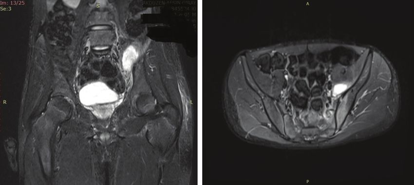
Figure 1. Thirteen years old male left psoas abscess (Case 1) coronal and axial preoperative MR images
MR: Magnetic resonance
253
İKSSTD 2021;13(3):252-7

therapy, therefore, microorganism growth did not occur in                          Patient’s leukocyte count was 23.13 k/mm³ and CRP level
the culture taken during the operation. He was followed with                       was 227 mg/L at admission. It was considered as primary
antibiotherapy for an appropriate period after surgery and                         psoas abscess because there was no known additional
was discharged without any complications after 34 days of                          disease (Figure 2). He operated under general surgery due
hospitalization. In the postoperative follow-up, the patient’s                     to gastric perforation in the same session. MSSA had grown
CRP level was 1.04 mg/L and white blood cell (WBC) level                           in the culture taken during the operation of the patient. He
was 7.28 k/mm³.                                                                    was admitted to general surgery service postoperatively and
                                                                                   discharged from the general surgery department on the
Case 2
                                                                                   8th postoperative day. In the postoperative follow-up, the
This patient was a male student (19 years) and presented                           patient’s CRP level was 8.63 mg/L and WBC level was 7,49
with low back, groin and hip pain, high fever and difficulty                       k/mm³.
in walking. He had diffuse variable immunodeficiency
and lumbar disc hernia and the diagnosis of ankylosing                             Case 4
spondylitis for a period of 1 month and during the follow-                         This patient was a male homeless (51 years)and presented
up with sulfasalazine treatment. The patient consulted                             with low back, groin and hip pain, high fever, nausea,vomiting
for aggravation of the clinic and worsening of the general                         and general condition disorder. Patient’s leukocyte count was
condition. He was admitted to anesthesia and reanimation                           13.28 k/mm³ and CRP level was 348 mg/L at admission. He
intensive care unit due to general condition disorder and                          had type II diabetes mellitus and Crohn’s Disease as additional
sepsis and systemic antibiotherapy was started. Patient’s                          diseases. He had also flexion contracture of the hip and
leukocyte count was 19.43 k/mm³ and CRP level was 279                              erythema of the skin (Figure 3). Percutaneous drainage was
mg/L at admission . He was operated twice during the                               performed by interventional radiology to this patients and
intensive care stay. Methicillin-sensitive staphylococcus                          he was admitted to our service. Streptococcus agalactiae had
aureus (MSSA) had grown in the culture taken during the                            grown in the culture taken during surgery. He was operated
operation of the patient. The patient’s general condition,                         from different incisions (ilio-inguinal and lumbar) in two
clinical findings and laboratory tests improved gradually,                         different sessions because of the failure to control the focus of
and he was ex - as a result of sudden cardiac arrest of                            infection and he was discharged with health after a total of 29
unknown cause (total hospital stay 24 days) on the fourth                          days of follow-up. In the postoperative follow-up, the patient’s
day of admission.                                                                  CRP level was 6.08 mg/L and WBC level was 6,41 k/mm³.

Case 3                                                                             Case 5
This patients was male worker (21 years) and presented                             This patients was a male worker (64 years). He presented
with low back, groin and hip pain and difficulty in walking.                       with low back, groin and hip pain and difficulty in walking. As
 Table 1. Demographic distribution of patients
                                        Patient 1      Patient 2                      Patient 3          Patient 4              Patient 5
 Age                                    13             19                             21                 51                     64
                                                       Immunodeficiency and                              Type 2 diabetes and    Laminectomy and
 Comorbidity                            -                                             -
                                                       lomber disc hernia                                Crohn’s disease        posterior fusion
 Job                                    Athlete        Student                        Worker             Homeless               Worker
 Length of hospitalization (day)        28             24                             8                  29                     35 + 52

 CRP turbidimetric (mg/L)               160            279                            227                348                    221

 Leukocyte (k/mm³)                      13.54          19.43                          23.13              13.28                  11.14

                                                                                                         Streptococcus
 Microorganism                          -              MSSA                           MSSA                                      MRSA
                                                                                                         agalactiae

 Time until Diagnosis                   3              30                             8                  15                     30

 MSSA: Methicillin-sensitive staphylococcus aureus, MRSA: Methicillin-resistant staphylococcus aureus

254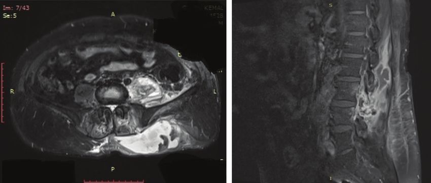
Hançerli et al. Report of Five Cases and Literature Review of Psoas Abscess

additional diseases he had L4-L5 laminectomy, discectomy           joint debridement. When the patient’s clinical and laboratory
and posterior instrumentation about 5 years ago. Other             findings improved during the follow-up, the patient had
finding was diffuse edema in the lower extremity. Patient’s        hip pain in his mobilization. The fourth operation was
leukocyte count was 11.14 k/mm³ and CRP level was 221              performed and the spacer was removed and the hip was left
mg/L at admission (Figure 4). Percutaneous drainage was            to girdlestone arthroplasty. The patient was discharged after
performed by interventional radiology to this patient than         the last operation (52 days). In the postoperative follow-up,
admitted to our service. Methicillin-resistant staphylococcus      the patient’s CRP level was 3.40 mg/L and WBC level was
aureus (MRSA) had grown in the culture taken during the            6.44 k/mm³.
operation of the patient. After psoas abscess was drained
with open surgery in this patient, septic arthritis developed      DISCUSSION
on the same side hip joint and the hip joint was debrided          Although psoas abscesses were diagnosed rarely, the
with open surgery (first hospitalization period 35 days).          incidence of these cases is reported higher in recent years
The patient recovered from the clinic and was discharged           due to the improvements in radiological imaging methods
after 22 days of outpatient follow-up. In the third operation,     and the fact that these tests are used more frequently by
antibiotic spacer was applied by femoral head resection and        clinicians (7). We think that the Although in the literature it

  Figure 2. Twenty one years old male right psoas abscess (Case 3) axial preoperative MR images
  MR: Magnetic resonance

  Figure 3. Fifty one years old male left psoas abscess (Case 4) axial and sagittal preoperative MR images
  MR: Magnetic resonance

                                                                                                                               255
İKSSTD 2021;13(3):252-7 Figure 4. Sixty four years old male left psoas abscess (Case 5) coronal and axial preoperative MR images MR: Magnetic resonance has been reported that 70% of psoas abcess patients are Crohn’s disease, while the other two had spinal disease under the age of 20, recent studies have identified the mean history. age above 40 years (7). Also psoas abcess is seen more often The pathogen microorganism is staphylococcus aureus in men (8). In the present study all patients were men and in 80% of primary psoas abscesses have been reported. mean age of patients was 33,6. Enteric bacteria are usually responsible for secondary Side pain and fever are the most common symptoms in psoas abscesses (13). Staphylococcus aureus was found to psoas abcess (9). All of patients in the present study study had be the causative agent in three patients in this study(one side pain and three of patients had high fever. In accordance patient MRSA, two patients MSSA). In one of our patients, with the literature, leukocytosis, high sedimentation rate and streptococcus agalactia was found as active microorganism, anemia were common laboratory findings in our patients.In whereas in the other patient, no agent was produced in the diagnosis of psoas abcess CT has a high sensitivity of up culture due to intensive antibiotic use. to 100%. CT can show the location and depth of the lesion Broad-spectrum antibiotics and drainage percutaneously and also can show the exact sizes (10). In this study CT was or open surgery is the primary treatment of choice for applied to all patients except 13-year-old patient. In order to psoas abcess (1,7,10). Although percutaneous drainage is show the soft tissue distribution of abscess after CT, MRI was the primary treatment option today, it was first described applied to all patients. in 1984 (14). There are very few reports showing that only Psoas abcess has been mostly reported unilaterally in the cases of psoas abcess respond to antibiotic treatment (4,15). literature (95-97%) (8). All patients in the present study In general, open drainage with a more limited application were also unilateral. Although right side involvement was is recommended when percutaneous drainage fails (8). reported in the literature, all but one patient were involved Although percutaneous drainage is the most effective on the left side (8,11). treatment, open drainage can achieve high success rates Among all psoas abscesses, 30% is known as primary in worsened patients requiring fast and exact response. and 70% is as secondary abcess. The most common Percutaneous drainage was applied to 80% of patients, cause of primary psoas abscess is hematogenous spread, followed by open drainage in all patients in this study. Crohn’s disease is the most common cause of secondary Therefore, open drainage should be the first treatment of psoas abscess (60%) (12). Other reasons are appendicitis choice for these patients. Patients whose general health (16%), ulcerative colitis, diverticulitis, colon cancer (11% condition have deteriorated and delayed diagnosis were together) and vertebral osteomyelitis (10%) in secondary referred to our tertiary hospital. This may be the reason psoas abcess (12). In this study, 40% of patients had primary why open drainage is applied to all patients in our clinic. and 60% had secondary psoas abscess (7,8). One of the Psoas abscess is a disease with fatal consequences. Mortality patients with secondary psoas abscess in the study had rate is 2,4% in primary psoas abscess and this rate may 256
Hançerli et al. Report of Five Cases and Literature Review of Psoas Abscess

increase up to 18.9% in secondary psoas abscess (6). In this                  2.   Mynter H. Acute psoitis. Buffalo Med Surg J. 1881;21:202-10.
study, one of the patients died and the exact cause of death                  3.   Ataus S, Alan C, Onder AU, Mihmanli I, Talat Z, Yalcin V. Psoas abscess.
                                                                                   Cerrahpaşa J Med. 2003;31:89-93.
of this patient could not be determined.
                                                                              4.   Mallick IH, Thoufeeq MH, Rajendran TP. Iliopsoas abscesses. Postgrad
We think that early diagnosis, effective drainage and                              Med J. 2004;80:459-62. doi: 10.1136/pgmj.2003.017665.
appropriate antibiotherapy are the key to manage this                         5.   Goldberg B, Hedges JR, Stewart DW. Psoas abscess. J Emerg Med.
                                                                                   1984;1:533-7.
disease which is not easy to diagnose and treat. However, it
                                                                              6.   Garner JP, Meiring PD, Ravi K, Gupta R. Psoas abscess-not as rare as we
should be kept in mind that serious complications, morbidity                       think? Colorectal Dis. 2007;9:269-74. doi: 10.1016/0736-4679(84)90007-
and mortality may occur in delayed cases.                                          6.
                                                                              7.   Bodakci MN, Hatipoglu NK, Daggulli M, et al. Etiological factors
Ethics                                                                             of psoas abscesses. J Clin Exp Invest. 2014;5:59-63. doi: 10.5799/
Informed Consent: Obtained.                                                        ahinjs.01.2014.01.0360
                                                                              8.   Tabrizian P, Nguyen SQ, Greenstein A, Rajhbeeharrysingh U, Divino
Peer-review: Externally peer reviewed.                                             CM. Management and treatment of iliopsoas abscess. Arch Surg.
                                                                                   2009;144:946-9. doi: 10.1001/archsurg.2009.144.
Authorship Contributions                                                      9.   Chern CH, Hu SC, Kao WF, Tsai J, Yen D, Lee CH. Psoas abscess: making
Concept: C.Ö.H., H.B., C.E.Ü., A.A., C.E., Design: C.Ö.H., H.B.,                   an early diagnosis in the ED. Am J Emerg Med. 1997;15:83-6. doi:
                                                                                   10.1016/s0735-6757(97)90057-7.
C.E.Ü., A.A., C.E., Data Collection or Processing: C.Ö.H., H.B.,
                                                                              10. Turunç T, Demiroglu YZ, Colakoglu S. [Retrospective evaluation of 15
C.E.Ü., A.A., C.E., Analysis or Interpretation: C.Ö.H., H.B.,                     cases with psoas abscesses.] Mikrobiyol Bul. 2009;43:121-5. (Turkish)
C.E.Ü., A.A., C.E., Literature Search: C.Ö.H., H.B., C.E.Ü., A.A.,            11. Bresee JS, Edwards MS. Psoas abscess in children. Pediatric Infect Dis J.
C.E., Writing: C.Ö.H., H.B., C.E.Ü., A.A., C.E.                                   1990;9:201-6. doi: 10.1097/00006454-199003000-00011.
                                                                              12. Ricci MA, Rose FB, Meyer KK. Pyogenic psoas abscess: worldwide
Conflict of Interest: No conflict of interest was declared by                     variations in etiology. World J Surg. 1986;10:834-43. doi: 10.1007/
the authors.                                                                      BF01655254.
                                                                              13. Santanella RO, Fishman EK, Lipsett PA. Primary versus secondary
Financial Disclosure: The authors declared that this study
                                                                                  psoas abscess. Presentation microbiology and treatment. Arch Surg.
received no financial support.                                                    1995;130:1309-13. doi: 10.1001/archsurg.1995.01430120063009.
                                                                              14. Mueller PR, Ferrucci JT, Wittenberg J, Simoene JF, Butch RJ. Iliopsoas
REFERENCES                                                                        abscess: Treatment by CT-guided percutaneous catheter drainage. AJR
1.   Gruenwald I, Abrahamson J, Cohen O. Psoas abscess: case report and           Am J Roentgenol. 1984;142:359-62. doi: 10.2214/ajr.142.2.359.
     review of the literature. J Urol. 1992;147:1624-6. doi: 10.1016/s0022-   15. KadambariD, Jagdish S. Primary pyogenic psoas abscess in children.
     5347(17)37650-4.                                                             Pediatr Surg Int. 2000;46:408-10. doi: 10.1007/s003839900329.

                                                                                                                                                       257
You can also read