SEEING BEYOND THE VISIBLE - FUTURE HEALTHCARE www.sensai.eu - Sens.AI
←
→
Page content transcription
If your browser does not render page correctly, please read the page content below
SEEING BEYOND
THE VISIBLE
FUTURE HEALTHCARE

www.sensai.eu

2   Sens.AI | 2018

    ABOUT FUTURE PROCESSING
    Our main goal and the central part of our operations is the
    use of software to offer solutions to business problems.

    ▪ The company was founded in 2000 by Jarosław Czaja
      (CEO).
    ▪ Revenues above the level of PLN 104 million (2017).
    ▪ 900 people.
    ▪ 18 years of experience.
    ▪ 150 clients in portfolio.
    ▪ R&D project management.
    ▪ Development of our own products.
3    Sens.AI | 2018

    OUR COMPETENCES

        10+
    YEARS OF EXPERIENCE
                                              69 EXPERTS                         SPECIALISTS                      SUPPORT

    Future Processing has more than 10   We have 69 experts specialized   There are machine learning and   Our experts will support the
         years of experience in the          in computer vision             medical imaging specialists    customer’s projects and R&D
        development of IT solutions                                                working for us             departments with their
      that support medical imaging                                                                          knowledge and competences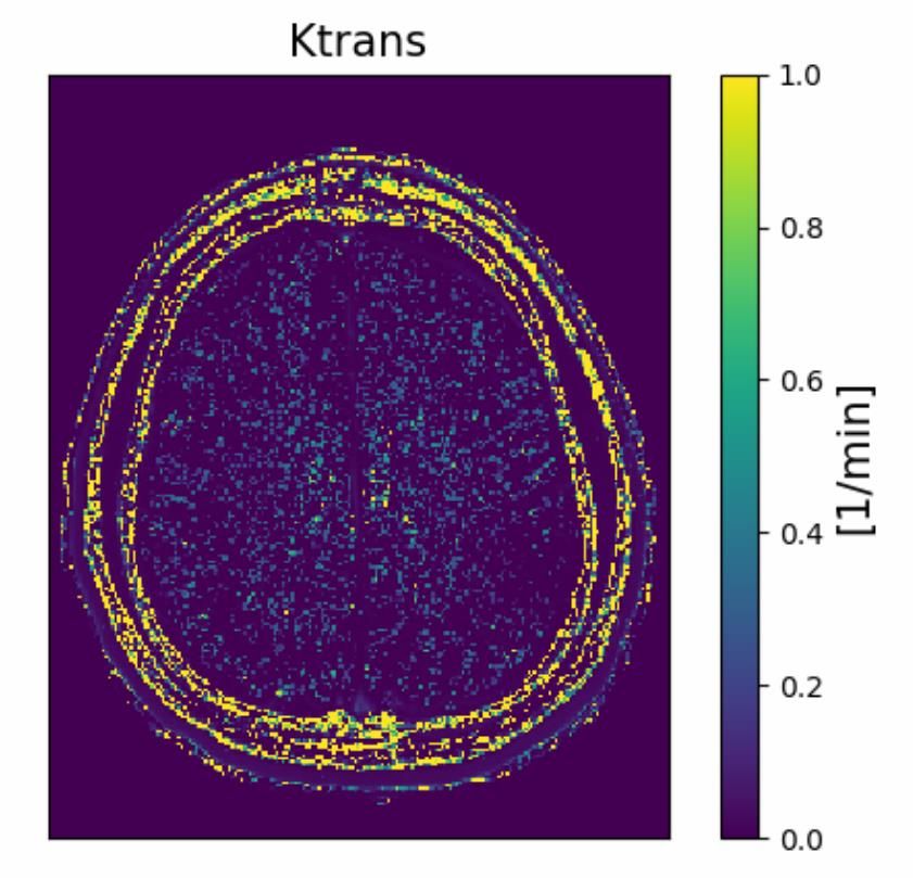
4   Sens.AI | 2018

    WE WORK IN ACCORDANCE WITH ISO
    Our specialists have knowledge of standards applicable in the
    medical industry.

    ▪ We meet the requirements of the ISO 13485 standard, as
      confirmed by the Certificate for the Management System
      according to ISO 13485:2016.

    ▪ Knowledge of standards ISO 13485 and 14971 and the
      ability to create software while complying with the 62304
      standard allows us to cooperate with institutions and
      authorities in the field of medical diagnostics.
5   Sens.AI | 2018

    Sens.AI

    (Enhancing the diagnostic efficiency
    of dynamic Contrast-enhanced
    imaging in personalised Oncology
    by extracting New and Improved
    Biomarkers)
    Seeing beyond the visible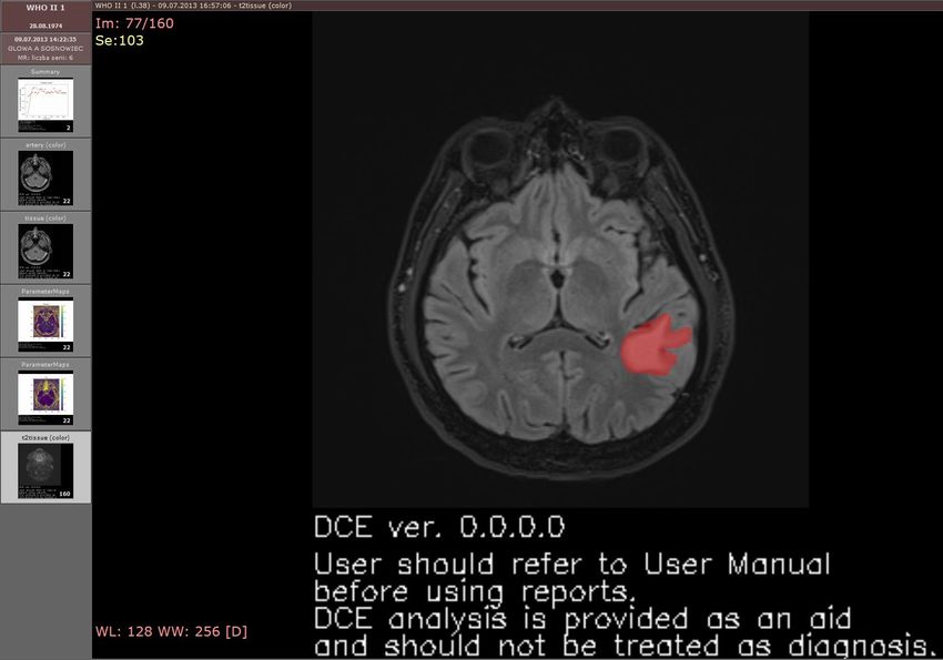
6   Sens.AI | 2018

    WHAT IS Sens.AI?
    Sens.AI is a system for comprehensive and automatic DCE (dynamic contrast-enhanced
    imaging) analysis.

    ▪ It is a tool supporting the diagnosis of brain lesions through the analysis of magnetic
      resonance images after contrast enhancement.

    ▪ Sens.AI analyzes sequences of medical images in search of relevant diagnostic
      information.

    ▪ The purpose of this is:
        • to support the diagnosis of brain lesions,
        • to improve the efficiency of diagnosis of patients with tumors,
        • to save time spent on manual segmentation and image analysis.
7   Sens.AI | 2018

    CLINICAL PARTNER
    Sens.AI is being created in close cooperation with:

                                Oncology Center – Institute named after Maria Skłodowska-Curie,
                                Branch in Gliwice
                                (Centrum Onkologii – Instytut im. Marii Skłodowskiej-Curie,
                                Oddział w Gliwicach)

                                Gliwice Center of Oncology (Centrum Onkologii)
                                is a multidisciplinary oncological center offering cancer patients
                                all the highly specialized methods of combination
                                therapy of all types of cancer that are recognized in the world.

                                The Sens.AI project is carried out with the Department of Radiology and
                                Imaging Diagnostics of the Oncology Center.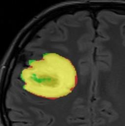
8   Sens.AI | 2018

    MULTIDISCIPLINARY TEAM
    Two teams of experts are working on the Sens.AI system:

                                 RESEARCH TEAM                        DEVELOPMENT TEAM
                                       (RESEARCH)

                           This team deals with the design and        Consisting of experienced software
                         analysis (theoretical and experimental)      engineers – this team works on the
                         of algorithms. It consists of experienced   implementation of the final product.
                            scientists (doctors of physical and
                                    technical sciences).
9 Sens.AI | 2018
DESCRIPTION OF THE SOLUTION
▪ It is a tool supporting the diagnosis of neoplastic lesions.
▪ Performing of analysis of medical images from magnetic resonance (MR)
after contrast enhancement.
▪ Following contrast-enhanced MRI, the images obtained are analysed by
Sens.AI. The system performs automated analysis of imaging data by
performing image segmentation and analysis of contrast flow in brain
tissue. After the cycle is completed, Sens.AI generates a clear and easy to
interpret report in the form of DICOM files.
▪ The entire analysis process is carried out without user’s intervention.10 Sens.AI |2018
INNOVATIVE ALGORITHMS
▪ Automatic and very fast DCE analysis of medical images is
possible thanks to the innovative algorithms created by
Future Processing using machine learning techniques,
especially deep learning.
▪ Algorithms, using machine learning techniques, allow
effective analysis of a series of medical images, obtained
during magnetic resonance imaging.
▪ The report (in DICOM format) includes segmented neoplastic
lesions in dynamic brain imaging, contrast flow graphs,
parametric maps and easy to interpret and analyze
biomarkers.11 Sens.AI | 2018
POSSIBILITY OF RISK ASSESSMENT
▪ Biomarkers, graphs and parametric maps of the brain can be
used to analyze the characteristics and stage of development
of the tumor, allowing risk assessment in patients with
cancer.
▪ Thanks to this, the medical staff receives support in the
process of diagnosing the patient.
▪ The medical staff interprets the results and makes the final
diagnosis.12 Sens.AI | 2018
WORKFLOW
MAGNETIC AUTOMATIC SEGMENTATION REPORT INTERPRETATION OF RESULTS
RESONANCE IMAGING AND ANALYSIS ON ANALYSIS BY A RADIOLOGIST13 Sens.AI | 2018
SEGMENTATION - EXAMPLE
Neoplastic lesion imaging in MR head image
J. Nalepa et al.: ECONIB: A deep learning-powered DCE tool for assessing brain tumor perfusion without region-drawing, RSNA
2018 (in review). Pixels correctly classified as tumor are highlighted in yellow, false negatives in green, and false positives in red.14 Sens.AI | 2018
PARAMETRIC MAPS - EXAMPLE
Ktrans – the first and most important DCE
parameter; the volume transfer constant for
gadolinium between blood plasma and the
tissue extravascular extracellular space (EES).
Like a chemical reaction rate, its units are given
in values of (1/time), such as min−1. In brief,
Ktrans reflects the sum of all processes
(predominantly blood flow and capillary
leakage) that determine the rate of gadolinium
influx from plasma into the EES.*
*References
Ferrier MC, Sarin H, Fung SH, et al. Validation of dynamic contrast-enhanced
magnetic resonance imaging-derived vascular permeability measurements using
quantitative autoradiography in the RG2 rat brain tumor model. Neoplasia 2007;
9:546-555. (Good correlation between Ktrans and autoradiographically measured
influx rate in a rat glioma model with high permeability).
Tofts PS. T1-weighted DCE imaging concepts: modelling, acquisition and analysis.
MAGNETOM Flash 2010; 3:30-35.15 Sens.AI | 2018
CONTRAST CONCENTRATION GRAPHS IN TUMOR TISSUE AND IN
AIF - EXAMPLE
Flow graphs
J. Nalepa et al.: ECONIB: A deep learning-powered DCE tool for assessing brain tumor perfusion without region-drawing, RSNA 2018 (in review).
Sample graphs of contrast saturation - orange dots indicate the average concentration of contrast in tumor tissue (the graph displays the model
adjustment), and pink dots – the average concentration of contrast in AIF (artery input function; the graph presents the model adjustment).16 Sens.AI | 2018
REPORT
Sample report
(in DICOM format)
generated by the Sens.AI
system.17 Sens.AI |2018
ADVANTAGES
▪ Automatic execution of both segmentation and analysis of
medical images for each patient.
▪ Time saving – the system eliminates the necessity of manual
segmentation and image analysis.
▪ DCE analysis and results in real time.
▪ Support for the diagnosis and prediction process. The results of
the algorithm are not affected by the error of the human eye.
▪ Processing and selection of data for analysis without user’s
intervention.
▪ Repeatability of results obtained during the segmentation and DEC
analysis conducted.
▪ Analysis and comparison of results possible with use of publicly
available DICOM file viewers.18 Sens.AI |2018
WHAT DO WE OFFER?
1. Sens.AI platform for easy integration with the hospital image
archiving and communication (PACS) system, as well as
algorithms for segmentation and DCE analysis of MR images after
contrast enhancement
2. Innovative algorithms for:
▪ segmentation of neoplastic lesions in the brain and
arteries from medical images coming from magnetic
resonance (MR) after contrast enhancement – for
customers who are looking for a modern tool for
segmentation,
▪ segmentation of various organs (imaged in various
modalities, such as MR, CT or PET),
▪ DCE analysis – for customers who already have a
segmentation tool and want to extend it with automatic and
fast DCE analysis.
3. Know-how regarding the application of machine learning
methods in medical applications.
4. Support of specialists in the field of machine learning
combining academic competencies with technological skills.THANK YOU
What can we do together?
Contact us and find out how
our specialists can support your
project.
Future Processing
ul. Bojkowska 37A
44-100 Gliwice, Poland
+48 32 438 43 06
sensai@future-processing.com
www.sensai.eu
www.sensai.euYou can also read