WELCOME State of the art and future of Ptychography: a follow-up to the previous Ptychography webinar - Dectris
←
→
Page content transcription
If your browser does not render page correctly, please read the page content below
WELCOME

   State of the art and future of Ptychography:
a follow-up to the previous Ptychography webinar

   WEBINAR
                                                  WIR SCHAFFEN WISSEN – HEUTE FÜR MORGEN
    XRD-CT: The Technique and Its Applications
    for Real-time Characterization of Functional
    Materials
    With Dr. Dubravka Sisak Jung from DECTRIS &
    Dr. Antony Vamvakeros from Finden Ltd.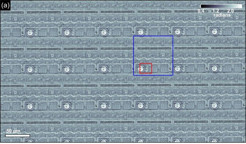
YOUR HOSTS TODAY

        Stefan Brandstetter                         Manuel Guizar Sicairos
        Head of Product Management                  Beamline scientist
        DECTRIS Ltd                                 PSI Paul Scherrer Institute
        stefan.brandstetter@dectris.com   WIR SCHAFFEN WISSEN – HEUTE FÜR MORGEN
                                                    manuel.guizar-sicairos@psi.ch
WIR SCHAFFEN WISSEN – HEUTE FÜR MORGEN

Manuel Guizar-Sicairos :: Beamline Scientist :: Paul Scherrer Institut

State of the art and future directions for ptychography

Dectris Application Webinar 2020

Who are we? The Coherent X-ray Scattering
               Group

Christian   Ana    Xavier   Zirui   Mirko    Manuel Johannes   Dmitry    Andreas Mariana
 Appel      Diaz   Donath   Gao     Holler   Guizar-   Ihli    Karpov    Menzel Verezhak
                                             Sicairos

 Funding

                                                         O. Bunk, G. Aeppli, H.-C. Stadler,
                                                         C. David, L. Heyderman, …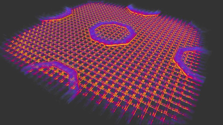
Swiss Light Source

         Beamlines of Soleil (Wikipedia)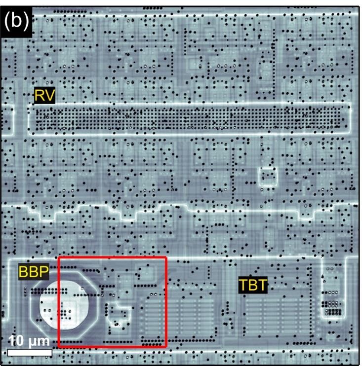
Ptychography (transverse translation diversity)
Moving the sample with respect to a known illumination pattern can provide
suitably diverse measurements

Makes phase retrieval more robust to stagnation, noise and ambiguities

Allows for extended samples, resolution is finer than the illumination and
translations
                                                                             Fourier intensity
                                                                             measurement

                Incident X-ray
                wave

                                    Moveable
              Complex-valued        object
              reconstruction

                                    H. M. L. Faulkner et al. Phys. Rev. Lett. 93, 023903 (2004)
Imaging throughput – The Eiger self portrait

          Sample 5 mm downstream of focus
          Beam at sample ~ 10 microns
          Scanning average step 3.5 microns

          Eiger as sample and detector

M. Guizar-Sicairos et al., Opt. Express 22, 14859 (2014)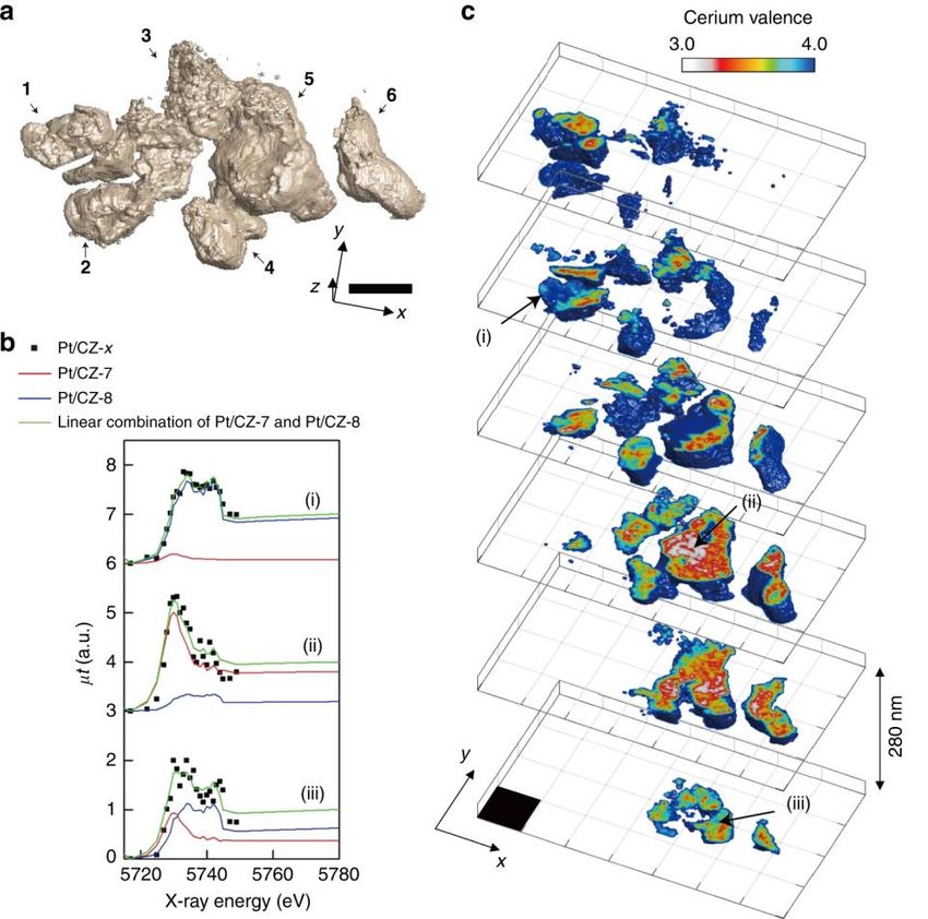
Imaging throughput – The Eiger self portrait

            98.4 Mpixel (13,028 x 7,556)
            Resolution 41 nm, 38.4 nm pixel
            > 25,000 resolution elements / second
            40 microsecond per resolution element

M. Guizar-Sicairos et al., Opt. Express 22, 14859 (2014)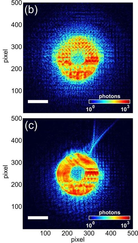
Time resolved ptychography

Cameron Kewish webinar addresses many topics in ptychography. Including
motivation for fast imaging of dynamic processes and a 2D example
In the future nanoscale 3D images could be pursued. New algorithms to deal with
ptychography on dynamic samples may be needed

                                             Time – Nanoscale 3D movies

                                           Evolution of dynamic samples,
                                           In situ temperature, compression, reduction

                                           Images from Fløystad et al., Adv. Eng. Mater. 17,
                                           545 (2015)

                                                                                               Page 9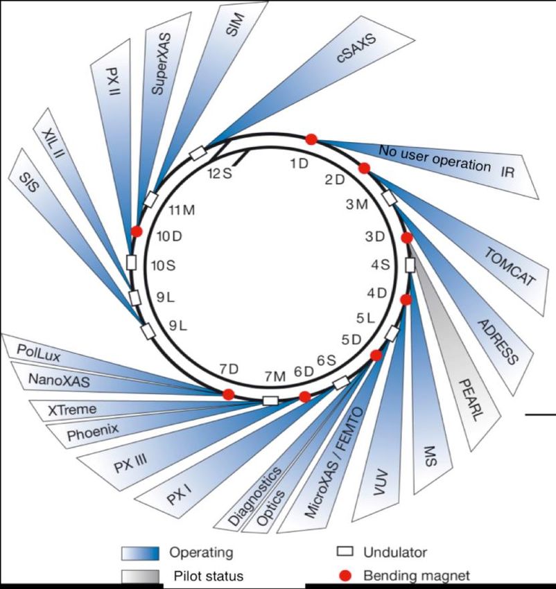
Reflection geometry

                 Forward geometry                    Reflection geometry                    Bragg reflection – High
                                                                                            sensitivity to crystalline lattice
                                                                                            deformations or defects

     Silicon-on-insulator nanostructure

     Strain component (220) reconstructed
     in 3D using Bragg ptychography
     combined with rocking curve

     Measurements at ESRF ID13 beamline

                                                                              Based on Chamard et al., Sci. Rep. 5, 9827 (2015).
                                                                              Under Creative Commons Attribution 4.0 International License.

Image based on https://en.wikipedia.org/wiki/Bragg%27s_law#/media/File:Braggs_Law.svg (CC BY-SA 3.0)                                 Page 10
Combining ptychography and fluorescence
Adding a fluorescence detector provides information about local elemental composition.
Deng et al., “Simultaneous cryo X-ray ptychographic and fluorescence microscopy of green
algae,” PNAS 112, 2314 (2015). https://doi.org/10.1073/pnas.1413003112
Almost every beamline that does ptychography has implemented or plans to implement this.
Works for 2D and 3D.
Deng et al., “Correlative 3D x-ray fluorescence and ptychographic tomography of frozen-
hydrated green algae,” Sci. Adv. 4, eaau4548 (2018). https://doi.org/10.1126/sciadv.aau4548
Both cannot be currently optimized simultaneously. Need faster detectors, improved
scanning, and vast computational resources. Efforts in this direction are undergoing.
Images from Deng et al., Sci. Rep. 7, 445 (2017). Under Creative Commons Attribution 4.0 International License.
Measurements in APS beamline 21-ID-D. Page 11Nanoscale 3D imaging
Combining ptychography and a sample rotation enables 3D imaging
Laminography geometry allows for extended flat samples to be imaged, enabling 3D zooming
into regions of interest and facilitating in situ and in operando studies
Ptychographic X-ray computed tomography
(PXCT)
Dierolf et al. “Ptychographic X-ray computed tomography at
the nanoscale,” Nature, 467 436 (2010)
Image modified from Donnelly et al. Nature 547, 328 (2017)
Ptychographic X-ray laminography (PyXL)
Holler et al., “Three-dimensional imaging of integrated
circuits with macro- to nanoscale zoom,“ Nat. Electron. 2,
464 (2019)Nanoscale 3D imaging
Example ptychographic laminography imaging metal layers and interconnections in an
integrated circuit at 20 nm resolution. Measurement in SLS, cSAXS beamline.
Holler et al, “Three-dimensional imaging of integrated circuits with macro- to nanoscale zoom,“ Nat. Electron. 2, 464 (2019) Page 13Beyond the depth of field limitation
Depth of field (DOF) arises due to diffraction. Affects lens-based
imaging, holography, etc.
Higher resolution results in smaller distance remaining in focus.
Limiting the volumes we can measure with very high resolution.
Multislice ptychography overcomes this limitation. Represents the
https://commons.wikimedia.org/wiki/File:DOF- object by several slices and taking into account propagation through
ShallowDepthofField.jpg
GNU Free Documentation License, Version 1.2
the object.
Maiden et al., “Ptychographic transmission microscopy in
three dimensions using a multi-slice approach,” JOSA A 29, 1606
(2012). https://doi.org/10.1364/JOSAA.29.001606
Optical demonstration of multislice tomography, glass tube with glass beads
30 mm
Images from Li & Maiden, Sci. Rep. 8, 2049 (2018). Under Creative Commons Attribution 4.0 International License. Page 143D chemical-state mapping
Combining X-ray spectroscopy (XANES) with 3D ptychography
Measurements with sampling the incident photon energy across an absorption edge
Additional to measuring the structure one obtains the valence or chemical state – functional
and structural imaging
Pt/CZ-x solid solution particles during
the oxygen storage process, measured
at Spring-8
3D nanoscale imaging of Ce valence 700 nm
state. 28 energies (5.727 – 5.744 keV).
Three-way exhaust catalysis is a key
reaction in automobiles
Challenge – How to scale up to larger
samples and more X-ray energies?
Images from Hirose et al., Commun. Chem 2, 50 (2019). Under Creative Commons Attribution 4.0 International License. Page 15Further reading F. Pfeiffer, “X-ray ptychography,” Nature Photon. 12, 9–17 (2018) Review article https://doi.org/10.1038/s41566-017-0072-5 C. Jacobsen, “X-ray Microscopy,” Cambridge University Press (2019) Covers many concepts and techniques for X-ray microscopy in 2D and 3D https://doi.org/10.1017/9781139924542 J. Rodenburg and A. Maiden “Ptychography” chapter in Springer Handbook of Microscopy pp 819-904 (2019) https://doi.org/10.1007/978-3-030-00069-1_17 M. Guizar-Sicairos and P. Thibault, “Ptychography: A solution for the “phase problem” finds countless applications,” Phys. Today (2021)
IF YOU WANT TO KNOW MORE
                                                   WIR SCHAFFEN WISSEN – HEUTE FÜR MORGEN
    Download slides

    Publications
    Webinar: Fast-Scanning Ptychography

    Contact Stefan Brandstetter or Manuel Guizar
    Sicairos directly at
    stefan.brandstetter@dectris.com
                                                                  For more webinars,
    and manuel.guizar-sicairos@psi.ch                                 please visit our
                                                                  dedicated webpage

    Visit dectris.com and psi.ch
Thank you andWIRsee you
SCHAFFEN WISSEN – HEUTE FÜR MORGEN
soon!
WEBINAR
XRD-CT: The Technique and Its Applications
for Real-time Characterization of Functional
Materials
With Dr. Dubravka Sisak Jung from DECTRIS &
Dr. Antony Vamvakeros from Finden Ltd.You can also read