SELF-SAMPLING FOR HPV TESTING IN PRIMARY CERVICAL SCREENING - DIVA
←
→
Page content transcription
If your browser does not render page correctly, please read the page content below
Digital Comprehensive Summaries of Uppsala Dissertations
                from the Faculty of Medicine 1641

                Self-sampling for HPV testing in
                primary cervical screening
                Including clinical and health economic aspects

                RIINA AARNIO

    ACTA
UNIVERSITATIS
 UPSALIENSIS    ISSN 1651-6206
                ISBN 978-91-513-0882-1
  UPPSALA       urn:nbn:se:uu:diva-405864
    2020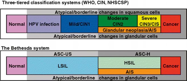
Dissertation presented at Uppsala University to be publicly examined in Sal IV,
Universitetshuset, Biskopsgatan 3, Uppsala, Friday, 17 April 2020 at 09:00 for the degree of
Doctor of Philosophy (Faculty of Medicine). The examination will be conducted in Swedish.
Faculty examiner: Professor Christer Borgfeldt (Lunds University, Department of Obstetrics
and Gynecology ).

Abstract
Aarnio, R. 2020. Self-sampling for HPV testing in primary cervical screening. Including
clinical and health economic aspects. Digital Comprehensive Summaries of Uppsala
Dissertations from the Faculty of Medicine 1641. 81 pp. Uppsala: Acta Universitatis
Upsaliensis. ISBN 978-91-513-0882-1.

Persistent infection with high-risk human papillomavirus (HPV) is a prerequisite for the
development of cervical cancer. HPV testing has higher sensitivity for high-grade cervical
intraepithelial neoplasia (CIN2+) than cytology, resulting in more effective screening. As HPV
testing also offers an opportunity for self-sampling, it could serve as an even more effective and
cost-effective method of cervical screening.
   First, we compared repeated self-sampling for HPV testing with Pap smear cytology in
detection of CIN2+ in primary cervical screening for women aged 30–49 years (n=36 390). We
found a more than twofold higher detection rate of CIN2+ and a fourfold higher detection rate
of CIN2 with self-sampling compared with cytology. However, no difference was seen between
the arms in the detection rate of CIN3+. It thus seems that CIN is detected at an earlier stage
with self-sampling than with cytology, but the impact of this needs to be further explored.
   Second, as management of HPV-positive women with normal cytology results is a challenge,
we wanted to evaluate the proportion of cases of histological CIN2+ in these women. In this
prospective study we performed LEEP and found that 15% (6/40) of the women had undetected
CIN2+. These findings can be used in counseling women about the risk of cervical cancer and
helping clinicians in decisions on management.
   Third, we performed a cost-effectiveness analysis on the same study population as in Study
I. Self-sampling for HPV testing resulted in a higher participation rate and more detected cases
of CIN2+ at a lower cost and was regarded as more cost-effective than Pap smear cytology
in cervical screening. These results can guide policy-makers when planning future screening
programs.
   Fourth, we compared self-sampling with sampling by medical professionals for HPV testing
in detection of CIN2+, using a combination of an FTA card as storage medium and a PCR-based
HPV test (hpVIR) in women aged 30–60 years (n=11 951). No difference in the detection rates
of histological CIN2+ was found between the arms.
   Taken together, self-sampling resulted in a higher participation rate than sampling by medical
professionals in cervical screening and that triage with repeated self-sampling resulted in high
compliance and detection rate of CIN2+. As repeated self-sampling for HPV testing was also
cost-effective, it could serve as an attractive alternative in the development of future cervical
screening programs. More research is needed on how to refine the management of HPV-positive
women by self-sampling only.

Keywords: HPV, self-sampling, cervical screening, CIN2+, cost-effectiveness

Riina Aarnio, Research group (Dept. of women´s and children´s health), Reproductive
biology, Akademiska sjukhuset, Uppsala University, SE-751 85 UPPSALA, Sweden.

© Riina Aarnio 2020

ISSN 1651-6206
ISBN 978-91-513-0882-1
urn:nbn:se:uu:diva-405864 (http://urn.kb.se/resolve?urn=urn:nbn:se:uu:diva-405864)
“Primum non nocere”

              To Mikko, Lukas, Ines and Ellen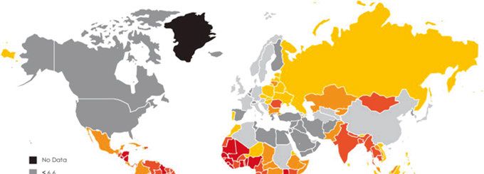
List of Papers

This thesis is based on the following papers, which are referred to in the text
by their Roman numerals.

      I      Gustavsson I, Aarnio R, Berggrund M, Hedlund-Lindberg J,
             Strand AS, Sanner K, Wikström I, Enroth S, Olovsson M,
             Gyllensten U. (2018) Randomised study shows that repeated
             self-sampling and HPV test has more than two-fold higher
             detection rate of women with CIN2+ histology than Pap smear
             cytology. British journal of cancer, 118(6):896-904

      II     Aarnio R, Wikström I, Gustavsson I, Gyllensten U, Olovsson M.
             (2019) Diagnostic excision of the cervix in women over 40 years
             with human papilloma virus persistency and normal cytology.
             Eur J Obstet Gynecol Reprod Biol X, 3:100042

      III    Aarnio R, Östensson E, Olovsson M, Gustavsson I, Gyllensten
             U. Cost-effectiveness analysis of repeated self-sampling for HPV
             testing in primary cervical screening: a randomized study.
             Manuscript.

      IV     Aarnio R, Isacson I, Sanner K, Gustavsson I, Gyllensten U,
             Olovsson M. Comparison of vaginal self-sampling and cervical
             sampling by medical professionals for the detection of HPV and
             CIN2+: a randomized study. Manuscript.

Reprints were made with permission from the respective publishers.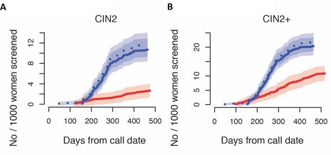
Contents

Introduction ................................................................................................... 11
   Cervical cancer ......................................................................................... 11
   HPV infection ........................................................................................... 11
      HPV ..................................................................................................... 12
      HPV life cycle ..................................................................................... 12
   Prevention................................................................................................. 13
      Primary prevention .............................................................................. 13
      Secondary prevention .......................................................................... 13
   HPV vaccination ...................................................................................... 13
      HPV vaccines ...................................................................................... 14
   Screening .................................................................................................. 15
      Consequences of HPV vaccination ...................................................... 16
   Cytology ................................................................................................... 17
      Classification of cytology and histology ............................................. 18
   Colposcopy ............................................................................................... 19
      Transformation Zone (TZ) ................................................................... 19
   Treatment of cervical precancerous lesions ............................................. 20
   HPV testing .............................................................................................. 22
      Triage in HPV primary screening ........................................................ 23
      HPV genotypes .................................................................................... 24
      HPV persistence .................................................................................. 25
      HPV self-sampling............................................................................... 26
   Health economics ..................................................................................... 27
      Cost-effectiveness analysis (CEA) ...................................................... 28
Aims .............................................................................................................. 29
Material and methods .................................................................................... 30
  Study population ...................................................................................... 30
     Studies I and III ................................................................................... 30
     Study II ................................................................................................ 30
     Study IV ............................................................................................... 30
  Ethics ........................................................................................................ 30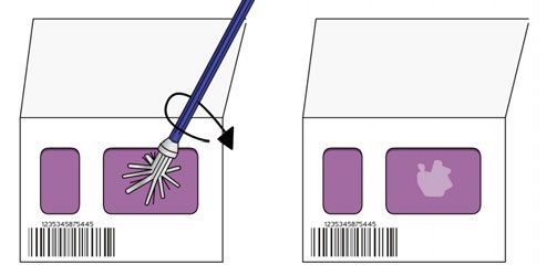
Methods .................................................................................................... 31
      Study I.................................................................................................. 31
      Study II ................................................................................................ 34
      Study III ............................................................................................... 34
      Study IV ............................................................................................... 36
Results ........................................................................................................... 38
  Study I ...................................................................................................... 38
  Study II ..................................................................................................... 40
  Study III ................................................................................................... 41
  Study IV ................................................................................................... 44
Discussion ..................................................................................................... 48
Future perspectives ....................................................................................... 56
Conclusions ................................................................................................... 58
  Study I ...................................................................................................... 58
  Study II ..................................................................................................... 58
  Study III ................................................................................................... 58
  Study IV ................................................................................................... 58
Summary in Swedish – Sammanfattning på svenska.................................... 59
Summary in Finnish – Yhteenveto suomeksi ............................................... 62
Acknowledgements ....................................................................................... 65
References ..................................................................................................... 67
Abbreviations AGC atypical glandular cells AIS adenocarcinoma in situ ASCUS atypical squamous cells of undetermined significance ASC-H atypical squamous cells, cannot exclude high-grade lesion CEA cost-effectiveness analysis CI confidence interval CIN cervical intraepithelial neoplasia CIN1 cervical intraepithelial neoplasia grade 1 CIN2 cervical intraepithelial neoplasia grade 2 CIN2+ cervical intraepithelial neoplasia grade 2 or more CIN3 cervical intraepithelial neoplasia grade 3 CIN3+ cervical intraepithelial neoplasia grade 3 or more CIS carcinoma in situ DNA deoxyribonucleic acid ECC endocervical curettage FDA (U.S.) Food and Drug Administration FTA Flinders Technology Associates HC hybrid capture HPV human papillomavirus ICER incremental cost-effectiveness ratio LBC liquid-based cytology LEEP loop electrosurgical excision procedure LLETZ large loop excision of the transformation zone NILM negative for intraepithelial lesion or malignancy Pap Papanicolaou PCR polymerase chain reaction PPV positive predictive value RCT randomized controlled trial SCJ squamocolumnar junction SIL squamous intraepithelial lesion SNOMED Systematized Nomenclature of Medicine TZ transformation zone VF vaginal fluid VLP virus-like particle
Introduction

Cervical cancer
Cervical cancer is the fourth most common cancer in women worldwide, with
over 550 000 new cases and 311 000 related deaths in 2018 (1). Cervical
cancer has significant differences in properties compared with many other
cancers. First, the majority of cases appear at younger ages (47% in women
aged 30%) and lowest in South Asia and North America (5–7%) (7). Prev-
alence is also higher at younger ages after sexual debut, being >20% in women
aged
(8). HPV is easily transmitted in both genders, mostly by mucosal contact, and
about 75% of sexually active individuals acquire the infection during their
lifetime. HPV infections are asymptomatic and most of them clear within two
years, but some infections may became latent or undetectable (9, 10). Persis-
tence of HPV is consistently and strongly associated with the risk of develop-
ing high-grade cervical intraepithelial neoplasia (CIN2+) (11), which in turn
brings an elevated risk of progression to cervical cancer (12). However, the
carcinogenic process of cancer to develop from incident HPV infection
usually takes time – approximately 5–10 years at a minimum and 20–25 years
on average. HPV also causes cancers of the oropharynx, anus, vulva, vagina
and penis and is today estimated to cause about 5% of all cancers globally
(13).
HPV
HPV is a double-stranded DNA virus
with a capsid which is 50–55 nm in
diameter, having icosahedral sym-
metry (14). The viral genome is cir-
cular and approximately 7900 base-
pairs long, including the following
regions: long control region, early
region and late region. While the long
control region regulates viral gene
expression and replication, the early
region encodes proteins E1, E2, E4,
E5 and oncoproteins E6 and E7,
required for viral gene expression,
replication and survival, and the late Figure 1. Papillomavirus Genome
Organisation. Reprinted from Doorbar J,
region encodes the capsid proteins Quint W, Banks L, Bravo IG, Stoler M,
L1 and L2. Broker TR, et al. The biology and life-
cycle of human papillomaviruses.
Vaccine. 2012;30 Suppl 5:F55-70.,
with permission from Elsevier.
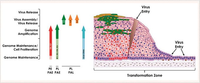
HPV life cycle
HPV infects by first entering the basal layer of the epithelium through a
microwound. The viral genome is maintained in these cells when genes E1
and E2 replicate episomes. Through expression of E6 and E7 the cells prolif-
erate and move outwards towards the epithelial surface. In the mid layers the
cells express all the early genes and the genome is amplified. In the upper
layers L1 and L2 proteins are made, allowing the packaging of the amplified
12viral genomes. It is likely to be a function of E4 that new virus particles are
then released from the epithelial surface in a productive infection. If this viral
gene expression is deregulated it can lead to high-grade intraepithelial neo-
plasia and if the viral genome is integrated into the host cell chromosome, it
can then lead to the development of cancer (15).
Figure 2. Life Cycle of High-Risk HPVs in Cervical Epithelium. Reprinted from
Doorbar J, Quint W, Banks L, Bravo IG, Stoler M, Broker TR, et al. The biology
and life-cycle of human papillomaviruses. Vaccine. 2012;30 Suppl 5:F55-70.,
with permission from Elsevier.
Prevention
Primary prevention
Prevention of a disease in individuals without the disease is called primary
prevention. In cervical cancer the issues are, for example, education and
prophylactic HPV vaccination of the population.
Secondary prevention
Prevention of a disease by interrupting its progression through identification
of early stages and then eliminating them is called secondary prevention. In
cervical cancer this is done by screening the population and further diagnosing
precancerous lesions in screening-positive individuals, and treating them.
HPV vaccination
In vaccine development the L1 gene is recombinantly expressed and then self-
assembled to virus-like particles (VLPs) containing no viral genome. These
VLPs constitute prophylactic vaccines which induce high levels of neutral-
izing antibodies in hosts.
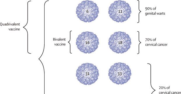
13Figure 3. HPV VLP types in the three HPV vaccines. Reprinted from Schiller JT,
Muller M. Next generation prophylactic human papillomavirus vaccines. The Lancet
Oncology. 2015;16(5):e217-25., with permission from Elsevier.
HPV vaccines
The first-generation prophylactic HPV vaccines were registered in 2006,
being a bivalent vaccine against high-risk HPV types 16 and 18, and a quadri-
valent vaccine including activity against even low-risk HPV types 6 and 11,
which cause genital warts. These vaccines have shown very high efficacy
against type-specific infections and precancerous cervical lesions when vac-
cinating HPV-negative young women (aged 15–26 years) (16). The bivalent
vaccine has also shown significant cross-protective effect against other high-
risk types, especially HPV31 and HPV45 (17, 18). Safety has been confirmed
in randomized controlled trials (RCTs) with over 73 000 participants that
showed no difference between intervention and control arms as regards mild
or severe systemic side effects (16), and by active surveillance after over 270
million vaccine doses given globally (18) during the last decade that have not
shown any serious or unexpected side effects (19).
A national vaccination program was started in 2007 in Australia, and today
at least 82 countries have introduced vaccination programs, but with different
strategies (19). In Sweden vaccination is school-based and was started in 2012
for girls aged 11 years. In 2014, a next-generation nonavalent HPV vaccine
against the same HPV types as the quadrivalent vaccine, and with additional
five high-risk types (31, 33, 45, 52 and 58) was registered. While the first-
generation vaccines are against the high-risk types that are responsible for
about 70% of all cervical cancer cases, the next-generation vaccine is against
14the high-risk types that are responsible for about 90% of all cervical cancer
cases. As the nonavalent vaccine prevents infection and precancerous lesions
related to virus types with similar efficacy as the quadrivalent vaccine, and
shows a non-inferior antibody response (20), it has been estimated being cost-
saving (21), and many countries are now switching to this alternative. Gender-
neutral vaccination, by herd effect, can give comparable protective effective-
ness even with low or moderate vaccination coverage (22); hence this is
already implemented in many countries and is planned in Sweden next
autumn.
Screening
The main principles of screening have been described by Wilson (23), includ-
ing facts that the disease should be important, have a recognizable latent stage
and the natural course should be understood. Secondly, there should be a
screening test that is suitable, acceptable, accurate, reliable, sensitive and
specific. Thirdly, the treatment should be effective and acceptable and there
should be a policy as to who should be treated. Fourthly, the diagnosis and
treatment should be cost-effective.
All these principles are partly fulfilled in cervical screening, and organized
screening with cytology has resulted in a major reduction in both the incidence
of cervical cancer and related mortality in developed countries (24). In
Sweden, for instance, the incidence has been reduced to about a half, with 540
new cases and 153 related deaths in 2018 (25). Still, the disease has not dis-
appeared despite screening for the last five decades, and the cancer incidence
has stagnated at a reduced level (26). National audits have shown that 45–64%
of all cancer cases are diagnosed among non-attenders, and these cancers were
also diagnosed at a more advanced stage (27, 28). In a population perspective,
non-adherence to screening invitations has been identified as the most
important risk factor of incident cervical cancer. This means that it is crucial
to make efforts to reach as high rate of participation in screening as possible.
In Sweden coverage of screening has stagnated at a level of about 75% of
women aged 23–70 years (29).
Continuous quality control of screening programs has led to identification
of some problems. Screening by means of cytology has been unable to prevent
cervical adenocarcinoma (30) and the overall low sensitivity of cytology is a
well-known drawback. Regular sampling at 2–5 year intervals has, however,
partly compensated for this weakness. Nevertheless, there has been an
increasing incidence of cervical cancer in Sweden in recent years, mostly
among women participating in screening with normal results in cytology (31),
indicating a problem with the sensitivity of the technique. The evidence that
HPV testing is more effective in reducing cervical cancer incidence compared
with cervical cytology (32) has led to implementation of HPV-based primary
15screening programs, a step that has not diminished the challenges in
organizing existing screening programs.
Developing countries are still meeting major problems in establishing
screening programs. The coverage of cervical screening in developing
countries is on average only about 20% and the elderly and poor women, with
the highest risk of cervical cancer, are least likely to be screened (33). There
is an urgent need for educated staff to take care of all aspects of a screening
program, not forgetting the education of a population, and cultural barriers. It
is estimated that over 80% of cervical cancer cases occur in developing
countries, where it accounts for 13% of all cancers in women (34). This
important point should not be forgotten in research and when developing
screening programs and strategies for the diagnosis and treatment of precan-
cerous lesions.
Figure 4. Cervical cancer, global map showing estimated age-standardized (world
standard) incidence rate per 100,000 in 2008 (all ages). Based on GLOBOCAN
2008. Reprinted from Forman D, de Martel C, Lacey CJ, Soerjomataram I, Lortet-
Tieulent J, Bruni L, et al. Global burden of human papillomavirus and related
diseases. Vaccine. 2012;30 Suppl 5:F12-23., with permission from Elsevier.
Consequences of HPV vaccination
Even though HPV vaccination is known to reduce cervical cancer incidence,
in near future there will still be a need for an appropriate screening program
that must be adapted to a vaccinated population. Although an existing vac-
cination program will reduce the incidence, there are several issues remaining
for attention. The currently used vaccines do not cover all the high-risk HPV
types, population-level vaccination coverage is not 100% and the population
with sexual debut before the era of vaccination will still exist for several
decades.
16The effect of vaccination on screening participation has been different in
different populations. In Sweden, the vaccinated population has showed
higher attendance to screening (35). Concerns have been raised about HPV
‘type replacement’, i.e. that non-vaccination HPV types would emerge after
vaccination, but this has not been seen in countries with the longest vaccina-
tion programs (36), probably because of HPV’s stable DNA, with a slow evo-
lution rate. However, the consequences of vaccination must still be closely
followed. While vaccination eradicates the major high-risk HPV infections,
the consequence in screening will be that a positive HPV-test result will be
less predictive of CIN3+, because of fewer high-risk HPVs detected (37). This
might allow longer screening intervals, and eventual additional triage
strategies such as genotyping or assays of other biomarkers could be
considered. Also, as HPV types 16 and 18 give rise to cervical cancer at
younger ages than the other high-risk types (38), screening in a vaccinated
population could be started later.
Cytology
The traditional cytological method in cervical screening has been
Papanicolaou-stained cytology (Pap smear) on a glass slide (39). Cytological
analysis has a high specificity (about 95%) but has been reported to have a
problem with low sensitivity (about 50%) in detecting CIN (40). One crucial
fact is that cytology is based on subjective analysis, with moderate interob-
server, intralaboratory and interlaboratory variations (41). Efforts in education
in morphological assessment of cytological samples, quality assurance of the
laboratories and investments in new technologies such as liquid-based cytol-
ogy (LBC) have been made during the last 50 years to increase the accuracy
of cytology. However, the sensitivity is still not higher than approximately
70% at best (41, 42) and LBC is neither regarded as more sensitive nor more
specific for detection of CIN2+ compared with conventional Pap smears (43).
However, LBC has the advantage that HPV testing can be performed on the
same sample, which can be adopted in different triage strategies.
Another, however minor, concern is the rate of false-positive test results,
for example, in connection with immature squamous metaplasia, parakeratosis
or inflammatory atypia. In settings with HPV testing as primary screening, the
observer’s knowledge of the woman’s HPV status might increase the false-
positive rate (44). To improve the sensitivity and reduce the rate of false-
positive results in cytological screening, continued quality control of cytolog-
ical laboratories is essential, not least because of steeply falling throughput
rates when primary HPV screening is introduced.
17Classification of cytology and histology
During the last few decades the terminology has been switching from the
three-tier CIN1–3 (Richart) system to the two-tier Bethesda classification
system (45) described in 1989, with the following cytological categories:
negative for intraepithelial lesion or malignancy (NILM); atypical squamous
cells of undetermined significance (ASCUS); atypical squamous cells, cannot
exclude high-grade lesion (ASC-H); low-grade squamous intraepithelial
lesion (LSIL); high-grade squamous intraepithelial lesion (HSIL); squamous
cell carcinoma; atypical glandular cells (AGC); adenocarcinoma in situ (AIS);
or adenocarcinoma (46).
Figure 5. Conceptual categorization of cytological findings in a Pap smear of the
cervix. CIS = carcinoma in situ. Reprinted from Herbert A, Bergeron C, Wiener H,
Schenck U, Klinkhamer P, Bulten J, et al. European guidelines for quality assurance
in cervical cancer screening: recommendations for cervical cytology terminology.
Cytopathology : official journal of the British Society for Clinical Cytology.
2007;18(4):213-9 with permission from John Wiley and Sons.
The advantage of the Bethesda system is that it is based on the existence of
two different forms of HPV infection, with productive infection leading to
low-grade SIL and transforming infection leading to high-grade SIL. Current
clinical management is mostly based on the two-tier system. This system was
created to provide effective communication from laboratory to clinic, to
facilitate cytology-histology correlation and to provide more reproducible
results. It also gave rise to the concept of ASCUS (47). The Bethesda system
is further applied in histological nomenclature. Here, even the CIN grade is
often included in the results. However, during the studies included in this
thesis the Swedish modification of CIN1–3 classification was used.
18Colposcopy
Colposcopy is the standard method to investigate the cervix in women with
atypical Pap smears and was first described by Hans Hinselmann in 1925. The
purpose of colposcopic examination is to identify diseased tissue for targeted
punch biopsy sampling for histological diagnosis. The procedure provides
illuminated magnification of the cervix and various solutions (3–5% acetic
acid and Lugol’s iodine) are applied for the evaluation.
Transformation Zone (TZ)
To describe and interpret colposcopic findings, colposcopists are recom-
mended to use the terminology of the International Federation of Cervical
Pathology and Colposcopy (48). Even this evaluation is subjective, with low
reproducibility (49), and different scoring systems, for example the Reid index
(50), have been developed for standardization. The latest modification, includ-
ing even the lesion size in scoring, is the Swede score system (Table 1). The
specificity of a score of ≥8 has been reported to be 90–95% for CIN2+, while
a score of ≤3–4 speaks against CIN2+ (51, 52). This can be interpreted as
recommending ‘see and treat’ management in cases of high scores and possi-
bly refraining from biopsies in cases of low scores. Archiving electronic
images taken during colposcopy in patient files is recommended.
Table 1. The Swede score system
0 1 2
Aceto uptake Zero or Shady, milky (not Distinct, opaque white
transparent transparent, not opaque)
Margins/surface Diffuse Sharp but irregular, Sharp and even, difference
jagged, "geographical" in surface level incl
satellites "cuffing"
Vessels Fine, Absent Coarse or atypical
regular
Lesion size 15mm or 3-4 quadrants
or endocervically
undefined
Iodine staining Brown Faintly or patchy yellow Distinct yellow
A crucial assessment is defining the location of the squamocolumnar junction
(SCJ) and the type of the TZ. The cervix is covered by both stratified non-
keratinizing squamous cells and a single-cell layer of columnar epithelium.
These two types of epithelium meet at the SCJ. The buffering action of the
mucus covering the columnar cells is interfered when everted columnar
epithelium is exposed to the acidic vaginal environment. This leads to the
destruction and replacement of the columnar epithelium by newly formed
metaplastic squamous epithelium. The metaplastic process mostly starts at the
19original SCJ and proceeds centripetally towards the external os through the
reproductive period to perimenopause. Thus, a new SCJ is formed between
the newly formed metaplastic squamous epithelium and the columnar
epithelium remaining everted onto the ectocervix and the TZ is the area
between the original and the new SCJ.
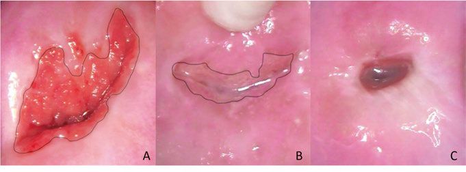
This immature metaplastic squamous epithelium in the TZ is sensitive to
persistent HPV infections and this is the site for transforming to atypical cells
and therefore important to assess. In TZ types 1 and 2 the TZ is fully visible.
In TZ type 3 the deeper limit is not visible and this is a common finding in
the postmenopausal period when the TZ often retracts into the endocervix (53)
(Figure 6).
Figure 6. Transformation zone (TZ) type 1 (A), TZ type 2 (B) and TZ type 3 (C).
Black line: new squamocolumnar junction. Images by the author.
Evaluation of TZ type 3 is a challenge. Performance of biopsy sampling
lacking the TZ is insufficient (54). It is also more difficult to obtain adequate
amounts of tissue from the endocervix by cytological sampling or curettage
for histology, as the sensitivity of endocervical cytobrush sampling in detect-
ing lesions varies between 44–93%, and is even lower with endocervical
curettage (ECC) for histological samples (55, 56); thus use of routine ECC is
not encouraged (57). Nevertheless, the sensitivity of colposcopy and biopsy
in detection of CIN2+ in women with abnormal cytology is around 70% (58,
59), and more than one biopsy is recommended to improve the accuracy (57,
59). Moreover, both the implementation of more sensitive HPV primary
screening and prophylactic HPV vaccination might have an impact on the
accuracy of colposcopy.
Treatment of cervical precancerous lesions
The procedures for treatment can be divided into ablative (cryotherapy, cold
coagulation, radical diathermy and laser ablation) and excisional (cold knife
20conization, laser conization, needle excision of the TZ and the loop electro-
surgical excision procedure (LEEP), called large loop excision of the transfor-
mation zone (LLETZ) in the UK. The excisional treatments have been devel-
oped from cold knife conization under general anesthesia to less invasive
procedures such as LEEP under local anesthesia, which is today the most
common procedure. The different treatments are similarly effective (60) but
still have adverse side effects such as infection or bleeding in the short term
and increased risk of late miscarriage and preterm labor (61), or cervical
stenosis (62) in the long term. These risks are higher in connection with more
radical excision techniques (cold knife and laser conization), and increase with
increased cone depth and when treatments are repeated.
The optimal management of women with histological CIN1 is surveillance,
since at least 70% of these lesions will resolve spontaneously and only very
few will progress (63). On the other hand, the recommended management of
histological CIN3 is excisional treatment because of the high risk of progres-
sion to cancer. Excisional treatment of CIN reduces the risk of invasive cervi-
cal cancer by 95% (64). However, it is well known that even in this group
management is mostly overtreatment, since only about 30% of women with
CIN3 develop invasive cancer in 30 years without treatment (65). When it
comes to histological CIN2, Swedish guidelines recommend excisional treat-
ment in women aged ≥25 years. However, the risk of progression to cancer in
this group is lower than in cases of CIN3, being only about 0.5% in two years
(66). During the same time period, the regression rate of CIN2 is 50% and the
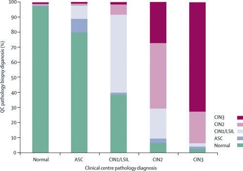
progression rate is 18% in an overall population, but in women agedFigure 7. Comparison of community pathology biopsy diagnoses with quality cont- rol pathology review diagnoses. Reprinted from Schiffman M, Castle PE, Jeronimo J, Rodriguez AC, Wacholder S. Human papillomavirus and cervical cancer. Lancet (London, England). 2007;370(9590):890-907., with permission from Elsevier. Prophylactic treatment of a precancerous lesion by a relatively simple proce- dure (LEEP) to avoid progression to cancer is acceptable in most cases. We cannot, however, predict which lesions would eventually become malignant if not treated, but we must keep in mind that any kind of invasive treatment is associated with adverse effects, fear, inconvenience and costs. Management should be based on careful selection of cases depending mainly on the grade of CIN, the type of the transformation zone and the age of the woman con- cerned. HPV testing The advantage of all molecular approaches in testing, such as HPV testing, is that the analysis is more objective, showing reduced variability and being less dependent on personnel compared with cytology. The first clinically validated commercial tests were the hybrid capture (HC)-based FDA (Food and Drug Administration)-approved HC2® test (QIAGEN, Gaithersburg, USA), and the polymerase chain reaction (PCR)-based GP5+/6+ PCR enzyme immunoassay, as they have shown good clinical performance in large randomized controlled trials (71-74). To have good clinical sensitivity and specificity for detection of CIN2+, a candidate test for screening purposes should be validated against either of these, according to guidelines (75, 76), and a more recent protocol for clinical validation of HPV tests, including genotyping, has been developed (VALGENT, Validation of HPV genotyping tests) (77). 22
HPV testing has a sensitivity for CIN2+ of about 95%, which has resulted
in at least a 50% rise in detection of CIN2+ lesions and a significant reduction
in the incidence of CIN3+ and invasive cervical cancer compared with cyto-
logical screening (78). As a consequence of the nature of HPV infection,
which frequently clears, a disadvantage of HPV testing is its specificity, which
is on average 6% lower compared with cytology (2), resulting in more screen-
ing-positive women needing follow-up. Also, the increase in detection of
CIN2 has been reported to be higher than the increase of CIN3+ (73, 79, 80),
raising concerns of overdiagnosis of self-clearing lesions. Swedish long-term
follow-up of primary HPV screening showed, however, the same cumulative
incidence of CIN2+ in both HPV and cytology arms, implying that the
improved sensitivity of HPV screening results in earlier diagnosis of CIN2+
rather than overdiagnosis (81). Other HPV tests such as the APTIMA mRNA
assay and the HC2 test at a higher viral-load cut-off point have shown higher
specificity, with a small loss in sensitivity (82, 83).
    Another important finding when including HPV testing in primary screen-
ing with cytology triage is the high negative predictive value for CIN2+ (84-
86), this effect being maintained even in primary screening with HPV testing
alone (87). This effect is also long-lasting, and screening intervals of six years
when using HPV testing are regarded as safe and effective, resulting in higher
cost-effectiveness.

Triage in HPV primary screening
Because of the relatively low specificity in HPV-based primary screening
there is a need for a triage strategy to find out which women are at most risk
of cancer development and thus need referral to colposcopy. Currently there
is no perfect triage and different algorithms are under research and develop-
ment. The RCTs presented so far (73, 79, 88, 89) have involved cytology
triage and this is therefore considered as a validated option. Consequently, in
current European guidelines, cytology is recommended for triage of HPV-
positive women (90). However, since the sensitivity of cytology is lower than
for the HPV test, HPV-positive women may have CIN2+ despite normal
cytology. These women still represent a group with a slightly higher risk of
CIN2+ (91, 92) and it is a challenge in developing appropriate follow-up
algorithms. One alternative is to use partial genotyping (HPV16/18 or other
high-risk type) (93, 94) with different follow-up intervals, and this algorithm
was recently implemented in Swedish guidelines.
    Other possible triage methods can be categorized into cytological or
molecular. Cytological methods include p16ink4a/Ki-67 dual immunostaining
(95, 96) and automated cytological evaluation (97). For these methods a cyto-
logical sample collected by professionals is needed and self-sampling cannot
be adapted. Molecular methods involving HPV testing with extended geno-

                                                                               23
typing (98), type-specific HPV viral load (99), viral and/or host gene methyl- ation (particularly high in HPV16 infection, cervical cancer and advanced CIN3) (100, 101), and altered microRNA expression (affecting tumor suppressors or oncogenes) (102) can also be adapted in connection with self- samples. Combinations of different methods including cytology can also be used, e.g. combining extended genotyping with cytology (103), and specific risk-score algorithms have been developed (104). All these methods, however, are still under evaluation and not yet in current clinical use. As HPV-negative women are at a very low risk of CIN2+ (105), repeated self-sampling for HPV testing to identify persistent infections could provide the highest protection against CIN2+. An alternative procedure for triage is simply repeating the HPV test after a couple of months. By way of this self- sampling strategy about 40% of women that are HPV-positive in their primary screening test have been found to clear the infection after 4–6 months (106), resulting in higher specificity for the detection of CIN2+ after the second sample. HPV genotypes Today over 200 different HPV types have been isolated and registered in The International Human Papillomavirus Reference Center. Of all the detected HPV genotypes about 40 are able to infect the genital tract. These can be divided into low-risk and high-risk types, where the low-risk types (e.g. 6 and 11) can cause genital warts (condylomata) while the high-risk types are associated with cervical cancer. In 2012 the International Agency for Research on Cancer defined 12 HPV types: 16, 18, 31, 33, 35, 39, 45, 51, 52, 56, 58 and 59 as high-risk types (107). All these types are included in the alpha-papillo- mavirus family and the types with highest risk are types 16, 31, 33, 35, 52, 58 from the alpha9 family and type 18 from the alpha7 family; they are all included in nonavalent vaccination. Moreover, type 68 is defined as probably carcinogenic and types 26, 53, 66, 67, 70 and 73 as possibly carcinogenic, with less evidence, and these last six types are usually not included in available HPV tests. Different types have different properties concerning prevalence, persis- tence and progression to CIN (108). HPV16 is described as the most common and persistent type, with a worldwide prevalence of about 3%. In total, 96% of all cervical cancer cases are attributable to one of the 12 high-risk types and type 68. The most carcinogenic type is HPV16, being associated with 60% of all cervical cancer. Moreover, HPV16 and 18 together are associated with 70% of all cervical cancer. Types 18 and 45 are not associated with a particu- larly high risk of CIN but are consistently related to adenocarcinoma (109) are therefore particularly important. With this background, genotyping for HPV16/18 is proposed as an alternative triage strategy in primary HPV 24
screening (94), and many commercial HPV tests report positivity for these
types, and the other high-risk types as one group.
   The level of type-specific viral load is often measured semi-quantitatively,
demanding specific characteristics of an HPV test. In serial measurements it
has been found that women with high viral loads have more persistent infec-
tions (110). The use of viral load in triage may therefore constitute a reasona-
ble strategy. A high viral load was first reported to be important as regards
HPV16 infections (111), but later a similar pattern was seen for the other geno-
types in the alpha9 family, showing a clear correlation between viral load and
CIN2+ (99). Developing management algorithms, however, is complex; every
genotype has a different viral-load level giving the same risk of CIN2+ and
more data is needed to establish the optimal titer thresholds for stratification
of screening-positive women. Multiple infections were earlier described as
being associated with a higher risk of CIN2+, but it rather seems that the risk
of CIN2+ in a multiple infection is equal to the risk associated with individual
genotype with highest risk (99, 109, 112).

HPV persistence
A type-specific persistent infection with high-risk HPV is a prerequisite for
the development of cervical cancer (3). Even if a persistent HPV infection is
a major risk factor of cervical cancer, very few HPV-positive women will
develop the disease (113). Most genital HPV infections are transient, with the
highest clearance in young women, and in a college population more than 90%
will have cleared the infection within 18 months (114). In a population of all
women aged >18 years about two thirds of HPV infections cleared within a
year (115). Studies on women aged >60 years have shown just over 60% HPV
persistence after 3.5 months (116) and about 55% HPV persistence after 5.5
months (117), indicating continuing clearance even in the elderly.
   There is still no consensus of opinion concerning the definition of persis-
tency of an HPV infection. In a Columbian prospective study on HPV persis-
tence, they proposed persistent infection to be defined as infection lasting
more than the median duration, for example, 9.5 months for HPV16 in women
aged >30 years (118). In a large meta-analysis, Koshiol et al. (11) remarked
that even testing intervals of ≤ six months produce strong summative relative
risks as regards the association between HPV persistence and CIN2+. It is
therefore suggested that repeat HPV testing at six months is a valuable way to
identify women at increased risk of cervical precancerous lesions and cancer.
   It is the type-specific persistency that increases a woman’s risk of CIN2+
or cervical cancer, but to accurately define an individual woman’s HPV per-
sistency is not possible without knowing the exact HPV type. This is the
advantage with an HPV test with extended genotyping, and, for example, in
‘test of cure’ it is useful to detect type-specific persistence, that is associated
with a high risk of recurrent CIN2+ (119).

                                                                                25
HPV self-sampling Highly sensitive molecular methods such as HPV testing offer the possibility of self-sampling where cytological analysis cannot be adopted (120). The advantages of self-sampling are several: the majority of women prefer it as a more convenient method (121-123) although some women are concerned about test accuracy and their ability to correctly carry out the procedure. Although complementary strategies such as offering invitations with timed appointments (124), reminder letters (125) and telephone calls (126) result in higher participation in screening (124), population coverage is still about 80% at most. Self-sampling for HPV testing has been shown to increase screening participation in non-attenders vs. other options (127-134). This strategy has also led to higher detection rates of CIN2+ than cytology-based screening (135) and offering self-sampling for non-attenders is recommended in current Swedish guidelines. In a large meta-analysis reduced sensitivity to detect CIN2+ in self-samples was noted when analyzed by way of signal-based assays, but no reduced sensitivity in the detection of CIN2+ in self-samples was reported when HPV testing was performed using amplification-based methods such as PCR (136, 137). A randomized study concerning a clinically validated PCR-based HPV test in a paired screen-positive design showed similar accuracy in self-samples and clinical samples in detection of CIN2+ and CIN3+ (138). Self-sampling has been carried out for the greatest number of years in the Netherlands, where self-sampling is available on request today and is planned to be introduced as a default option in screening in the near future (139). It has recently been proposed that all HPV tests used in connection with self-sampling should be validated for their accuracy and show agreement with clinician-collected sampling in a designated protocol (VALHUDES, Valida- tion of HPV assays and collection devices for HPV testing on self-samples and urine samples) (140). Every part of HPV testing of self-samples, including sampling material (vaginal fluid, urine), collection device (swab, brush, lavage, tampon), and storage material (liquid, dry), together with the used validated HPV analysis method should also be accredited. Urine sampling might be regarded as more acceptable in self-sampling than vaginal sampling (141) but has shown lower clinical sensitivity for CIN2+ than vaginal sampling (137, 142, 143). Different collection devices and storage media have shown similar results in accuracy (137), but dry storage material is preferred as regards price, safety aspects and easier mailing, where a flat medium is optimal. It is of importance that the self-sampling kit includes clear infor- mation of the sampling procedure, which should be easily understood and acceptable for the women. In the choice of test, the possibility of biobanking the samples should also be taken into account. 26
Self-sampling can be used in different strategies such as opt-in, direct mail-
ing, door-to-door or community campaigns. In an opt-in strategy women need
to confirm acceptance of receiving a self-sampling kit, or they pick up the kit
by themselves. This strategy has resulted in reduced participation (144-146)
in comparison with direct mailing. Door-to-door offering has resulted in a high
rate of participation in a low-resource setting but is not feasible in wide-scale
screening (133). Before implementing self-sampling for HPV-testing in
primary cervical screening, a careful pilot study should be carried out to assess
feasibility, the clinical accuracy of the combination of the considered HPV
test together with the sampling device and storage medium, not to forget the
costs, logistics, and population compliance. Also, not all women feel comfort-
able with self-sampling and the possibility of sampling by medical profession-
als should also remain available.

Health economics
Since one of the principles of screening is that the diagnosis and treatment of
the disease should be cost-effective, this is a point that should be validated.
Any possible changes in an organized screening program should be carefully
evaluated. As primary screening with HPV testing gives major reductions in
the number of cancer cases even with longer screening intervals and offers an
opportunity for self-sampling, it could be expected to be a cost-effective
screening alternative.
   The results of several studies around the world with somewhat different
settings support the notion that HPV-based screening is cost-effective vs.
cytological screening when applied among a population aged >30–35 years at
five-year intervals (94, 147-153). Self-sampling results in increased response
rates among non-responders vs. other options (127, 131-134, 154) and hence
results in increased coverage, which can also result in fewer and earlier-
detected cancer cases, followed by cost savings. Self-sampling for HPV test-
ing is one of the most effective and cost-effective interventions to improve
participation as regards non-responders in several countries (155-160). When
self-sampling is offered to non-responders some concerns about costs in
regard to switching have been raised, but the costs are compensated for if high-
level coverage is reached (157).
   When it comes to primary screening, a modelling study has shown that in
women aged ≥35 years with repeated vaginal self-sampling, HPV testing is
potentially cost-effective compared with conventional clinician-taken Pap
smear cytology even in maintained screening intervals (161). In low- and
middle-income countries self-sampling in primary screening could be cost-
effective if high-level coverage is achieved (162).

                                                                              27
Cost-effectiveness analysis (CEA) In a world with a continuous inflow of new medical technologies, interven- tions and treatments with rising costs, healthcare providers are in need of ways to evaluate the obtained benefits in relation to additional resources spent. A cost-effectiveness analysis is designed to allow decision-makers to clearly understand the tradeoffs of costs, harms, and benefits between alternative interventions and to combine those considerations into a single metric, the incremental cost-effectiveness ratio (ICER), which can be used to inform decision-makers (163). The ICER is defined as the ratio of the incremental difference in total cost to the incremental difference in effectiveness when comparing alternatives. ICERs can then be used to compare different inter- ventions to define which one provides greatest value for money. A low ICER can mean that intervention improves health at a small additional cost per unit of health. A negative ICER can mean either that the new intervention is less costly than the existing one or that the new intervention is less effective than the existing one. An intervention is ‘dominated’ if it is higher in cost and less effective than the comparator and is not of good value for money. In a CEA with a societal perspective all costs from formal and informal healthcare sectors and costs from the non-healthcare sector should be taken account, while a CEA with a healthcare perspective only includes formal healthcare- sector costs (costs for the patient and costs for a third-party payer [other than patient/healthcare provider]) (164). A cost-effectiveness analysis is validated by a sensitivity analysis of different effects and costs. 28
Aims

The overall aim of this work was to increase knowledge about the use of
self-sampling for HPV testing in primary cervical screening.

The specific aims of the studies were:

   I    To compare repeated self-sampling for HPV testing with Pap smear
        cytology in detection of CIN2+ in primary cervical screening.

  II    To evaluate the proportion of cases of histological CIN2+ after
        LEEP in women with persistent HPV infection and normal Pap
        smear results.

 III    First, to compare the cost-effectiveness of repeated self-sampling for
        HPV testing with Pap smear cytology in primary cervical screening.
        Second, to estimate the cost of treatment and follow-up of histo-
        logical CIN2+ in connection with these screening strategies.

 IV     To compare self-sampling and sampling by medical professionals
        for HPV testing in detection of CIN2+ and CIN3+ when using a
        combination of an FTA card as storage medium and a PCR-based
        HPV test.

                                                                           29
Material and methods Study population Studies I and III During 2013–2015 a total of 36 390 women aged 30–49 years (at the date of invitation) scheduled for regular screening invitation in Uppsala County, Sweden, were included. We excluded women with previous hysterectomy, current pregnancy or clinical test results (Pap smear cytology, HPV test or histology) relating to cervical cancer registered within one year before the date of invitation. The follow-up period was 18 months from the date of invitation. Study II From April 2013 until March 2016 we prospectively recruited 91 women aged over 40 years with persistent HPV infection without any abnormalities in cytology at the gynecological out-patient clinic, Uppsala University Hospital. We excluded women who had plans for future pregnancies, who could not understand the information in Swedish, and where LEEP was regarded as being technically difficult to perform. Study IV During March and April 2016 a total of 11 951 women aged 30–60 years (at the date of invitation) scheduled for a regular screening invitation in Uppsala County were included. After sampling, women with clinical test results (Pap smear cytology, HPV test or histology) relating to cervical cancer registered within one year prior to the start of the study period were excluded from the analysis. The women whose first HPV samples arrived at the HPV laboratory during 2016 were included in the analysis. The follow-up period was 18 months from the start of the study period. Ethics All clinical data were coded and analyzed anonymously. In Studies I, II and IV participants received oral and/or written information, and consent was 30
given. All study designs were approved by the Regional Ethics Committee,
Uppsala, Sweden (Dnr 2012/099 for Studies I and III, Dnr 2012/460 for Study
II, Dnr 2016/008 and Dnr 2019/929 for Study IV).

Methods
During the study period (Studies I–IV), the regular screening program in
Uppsala County was 3-yearly Pap smears for women aged 23–49 years and
5–yearly HPV tests for women aged 50–60 years. Women not attending
screening were recalled the following year.

Study I
By means of a computer-based allocation process the women were random-
ized in two groups, one to perform self-sampling of vaginal fluid (VF) for
HPV testing (n=17 997, HPV arm) and the other group to undergo screening
by Pap smear cytology (n=18 393, control arm).

HPV arm
Women in the HPV arm were sent an invitation including information on how
to perform the sampling at home, a sampling brush, an FTA (Flinders Tech-
nology Associates) card and a preaddressed return envelope. The FTA card
was returned by regular mail to the HPV lab at Uppsala University for HPV
testing. A reminder was sent to women who did not return their self-sample
within three weeks. Women who were HPV-positive in their first self-sample
were informed of the test result and told that they could contact a gynecologist
if they had any questions or symptoms. These women were sent a new kit in
3–6 months to repeat the self-sampling. Women who were HPV-positive in
two consecutive self-sampling tests were referred to colposcopy. HPV-
negative women in the first or second HPV test were referred back to the
regular screening program. Women who chose not to participate in the study
were returned to the regular screening for Pap smear sampling.

Control arm
Women in the control arm were managed according to the regular screening
program in Uppsala County during the study period where a midwife
performed cervical sampling for Pap smear cytology. Women with CIN2+
based on cytology were referred to colposcopy within a month, while women
with CIN1/ASCUS based on cytology were offered follow-up with HPV test
and Pap smear cytology after three months, according to the clinical routine.
All HPV-positive women and women with CIN2+ in follow-up cytology were
referred to colposcopy and eventual biopsies. HPV-negative women without

                                                                             31
You can also read


