Structures and methods - Understanding Nudibranchs and other Sea Slugs - The Book - David A. Mullins 2020 - Respiration - Nudibranch Domain
←
→
Page content transcription
If your browser does not render page correctly, please read the page content below
Understanding Nudibranchs and other Sea Slugs – The Book

                                                   Gills and other respiratory
                                                   structures and methods

David A. Mullins 2020 - Respiration                        1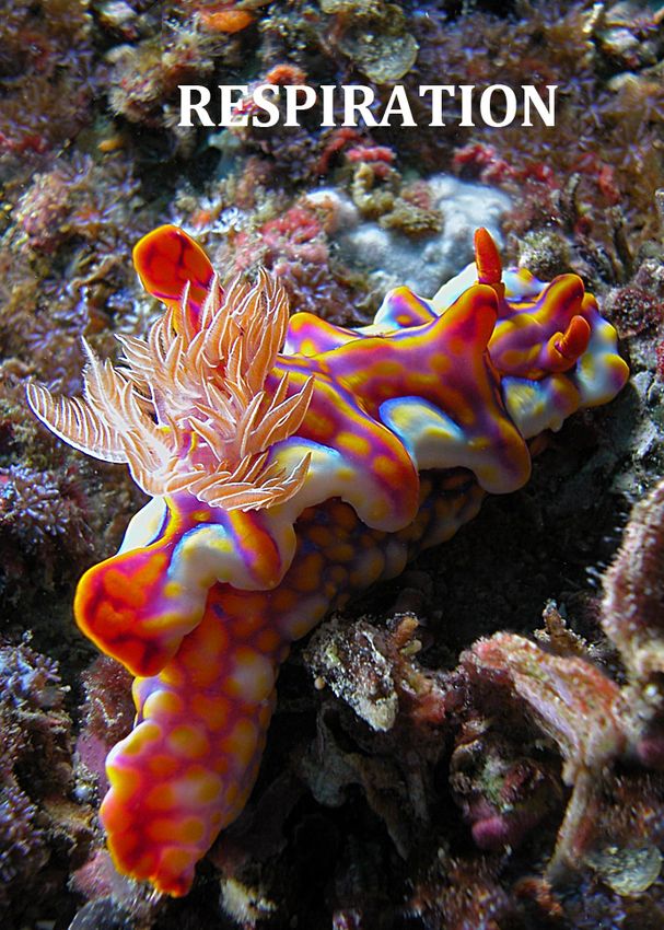
Understanding Nudibranchs and other Sea Slugs – The Book
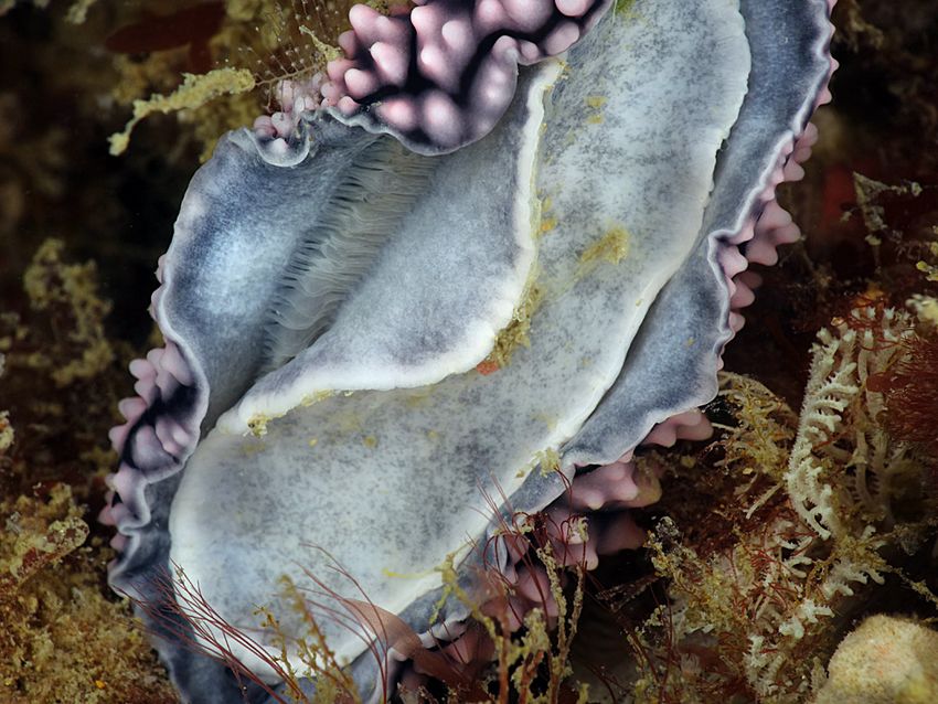
RESPIRATION
The term nudibranch literally translates as “naked gill” and that type of sea slug, with the large
flower-like gill exposed upon the back, is the type with which most people are familiar. This
exposed, expanded gill however is only one of several different structures or arrangements evolved
by sea slugs to respire, even amongst the true nudibranchs.
The exposed dorsal gill of Chromodoris magnifica plainly illustrates the origin and
meaning of the term nudibranch (naked gill) to describe these sea slugs.
All animals respire, requiring gas exchange with their environment in order that oxygen may be
taken up for metabolic processes and the resulting carbon dioxide, a waste product, removed.
Respiration and “blood” circulation go hand in hand unless the animal is small and simple with only
a few cell layers of thickness. In those animals gases are exchanged by diffusion through their body
wall. This is also the case sometimes with small animals that have a large surface area to body
volume and even those sea slugs with a gill also supplement gas exchange by cutaneous respiration.
The marine animal needs to extract dissolved oxygen from the surrounding water and this is much
more difficult than extracting it from air. Problems they face include a much slower diffusion rate
compared to air (by a factor of 10,000) and depending upon the temperature, water (1% O2
concentration) contains about 20 times less oxygen than air (21% O2 concentration). However, in
their favour, sea slugs have a low metabolic rate and are relatively passive compared to fish, for
example.
David A. Mullins 2020 - Respiration 2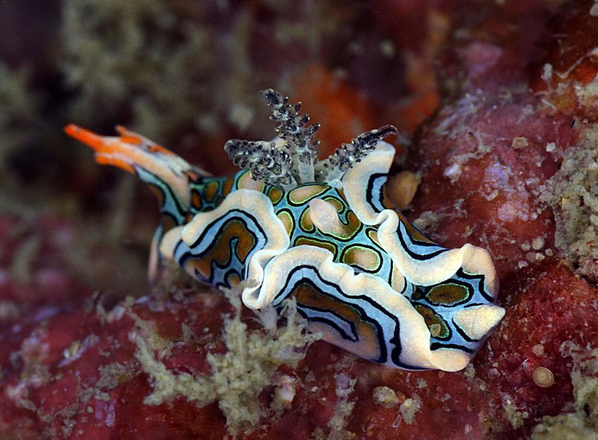
Understanding Nudibranchs and other Sea Slugs – The Book

      Respiratory structures have evolved to provide the necessary thin-walled, large surface area
      arrangement required for gaseous exchange in complex animals, together with a complementary
      blood supply to and from the structure. The blood is delivered and passed through these thin-
      walled structures, whereby diffusion of gases takes place due to the concentration gradient – CO2
      out and O2 in - and then recirculated in a system around the body, that in molluscs is termed an
      open system. (See explanatory box – Page 4.)

        The very small nudibranch Vayssierea felis has no gills        The dendronotid nudibranch Tritoniopsis elegans has
        or other respiratory structures and respires through its       lace-like secondary gill structures down the length of
        body surface.                                                  each side of its body for respiration.

        Phyllodesmium colemani, an aeolid nudibranch, makes            The phyllidiid nudibranch Phyllidiella pustulosa has
        use of the huge surface area provided by its cerata,           numerous respiratory leaflets situated between
        those many long projections of its dorsum.                     mantle and foot in the groove of the hyponotum.

      In sea slugs these structures, other than a “typical” gill, may be as simple as thin-walled outgrowths
      on the surface of the mantle serviced by “blood” vessels e.g. the multifunctional finger-like cerata of
      aeolids, the simple or highly branched lateral appendages of the dendronotids or the secondary gill
      leaflets of the phyllidiids and Armina nudibranchs situated under the mantle in the hyponotum. The
      fine nature of these structures is possible in an external situation because they are supported and
      separated by the surrounding water. There is even a great variation in gill shape, arrangement and
      size amongst those with a “typical” gill, from greatly expanded to almost undetectable, from simple
      to complex and whether it is able to be contracted or retracted or not. Even those that possess a gill
      or secondary branchial structures are believed to also utilize their body surface that includes that
      now enlarged and exposed mantle expanse, for gaseous exchange.

David A. Mullins 2020 - Respiration                                3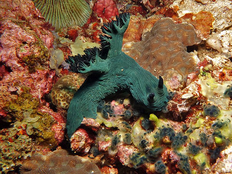
Understanding Nudibranchs and other Sea Slugs – The Book

                                      The Open Circulation of Sea Slugs
           Nearly all molluscs have what is called an “open system” of “blood” circulation, the exception being
           the highly active cephalopods such as the octopus and squid.

           In a closed circulation system the blood is transported around the body entirely enclosed in vessels,
           the arteries, veins and capillaries, both to and from the tissues, unidirectionally under reasonable
           pressure to enable diffusion through the capillaries. An open circulatory system however only uses
           closed vessels for part of the journey and the circulation is low pressure and cannot be called
           directional for much of its path. Sea slugs and nudibranchs have blood-filled body cavities or sinuses
           called haemocoels, in which the tissues and organs are bathed directly in the blood, to be oxygenated,
           receive nutrients and remove wastes and carbon dioxide.

           The blood of sea slugs is more correctly called haemolymph as, by operating in the open system, it is
           comprised of both blood and interstitial fluid or lymph fluid, there being no separation between the
           two. The oxygen binding protein is haemocyanin, a copper compound that can carry one oxygen
           molecule rather than the iron compound, haemoglobin of most vertebrates, which can carry four.
           Haemocyanin is suspended directly in the haemolymph fluid and does not require carrying by blood
           cells. Deoxygenated haemolymph is virtually colourless but acquires a bluish tint when oxygenated.

           Most sea slugs possess a two-chambered heart with an auricle and a ventricle. To generalize: Vessels
           carrying oxygenated haemolymph from the gills and lateral sinuses of the notum, plus from the
           kidney, empty into the first chamber of the heart, the auricle, that pumps it into the second chamber,
           the ventricle, that pumps it through the aorta/s that then progressively divide into many finer vessels
           around the body thus distributing haemolymph into a network of open arterial sinuses or
           haemocoels that bathe the organs and tissues. Muscular movements by the sea slug also assist in
           moving the haemolymph around the body and mixing it through the haemocoels. Haemolymph drains
           from the sinuses into vessels that conduct it separately to the gills and the kidney thus completing the
           open circulation.

           Additionally, the haemocoels perform a hydrostatic skeleton function giving the sea slugs shape and
           form. By acting in opposition to contractile musculature, the gills, rhinophores, cerata, proboscis and
           penis, for example, can be everted or expanded. Research has shown that most molluscs assist these
           functions by controlling the action of the heart and therefore the haemolymph pressure. The heart
           can be slowed down or even stopped for periods of time.

           The more primitive sea slugs have separate anterior and posterior haemocoels whereas the true
           nudibranchs have lost the separation between the two producing a single large and continuous
           haemocoel.

           The operation of an open circulation system is less energy dependent than that of a closed system but
           cannot transport comparable volumes and is less efficient there being no systematic way of passing
           haemolymph through the gills for oxygenation. It should also be noted that the haemolymph leaving
           the kidneys goes directly to the heart without traversing the gill. The open system is thus more suited
           to animals of low metabolic function, smaller size and possessing the capability of also absorbing
           oxygen through their integument.

           As we have seen, some sea slugs do not possess a gill and strange to relate some do not have a heart.
           Species of the Sacoglossa genus Alderia for example, do not possess a heart and the haemolymph
           circulation is achieved by rhythmic pulsations of its cerata on alternating sides of the body thereby
           performing as secondary hearts.

                                                            -------

David A. Mullins 2020 - Respiration                          4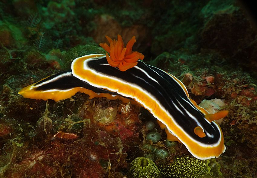
Understanding Nudibranchs and other Sea Slugs – The Book

      The primitive sea slugs possess what is often referred to as a ctenidium, an outgrowth of the mantle
      made up of a series of thin filaments of tissue attached to each side of a central axis, that resemble
      the teeth of a comb. Its surface is often covered with cilia to facilitate water movement. This is their
      respiratory structure, analogous to a gill, inherited from the mantle cavity of their shelled
      ancestors. One evolutionary theory put forward is that the ctenidium was originally just to provide
      a water current in the mantle cavity for feeding and excretory purposes. Over time it enlarged and
      vascularized to develop into an organ of respiration. The gill on the back of the dorid nudibranchs is
      argued by some, to be the direct descendant, though highly differentiated, of the ctenidium due to
      its position, relationship with the anus, innervation and “blood” circulation. Other authorities
      though, consider it to be a completely new adaptive organ and refer to them as secondary (anal)
      gills arguing that dorid veliger larvae detort (see Development Chapter) just prior to
      metamorphosis losing their primary gas exchange organ or ctenidium and grow new “secondary”
      gills, without cilia, on the mid to posterior region of the dorsum. The debate appears, as yet, to be
      unresolved.

        Japonacteon suturalis belongs to a primitive                   Bullina lineata has a thinner more inflated shell
        group of sea slugs whose lineage diverged early.               but the ctenidium is still retained within the
        It has a thick shell and retains the ctenidium                 mantle cavity.
        deep in the mantle cavity.

      Micromelo undatus, along with its relatives shown              The highly modified Hydatina physis with all of its
      here, has large posterior lobes to its headshield. These       lobes and extensions displayed. The ctenidium can
      protect the opening to the mantle cavity, diverting silt       sometimes be seen protruding out from under the
      and debris away from the opening stopping                      right side of the shell but protected by the folded up
      congestion of the ctenidium when it burrows.                   parapodium.

David A. Mullins 2020 - Respiration                              5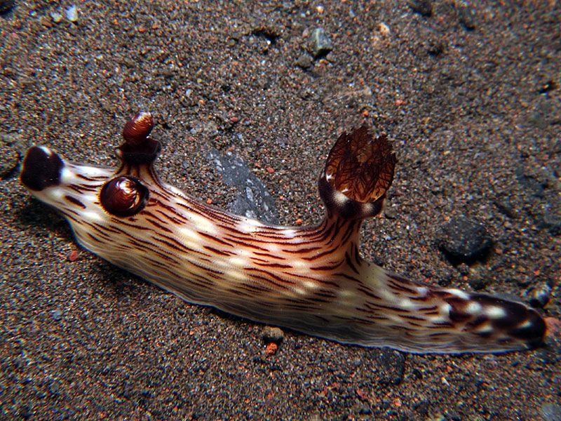
Understanding Nudibranchs and other Sea Slugs – The Book

           The Different Gills and Secondary Respiratory Structures
      The descriptions here of the gills or secondary gills of different sea slug groups are necessarily
      generalized for there are many variations on a theme within those groups. An overview is given but
      space does not permit an exhaustive list of the anatomy of every genus. Some minor deviating
      groups are not included.

      The Head-shield Slugs - Cephalaspidea, Acteonoidea and Ringiculoidea
      Most of the head-shield slugs have a simple plicate gill located in the enclosed mantle cavity, where
      present, or more exposed where that cavity is opened or reduced or formed into lobes. In most
      instances the gill is not readily observable. An example of a more exposed gill in the head-shield
      slugs is that of the Gastropteridae where the mantle cavity is reduced or even absent such that the
      gill although usually small is located between the right parapodium and the body proper. In the
      genus Sagaminopteron however the gill is large with long processes and is often readily visible
      along the right side. Those species with a gill enclosed or semi-enclosed in the mantle cavity or
      tucked under the parapodial fold, siphon water through and over the gill from front to back.

     Most species of Gastropteridae have a small gill            Sagaminopteron psychedelicum also has a large gill
     but Sagaminopteron ornatum has a large                      that is distinctive with white flecks on a dark
     translucent gill that is often visible above the edge       translucent background, and is carried high above
     of the right parapodium.                                    the parapodia.

     The gill of most Runcinidae is located posteriorly          In species of Aglajidae, such as Chelidonura amoena
     on the right side near the anus and is often visible        shown here, the gill is contained in the mantle cavity
     as exhibited by this Runcina fijiensis specimen.            the opening of which is protected by the large tightly
                                                                 wrapped right parapodium.

David A. Mullins 2020 - Respiration                          6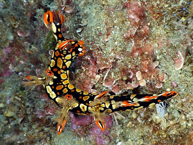
Understanding Nudibranchs and other Sea Slugs – The Book

      The Sap-sucking Slugs – Sacoglossa
      As discussed earlier the Sacoglossa have a wide diversity of body shape and we can therefore
      expect a similar diversity of respiratory methods and structures. In general the more primitive
      shelled forms have a gill in the mantle cavity under the shell not unlike the head-shield slugs.
      However in the Sacoglossa it differs in structure, being epithelial folds of the mantle roof and
      composed of numerous simple lamellae bearing cilia for water current generation. This includes the
      unusual bivalved sacoglossans of the Juliidae family. The flattened, leaf-like forms, such as Elysia
      and Thuridilla of the Plakobranchidae do not possess a gill. In saying they are flattened most
      actually carry their leaf-like parapodia in an upright position towards the mid-line, closed or semi-
      closed. These folded extensions of the foot on each side of the body create a huge surface area for
      gaseous diffusion and many carry a complex network of vessels.

       This undescribed species of Lobiger carries a gill              The large surface area of the parapodia of many
       under its shell but also utilizes the surface area of the       sacoglossans assists gas diffusion and are serviced by a
       large processes for diffusion.                                  network of vessels visible here in Elysia tomentosa.
      Those without parapodia, for example Bosellia, are essentially just flattened but respire in the same
      manner, through the broad surface of their body. The sacoglossans with processes on their dorsum,
      commonly called cerata, also do not possess a gill per se. Instead they make use of the large surface,
      the projecting cerata provide, to respire. This is why the cerata are often referred to as secondary
      gills. Cyerce and Polybranchia, for example, have flattened leaf-like or thicker cushion-like cerata,
      whilst Costasiella, Ercolania, Stiliger and Hermaea, to name just a few, have fusiform cerata of
      varying length, girth and number.

       The many fusiform cerata on the back of Costasiella              Cyerce elegans does not have a true gill but the
       kuroshimae provide a large surface area for the                  large balloon-like cerata crowded on its back act as
       exchange of gases.                                               secondary gills to provide sufficient oxygen.

David A. Mullins 2020 - Respiration                                7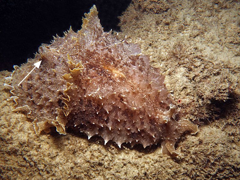
Understanding Nudibranchs and other Sea Slugs – The Book

      Some species of Sacoglossa carry functional kleptoplasts (sequestered chloroplasts from their diet)
      that produce oxygen as a by-product of photosynthesis. This can be a two-edged sword however if
      the production of oxygen is not moderated. In fact it has been suggested that their secondary
      processes act as a “reverse gill” to remove excess oxygen. (See Kleptoplasty in Solar Powered
      Chapter).

      The Sea Hares – Anaspidea
      Most Sea Hares have a thin remnant of shell that lies under the skin in nearly all species (partly
      exposed in a couple). The plicate gill lies across the reduced mantle cavity as a tufted crescent or
      fan, depending on filament size, partially protected by the shell but otherwise by the parapodia that
      fold up over the mantle and are sometimes fused along the mid-line in which circumstances they
      form a parapodial cavity. Water circulation is facilitated by entry anteriorly though a gap in the
      parapodia and expulsion via a posterior siphon of varying size.

       The partially exposed shell remnant of the sea hare               The parapodial flaps of Dolabella auricularia are
       Aplysia nigrocincta (arrowed) covering the mantle                 joined at the midline. An exhalent siphon (arrowed)
       cavity and gill. This species is one of the few sea               is visible in the centre of the disc-like posterior
       hares to have an exposed shell.                                   “shield” that houses a flat shell within its tissues.

      The Umbrella-shell Slugs - Umbraculoidea
      In this order the external shell is much reduced to a limpet shape or just a flattened conical dorsal
      cap. There is no mantle cavity at all for the bipinnate gill and therefore it sits under the lip of the
      shell on the right side between the mantle and the foot. In Umbraculum the large gill is attached for
      most of its curved length and is not readily visible because it is accommodated in a groove-like
      cavity formed by the overlapping margins of the mantle and the foot but will sometimes be
      extended. In Tylodina it is only attached via the anterior half and is often seen protruding laterally.

                      The free, unattached posterior portion of the gill of Tylodina corticalis (arrowed) is
                      seen here protruding from underneath its limpet-like shell on the right side.

David A. Mullins 2020 - Respiration                               8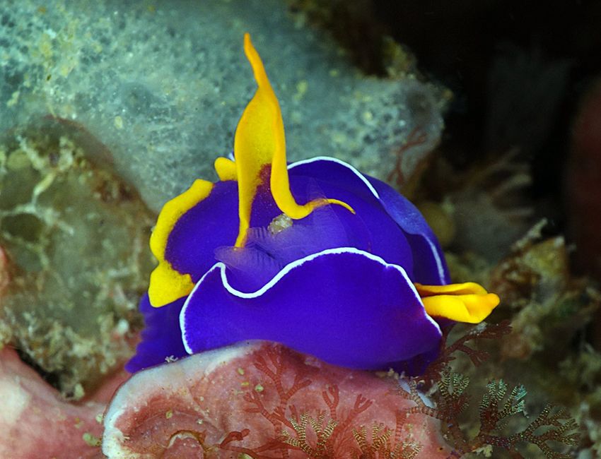
Understanding Nudibranchs and other Sea Slugs – The Book

          A peek under the shell of Tylodina corticalis reveals       Umbraculum umbraculum has the gill wrapped
          the gill and its bipinnate nature. The colour closely       around the right side situated in a groove between
          matches the rest of the body and foot. There are            the underside of shell and the foot and is attached
          about 12 feathery pinnae on each side of the rachis.        for most of its length.

      The Side-gilled Slugs – Pleurobranchoidea
      None of the species in this group possess an external shell and the mantle in most instances has
      expanded to cover the entire body, and even further, thus creating an overhanging skirt. The
      bipinnate gill resides down along the right side (hence the name - side-gilled slugs) between the
      underside of the mantle skirt and the foot. It is sometimes visible in certain species, for example
      species of Pleurobranchaea, where the reduced mantle of this genus can cause the gill to be exposed
      at times. The gill rachis may be either smooth or tuberculate. Those tubercles may be located at the
      base of the rachis and also sometimes along its axis at pinnae junctions depending upon species. It
      is normally attached anteriorly for more than half its length with the free posterior portion
      muscular and able to articulate. The central posterior edge of the mantle emarginates, forming up
      into a “funnel” to act as an exhalent siphon, thereby facilitating water flow across the gill in
      Pleurobranchus and Euselenops for example, but not so in others such as Berthella or Berthellina.

       The gill of the side-gill slug Berthella martensi is            A ventral view of Berthella stellata showing how the
       revealed here because segments of the mantle that               gill lies along the right side protected between the
       normally cover it have been autotomized.                        underside of the mantle and the foot.

David A. Mullins 2020 - Respiration                               9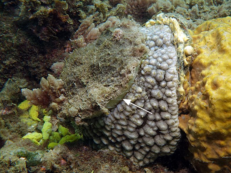
Understanding Nudibranchs and other Sea Slugs – The Book

      The True Nudibranchs - Nudibranchia
      The majority of these slugs has an exposed gill or exposed secondary gill processes. This discussion
      will make more sense if we break the nudibranchs up into the two clades, the dorids and
      cladobranchs, each with three sub-groups as presented previously in the Anatomical Overview.

      The Dorid Nudibranchs - Doridoidea Clade
      Most of the dorid nudibranchs, with some exceptions, e.g. the Phyllidiidae, can be recognized by the
      typical circle of gill branches on their dorsum that surround (or are located in front of) the anus the
      opening of which is situated on the tip of a short papilla. There are three sub-groups as follows, but
      be aware that these are old, traditional, but still convenient groupings for practical purposes.

       The phanerobranch dorid               The cryptobranch dorid               The porostome dorid Phyllidiopsis
       Nembrotha milleri – gills cannot      Hypselodoris apolegma – gills can    shireenae – secondary gill leaflets under
       retract into pocket.                  retract completely into pocket.      mantle in groove of hyponotum.

      - Phanerobranch Dorids – Phanerobranchia
      The gills of Phanerobranch Dorids (Phanerobranch = visible gill) cannot be retracted into a distinct
      pocket. In most the gill is inserted directly on the dorsum while some have sheaths into which the
      gill can partially contract but these are not pockets and cannot close over the gill. Depending on
      species there are a variable number of gill plumes of pinnate (simple), bipinnate or tripinnate
      composition. Many of the phanerobranchs possess structures or appendages around or in
      proximity to their gills for protective purposes. Apart from the physical barrier these might present
      to a potential predator some structures also carry chemical deterrents of a noxious or toxic nature.

       The six separately inserted gills of Hexabranchus              A juvenile Periclimenes imperator shrimp lives
       sanguineus encircle the anus. They can contract but            within the protection afforded by the feathery
       not retract under the mantle surface.                          gills of Hexabranchus sanguineus.

David A. Mullins 2020 - Respiration                          10
Understanding Nudibranchs and other Sea Slugs – The Book
Nembrotha kubaryana displays its bushy gills. This specimen of Nembrotha purpureolineata has
There are five tripinnate gill leaves often carried three (max. of five) non-retractile, mulitpinnate gill
curled up as illustrated here. leaves.
The small purple–tipped gill leaves of Tambja kava Tambja morosa carries from three to five well-
form a semicircle anteriorly around the anus. developed tripinnate gill leaves that are non-retractile.
The circular gill pattern of Aegires flores is A close-up view of Aegires flores (whole animal left)
apparent here with the paddle-shaped protective showing the small tripinnate gill leaves dwarfed by
tubercles. the protective ring of tubercles.
Some examples of phanerobranch dorid gill arrangements
David A. Mullins 2020 - Respiration 11Understanding Nudibranchs and other Sea Slugs – The Book
Aegires serenae has three exceedingly large Aegires gardineri has a more compact but closer
protective appendages in front of and arching over protective coverage of the gill.
the gill.
Gymnodorids have a variety of gill types but all are Gymnodoris amakusana has the gill branches
unprotected. Here Gymnodoris citrinus displays its arranged in a transverse linear row anterior to the
complete circular arrangement around the anus. anus.
Trapania japonica has a yellow extra-branchial This undescribed species of Thecacera has two
process on each side of the gill. extremely long extra-branchial appendages
protecting the gill.
Further examples of phanerobranch dorid gill arrangements
some with protective appendages
David A. Mullins 2020 - Respiration 12Understanding Nudibranchs and other Sea Slugs – The Book
- Cryptobranch Dorids – Cryptobranchia
The gill of the Cryptobranch Dorids (crypto branch = hidden gill) is able to not only contract but
also to actually retract completely into a distinct and common branchial pocket beneath the mantle
surface that can be closed off. Depending on species, there are a variable number of gill plumes of
pinnate (simple), bipinnate, tripinnate or even quadripinnate composition. Some of these
nudibranchs have been observed to vibrate their gill plumes, an action considered to improve
efficiency by increasing the water flow over them. A very few species in this group also possess
significant structural protection for their gills. Some of these structures also carry glands containing
noxious/distasteful compounds. The appearance of some cryptobranch gills bears an uncanny
resemblance to opened octocoral polyps.
A sequence of images of the cryptobranch dorid Diversidoris sp.
showing the entire retraction of the gill into a common pocket
beneath the mantle.
The gill circle of Hypselodoris obscura is open In this view of Goniobranchus sp. both the anal papilla
posteriorly. Here the detail of the gill and the central (arrowed lower) and the kidney duct (arrowed upper)
location of the anal papilla can be seen. are visible in the centre of the gill circle.
David A. Mullins 2020 - Respiration 13Understanding Nudibranchs and other Sea Slugs – The Book
A composite image of Goniobranchus aureopurpureus A composite image of Jorunna sp. showing the gill
showing gill retraction and the raised edge to the gill displayed, and then completely withdrawn.
pocket.
Ceratosoma tenue has a protective horn posterior to In this image of Ceratosoma tenue there is evidence
the gill that arches forwards over the top. The horn of a fish strike to the protective horn. The fish will
carries glands of noxious/distasteful chemicals. not return for a second bite. Gill saved.
Miamira magnifica bears a protective appendage in Miamira alleni has several lobe-like appendages,
front of the gill. The genera Ceratosoma and Miamira some with distasteful defensive glands, surrounding
are the only cryptobranchs with physical protective the gill.
structures.
Examples of cryptobranch gill retraction and the protective appendages of the
Ceratosoma and Miamira genera
David A. Mullins 2020 - Respiration 14Understanding Nudibranchs and other Sea Slugs – The Book
Jorunna rubescens has a tall sheath The gill sprouting out of the tall gill The “volcano” of Hypselodoris
to the gill pocket. pocket of Hypselodoris krakatoa. krakatoa.
Named after the volcano.
Goniobranchus leopardus has gills The blood red gills of Ardeadoris The long thin tapering gills of
that are triangular in section rubroannulata. Ardeadoris egretta are almost round
arranged in an arc around the anus. in cross-section.
Goniobranchus fidelis with its gill The expanded feeding polyps of The distinctive inward curving goblet
fully displayed. The gill appears not some octocorals look somewhat shape of the gill of Actinocyclus
unlike an expanded octocoral polyp. similar to some cryptobranch gills. verrucosus.
Doriprismatica atromarginata The large but sparse pinnate gill of The gill of Goniobranchus
showing damage to both sides of the Taringa halgerda. collingwoodi is a circle, open
mantle. Continued predation posteriorly, with the ends spiraling
prevented by noxious glands. inwards.
A selection of cryptobranch gills
David A. Mullins 2020 - Respiration 15Understanding Nudibranchs and other Sea Slugs – The Book
- Porostome Dorids – Porostomata
This group is defined by its suctorial method of feeding. They lack a radula and feed by secreting
digestive enzymes upon their food and sucking up the partially digested tissue. The dendrodorids
(Family Dendrodorididae) have the same gill arrangement and branchial pocket as the
cryptobranch dorids. The phyllidiids (Family Phyllidiidae) are different to all of the other dorids in
not possessing a typical gill arrangement on the dorsum. Instead they have secondary gill leaflets
situated ventro-laterally between the mantle and foot, in the hyponotum groove. These secondary
gills occupy this groove right around the entire animal excepting for interruption at the mouth and
genital region. The leaflets are flat, triangular in shape attached along the long edge and have a
rounded apex, alternating large and small. Nevertheless, the phyllidiids are still considered
“cryptobranch dorids” in so far as most retain a cavity that surrounds the anus thought to be
homologous with the gill cavity of the cryptobranchs, even though the typical gill has been lost.
Most also retain the posterior dorsal location of the anus with a few having a ventral location
instead.
Arrowed above is the salmon pink coloured anal papilla of The secondary gill leaflets of Phyllidiopsis krempfi,
Phyllidiopsis shireenae. In almost all other dorids, apart usually well hidden, are visible here, located ventrally
from the phyllidiids, we would expect to also see a gill between the underside of the mantle and foot across
arrangement closely associated with the anus, either the groove of the hyponotum.
encircling or anterior thereto.
A close up view of the secondary gill leaflets situated The Dendrodorididae have gills that are similar to
under the mantle of Phyllidia varicosa. The alternation the cryptobranchs being able to withdraw under the
of big and small leaflets can be seen as well as the mantle into a common pocket. Dendrodoris
triangular shape and rounded apex. arborescens shown here has its gill partially
withdrawn with the rim of the pocket visible.
David A. Mullins 2020 - Respiration 16Understanding Nudibranchs and other Sea Slugs – The Book
In those species that are able to contract or retract the gills this action is performed by retractor
muscles acting against the hydrostatic fluid pressure that provides a “skeleton” giving the slug its
form and shape. Once the muscle relaxes the internal fluid pressure pushes the gill back out.
The Cladobranch Nudibranchs - Cladobranchia Clade
None of the nudibranchs in this clade possess what is termed a “typical” gill on the dorsum. The
premise for this clade is the possession of a branched digestive gland. Again there are three groups.
Currently the Arminina and Dendronotina are not considered natural groupings.
The arminid nudibranch The dendronotinid nudibranch The aeolid nudibranch
Dermatobranchus funiculus – no Marionia sp. – secondary gill Phyllodesmium undulatum –
respiratory structures on the dorsum. structures on the dorsum. multifunctional cerata on dorsum act
as secondary gill structures.
- The Arminid Nudibranchs – Arminina
The arminids exhibit two main body forms, those with a broad and long tapering body on which
most carry a series of longitudinal ridges and those with cerata on the notum. In the former, Armina
possess ventral secondary respiratory leaflets in the hyponotum, between mantle and foot while
Dermatobranchus does not, seemingly reliant on diffusion into its flat body across its broad ridged
notum and body side walls under the mantle. Janolus and Madrella are representatives of the
second body form where the notum is usually crowded with cerata that, with the exceptional
surface area they provide, are able to act as secondary respiratory agents.
An undescribed species of Armina. Armina have no All the Dermatobranchus lack any respiratory
gill on the dorsum but have secondary respiratory structures, even the hidden secondary gill leaflets of
leaflets located in the hyponotum between their sister genus the Armina. Dermatobranchus
underside of mantle and foot. rubidus shown above.
David A. Mullins 2020 - Respiration 17Understanding Nudibranchs and other Sea Slugs – The Book
An undescribed species of Janolus showing the dense Madrella ferruginosa has long cerata on the dorsum
coverage of cerata, even in front of the rhinophores, performing a respiratory function. They are often
that act as secondary respiratory structures. carried tightly curled up.
- The Dendronotinid Nudibranchs – Dendronotina
All dendronotinids have dorsolateral appendages down the sides of the body. These may be simple
or complexly branched, lobed, nodular or tuberculate. All are believed to act as secondary
respiratory structures. Additionally some carry or have embedded close by other more finely
branched processes that act specifically as gills. The tritoniids, for example Tritoniopsis, have very
fine delicately branched processes that are referred to as dendritic gills. Bornella has dorsolateral
cerata-like processes that protect one or two gills embedded at their base. Some species of Doto and
Kabeiro bear small simple gills at the base of the larger nodular cerata. Scyllaeidae, for example
Notobryon, have the mantle ridge formed into one or two large lobes that bear tufts of dendritic
gills on the surface of the inner face. The single species of the Tethys genus has a pair of small
secondary respiratory structures at the base of each of the several large lobed cerata while the
closely related Melibe have scattered branched filaments upon the external (sometimes also the
internal) faces of those cerata and sometimes also found on the body proper that are considered by
some workers to have a respiratory function.
The dendronotinid Bornella anguilla, displaying the A close-up image showing detail of the secondary gills
pairs of lateral flap-like appendages protecting the of Bornella anguilla and how they are protected by
secondary gills located at their bases on the medial being on the inside surface the colourful lateral
sides. appendages.
David A. Mullins 2020 - Respiration 18Understanding Nudibranchs and other Sea Slugs – The Book
The plain bifid dorsolateral appendages of Long thought an aeolid, the dendronotinid
Marianina rosea assist with it respiration needs. Embletonia gracilis has cerata with up to four blunt
fingers on their terminations. The cerata act as
secondary respiratory structures.
Notobryon wardi has two large lobes projecting Some authorities claim that the fine filaments on the
from each side of its mantle providing protection cerata of species of Melibe act as secondary respiratory
for the dendritic gill-like processes that arise on structures. Melibe viridis above however has tubercles
their medial side. rather than filaments on both surfaces of its large
cerata.
The fine lace-like dorsolateral structures of The secondary gill structures along both notal edges of
Tritoniopsis elegans held up or out from the edges of Marionia are more tufted with robust stems. The
its mantle act as its gills. The “gills” may vary in stems divide into a number of branches and subdivide
colour in this species from white to orange to blue. several more times to produce fine terminations.
Some examples of the secondary gill structures of dendronotinids
David A. Mullins 2020 - Respiration 19Understanding Nudibranchs and other Sea Slugs – The Book
A closer view of the structure of Marionia gills An undescribed species of Lomanotus. The notal
showing the tree-like appearance with many ridge carries many small papillae down along its
subdivisions from each stem. length, transparent (arrowed) in this species.
Dendronotus noahi displaying its branched lateral A close up image of the processes of Dendronotus
processes. As well as acting in a respiratory noahi showing the detail.
function the dark digestive gland can be seen to
penetrate as well.
Doto ussi showing the dense presentation the This undescribed species of Kabeiro bears many
nodulose cerata create on its dorsum. Some species large cerata covered with low white tubercles. The
of Doto have fine respiratory filaments at the base secondary gill structures of Kabeiro are either
of their cerata on the medial face but they are not prominent due to cerata spacing, or absent.
readily visible.
More examples of dendronotinid secondary gill structures
David A. Mullins 2020 - Respiration 20Understanding Nudibranchs and other Sea Slugs – The Book
- The Aeolid Nudibranchs – Aeolidina
The aeolids are elongate nudibranchs that do not have gills but carry many cerata on their notum.
These perform a number of functions that includes, due to their large surface area, thin skin and
vascularization, that of a secondary respiratory ability. The thinness of the body wall is also of
assistance for additional whole-of-body surface gaseous diffusion. The cerata are usually well
organized into regular bilateral groups down both sides of the body although the arrangement can
vary. Some species carry many more than others. While some carry their cerata laid flat many bear
them upright. Cerata present in many different shapes and sizes. Many are tubular and taper to the
apex. Others are inflated or flattened or curved. Most are smooth but some have papillae or
tubercles on their surface. A branch of the digestive gland extends into each ceras and is often
visible in those with transparent skin. The cerata are sacrificial, the animal being able to autotomise
one or many to create a distraction if attacked. (See Defense chapter for a discussion on that
aspect.)
Flabellina lotos carries its cerata in the vertical Species of Cerberilla carry their cerata laid flat upon
position. The branch of the digestive gland within is the dorsum, a possible burrowing adaptation, as
clearly visible. shown by Cerberilla annulata above.
Phyllodesmium hyalinum carries its sparse cerata The long and profuse cerata of Phyllodesmium
out to the sides of the body. It is known to readily colemani present as a seemingly tangled
autotomise cerata when disturbed. arrangement. They are tubular and smooth.
----------
David A. Mullins 2020 - Respiration 21You can also read