The future of anatomic pathology: deus ex machina? - Journal of ...
←
→
Page content transcription
If your browser does not render page correctly, please read the page content below
Editorial Commentary
                                                                                                                              Page 1 of 5

The future of anatomic pathology: deus ex machina?
Robin Lloyd Dietz, Liron Pantanowitz

Department of Pathology, University of Pittsburgh Medical Center, Pittsburgh, PA, USA
Correspondence to: Robin Lloyd Dietz, MD. Department of Pathology, University of Pittsburgh Medical Center, Pittsburgh, PA, USA. Email:
dietziirl@upmc.edu.
Provenance: This is an invited Editorial commissioned by Assistant Editor Mingyu Chen (Department of General Surgery, Sir Run-Run Shaw
Hospital, Zhejiang University, Hangzhou, China).
Comment on: Van Es SL. Digital pathology: semper ad meliora. Pathology 2019;51:1-10.

Received: 09 February 2019; Accepted: 25 February 2019; Published: 12 March 2019.
doi: 10.21037/jmai.2019.02.03
View this article at: http://dx.doi.org/10.21037/jmai.2019.02.03

This editorial is in response to the article on digital                   deep learning algorithms in the near future AI is anticipated
pathology published by Van Es (1), which is in turn a                     to do much more with WSI than merely provide precise
response to the articles published by Eric F. Glassy (2) and              measurements.
Thomas James Flotte (3). Our goal is to add what we feel                     CAD can use several AI techniques, depending on the
are pertinent historical details and offer our perspective                type of problem. Generally, AI uses computer systems
concerning the emerging role of digital pathology in                      to mimic natural human intelligence (6). Machine
anatomic pathology.                                                       learning represents a branch of AI that involves building
   Digital pathology is the study and practice of pathology               mathematical models on training data and applying these
primarily based on whole slide images (WSI), but also on                  models on newly observed data to better understand
static and dynamic (live-feed) images. WSI (sometimes                     the new data and accordingly make predictions. Deep
referred to as virtual microscopy or eSlides) are acquired                learning, also a branch of AI, uses neural networks as
by stitching together hundreds to thousands of individual                 estimators to determine feature extraction strategies from
microscopic pixel-based images generated by a tiled-                      data, rather than relying on user intuition (7). Researchers
or line-based whole slide scanner (4). These images                       have demonstrated much success to date applying both
are organized to form a pyramidal structure with low-                     techniques to WSI (8,9). Mass generation of large WSI
magnification images at the tip of the pyramid and high-                  datasets has been essential for applying AI in digital
magnification images at the base. WSI can either be viewed                pathology. The use of AI in digital pathology will no doubt
directly or with the enhancement of computer software.                    increase as WSI gets used more by many labs around the
When software is used to enhance the experience of the                    world for primary diagnosis and more imaging data is
pathologist viewing WSI including image manipulation,                     generated.
it is referred to as computer assisted diagnosis (CAD).                      The concept of applying AI to solve problems in
CAD involves different image processing techniques (e.g.,                 pathology is not new. Indeed, almost two decades ago
segmentation, feature extraction, dimension reduction) and                computer-assisted technology was adopted by the
when coupled with artificial intelligence (AI) can reduce the             cytopathology community to automate screening of
time needed to arrive at a diagnosis, improve accuracy, as                Pap tests. Although such an automated workflow for
well as decrease intra- and interobserver variability (5). As             cytopathology had been desired since the early 1950’s, it was
Van Es noted, digital pathology has been accepted as equal                not until 1995 that the Papnet and AutoPap automated Pap
(or at least non-inferior) to traditional light microscopy for            test screening systems employing digital imaging technology
diagnostic accuracy, and may even be better than traditional              were approved by the U.S. Food and Drug Administration
microscopy for taking some microscopic measurements (1).                  (FDA) (10). A key lesson to be learned from these early
However, with the introduction of disruptive tools such as                computer-assisted automated Pap test screening systems

© Journal of Medical Artificial Intelligence. All rights reserved.           jmai.amegroups.com                 J Med Artif Intell 2019;2:4
Page 2 of 5 Journal of Medical Artificial Intelligence, 2019
is that standardizing pre-imaging steps (e.g., uniform teaching hospital. Despite having proven performance and
specimen fixation and staining and creating a flat monolayer more than a 30-year head start in their field of automated
from liquid-based cytology samples) using equipment diagnosis, today not many “robot” clinicians can be found
and reagents from the same vendor that performs image rounding the wards (16). Even after the clinical effectiveness
acquisition and analysis is important. A lack of uniformity of an AI system is proven, AI systems still face the challenge
in the workflow and too much heterogeneity will result in of integration into clinical care. This may prove to be
variability that can hamper digital pathology image analysis difficult considering that most AI algorithms process data
results (11). Recently the College of American Pathologists in a “black box” in which developers and users do not know
(CAP) and National Society for Histotechnology (NSH) how computers arrive at conclusions (15). Several other
have started a Whole Side Quality Improvement Program. challenges to implementing AI solutions in digital pathology
In this program participating laboratories are asked to have been described, which must be weighed against their
subject their paired glass and digital slides for quality benefits when considering their potential application in
grading (12). While this is a step in the right direction for clinical practice (Table 1) (18).
ensuring quality of digital images in surgical pathology, While laboratories have long sought automated methods
there is currently no standardized approach worldwide for for anatomic pathology, there was only relatively recently
preprocessing (e.g., tissue fixation time) and postprocessing renewed excitement with the 2017 FDA approval of the
steps (i.e., AI algorithm analysis) in anatomic pathology. Philips IntelliSite Pathology Solution (PIPS) for primary
Several pathology laboratories have adopted WSI review and interpretation of formalin fixed surgical
technology and already gone “fully digital” for primary pathology specimens (19). This long-awaited approval
diagnosis (13). However, the transformation to WSI is not encouraged a global uptick in the number of whole slide
yet economically feasible for all labs. Newer commercial scanning operations and AI start-up companies to process
systems are also attempting to improve the throughput, the massive amount of data being generated by slide
scalability, interoperability and accuracy of digital pathology digitization. Pathology AI start-up companies have focused
platforms including plug-in image algorithms that not only on making diagnoses (20), but also on screening,
facilitate automation in the laboratory. It is unclear how quality assurance, prognostication, and even discovery. For
many laboratories are actually using AI for routine case example, patterns of lymphocyte infiltration have been
sign-out apart from some that have adopted AI for cost- shown to provide prognostic and therapeutic information
efficiency (e.g., Lumea) and quantitative image analysis of for patients. Applications that are easy to use, financially
immunohistochemical stains (14). If pathologists do not sustainable, perform well and make a positive impact are
embrace innovative ways to improve their methods such more likely to be successfully adopted by pathologists (18).
as is offered by digital pathology, in a way that is palatable A suite of “killer” AI applications with proven clinical utility
to all pathology groups, we will likely get left behind and is needed to promote the adoption of digital pathology and
at worst marginalized by our clinical colleagues in the associated AI in anatomic pathology (Figure 1).
emerging era of AI. We agree with Thomas James Flotte that surgical
AI is a tool, and like most tools works best in certain pathologists will need to play a critical role in the
situations and in the hands of trained users. AI works best development of successful AI applications (3). In addition
for identifying patterns in large, high-dimensional datasets to curating data and providing annotations, pathologists
that meet certain criterion standards. Like any other need to be responsible for validating that these applications
laboratory test, AI should be clinically validated against are necessary and that they work, verifying their accuracy
current quality standards to ensure clinical effectiveness and and safety, as well as encourage their integration into
safety in practice (15). Expert clinical support tools have routine workflow. The vast majority of practicing anatomic
been in development since at least the 1970’s, including pathologists will not need to be experts in computer science
MYCIN, CASNET, CADUCEUS, and INTERNIST-1 or informatics, but when clinically using AI tools should
(16,17). These expert systems were designed to encode understand their limitations and the implementation
the diagnostic clinical expertise of physicians and quickly process required for them to be used in daily practice.
produce diagnoses or treatment options based on input Implementing an AI program that is later found to have
data. The diagnostic performance of INTERNIST-1 was done harm to patients could drastically set back the field of
found to be qualitatively similar to clinicians at an academic AI (15), which is why we must proceed cautiously during
© Journal of Medical Artificial Intelligence. All rights reserved. jmai.amegroups.com J Med Artif Intell 2019;2:4Journal of Medical Artificial Intelligence, 2019 Page 3 of 5
Table 1 Challenges and opportunities of artificial intelligence (AI) in digital pathology (18)
Pro or Con Explanation
Challenges
Lack of labeled data High-quality labelled images are essential for training AI algorithms
Pervasive variability Histologic patterns of basic tissue types are variable and thus difficult for AI to learn
Non-boolean nature of diagnostic tasks Pathology diagnoses are complex, requiring contextual knowledge and experience. AI
algorithms are better with binary (yes/no) decisions
Dimensionality obstacle Large pathology images need to be broken down into small “patches” or image tiles, which
can cause a loss of crucial information
Turing test dilemma Human pathologists should have the final word, even with AI assistance
Uni-task orientation of weak AI Current “weak AI” can only work on single highly specific tasks, and is not able to multitask
or function at a level of human intelligence
Affordability of required computational Expensive graphics processing units required to train deep learning algorithms could be a
expenses limiting factor for many laboratories
Adversarial attacks Deep artificial neural networks can be fooled by a targeted manipulation of a very small
number of pixels (the adversarial attack), misleading AI
Lack of transparency and interpretability Neural networks are considered to be a “black box”
Realism of AI AI is difficult to implement in pathology. Successful AI tools should by easy to use, financially
feasible, and performance-proven
Opportunities
Deep features Transfer learning from other domains can provide features for medical images
Handcrafted features Computer vision can still be helpful
Generative Frameworks Naïve Bayes, restricted Boltzmann machines, and generative adversarial networks are
generative methods that focus on learning to produce data without making any decisions
Unsupervised learning May be possible to extract data from images that are not annotated
Virtual peer review Systems can find similar cases in an archive for pathologist to compare
Automation Laborious and complex tasks can be simplified
Re-birth of the hematoxylin and eosin The ability to extract complex information from scanned H&E stained slides, coupled with
(H&E) image other laboratory tests, could lead to new diagnostic, theranostic and prognostic information
this dawn of AI in digital pathology. anticipate that regulatory bodies will in the near future
The title of this editorial follows the theme of Latin approve deep learning techniques that arrive at a diagnosis
phrases used in the titles of previous articles. “Deus ex through a “black box.” Although plausible, it would be
machina” is a story plot event in which the author invents a difficult and somewhat strange to reduce the whole of
contrived solution that resolves a main conflict and abruptly anatomic pathology to a point-of-care test where any type of
ends the story. We do not think that digital pathology and specimen is put into a single machine to instantly yield the
AI are the deus ex machina for anatomic pathology. In the best possible diagnosis. We will have to wait and see and, in
next decade we will probably only see augmented/assisted the interim, learn from our colleagues in other clinical fields
computer diagnosis with restrained independent or so- who are also struggling to deploy AI for clinical work.
called strong AI that completely replaces pathologists. We
© Journal of Medical Artificial Intelligence. All rights reserved. jmai.amegroups.com J Med Artif Intell 2019;2:4Page 4 of 5 Journal of Medical Artificial Intelligence, 2019
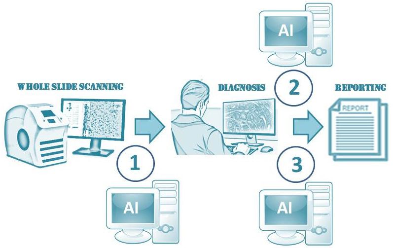
Figure 1 Proposed AI workflow in anatomical pathology. (1) AI tools that operate on scanned digital images before they are accessed by a
pathologist can perform screening and pre-diagnostic work. (2) AI tools that run when initiated by a pathologist can help perform mundane
tasks (e.g., quantification), confirm diagnoses, or determine prognosis. (3) AI applications can also be run on digital cases after they have
been signed out to perform QA (e.g., cytology-histopathology or radiology-pathology correlation). AI, artificial intelligence.
Acknowledgements 6. Stead WW. Clinical implications and challenges
of artificial intelligence and deep learning. JAMA
None.
2018;320:1107-8.
7. VanderPlas J. Python data science handbook: essential
Footnote tools for working with data. 1st ed. Sebastopol, CA:
O’Reilly, 2017:513.
Conflicts of Interest: The authors have no conflicts of interest
8. Gurcan MN, Boucheron LE, Can A, et al.
to declare.
Histopathological image analysis: a review. IEEE Rev
Biomed Eng 2009;2:147-71.
References 9. Janowczyk A, Madabhushi A. Deep learning for digital
1. Van Es SL. Digital pathology: semper ad meliora. pathology image analysis: a comprehensive tutorial with
Pathology 2019;51:1-10. selected use cases. J Pathol Inform 2016;7:29.
2. Glassy EF. Digital pathology: quo vadis? Pathology 10. Icho N. The automation trend in cytology. Lab Med
2018;50:375-6. 2000;31:4.
3. Flotte TJ, Bell DA. anatomical pathology is at a crossroads. 11. Pantanowitz L, Liu C, Huang Y, et al. Impact of altering
Pathology 2018;50:373-4. various image parameters on human epidermal growth
4. Farahani N, Parwani A, Pantanowitz L. Whole slide factor receptor 2 image analysis data quality. J Pathol
imaging in pathology: advantages, limitations, and Inform 2017;8:39.
emerging perspectives. Pathology and Laboratory 12. Newitt VN. Program zeroes in on histology digital scan
Medicine International 2015;7:23-33. connection. CAP Today. 2018:P4.
5. He L, Long LR, Antani SK, et al. Computer-assisted 13. Fraggetta F, Garozzo S, Zannoni GF, Pantanowitz L, Rossi
diagnosis in cervical histopathology. SPIE Newsroom ED. Routine Digital Pathology Workflow: The Catania
2010. Doi: 10.1117/2.1201011.003358. Experience. J Pathol Inform 2017;8:51.
© Journal of Medical Artificial Intelligence. All rights reserved. jmai.amegroups.com J Med Artif Intell 2019;2:4Journal of Medical Artificial Intelligence, 2019 Page 5 of 5
14. MSN. LUMEA Technology Overview by Dr. Matthew O. acquisition capabilities of the MYCIN system. Comput
Leavitt, Founder. Available online: https://www.msn.com/ Biomed Res 1975;8:303-20.
en-us/health/medical/lumea-technologyoverview-by-dr- 18. Tizhoosh HR, Pantanowitz L. Artificial intelligence and
matthew-o-leavitt-founder/vi-BBRUSen digital pathology: challenges and opportunities. J Pathol
15. Maddox TM, Rumsfeld JS, Payne PRO. Questions for Inform 2018;9:38.
artificial intelligence in health care. JAMA 2019;321:31-2. 19. FDA allows marketing of first whole slide imaging system
16. Miller RA, McNeil MA, Challinor SM, et al. The for digital pathology. 2017 April 12: FDA new release.
INTERNIST-1/QUICK MEDICAL REFERENCE 20. Komura D, Ishikawa S. Machine learning methods
project--status report. West J Med 1986;145:816-22. for histopathological image analysis. Comput Struct
17. Shortliffe EH, Davis R, Axline SG, et al. Computer-based Biotechnol J 2018;16:34-42.
consultations in clinical therapeutics: explanation and rule
doi: 10.21037/jmai.2019.02.03
Cite this article as: Dietz RL, Pantanowitz L. The future
of anatomic pathology: deus ex machina? J Med Artif Intell
2019;2:4.
© Journal of Medical Artificial Intelligence. All rights reserved. jmai.amegroups.com J Med Artif Intell 2019;2:4You can also read