THE LIVERPOOL OCULAR ONCOLOGY CENTRE - A guide for patients Bertil Damato - 6th Edition
←
→
Page content transcription
If your browser does not render page correctly, please read the page content below
THE
LIVERPOOL OCULAR ONCOLOGY CENTRE

          A guide for patients

            Bertil Damato

                                 6th Edition
THE LIVERPOOL OCULAR ONCOLOGY
CENTRE
A guide for patients

Bertil Damato

                       The oncology team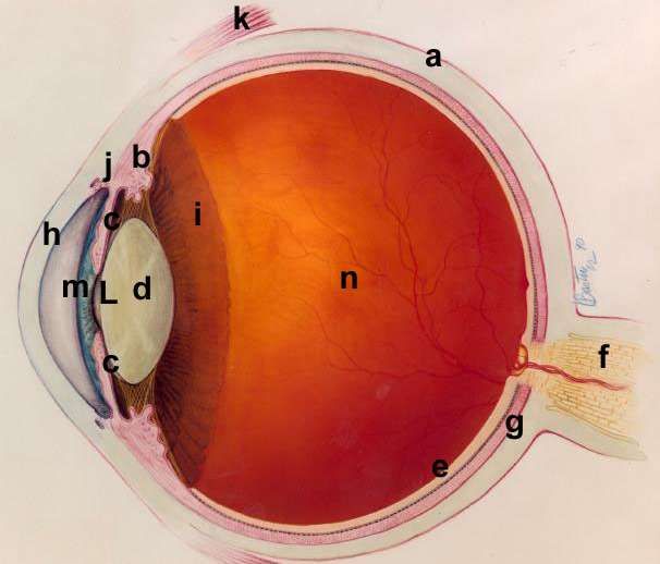
_____________________________________
Contents

Part 1: Overview
       Introduction                           2
1.     The ocular oncology centre             4

Part 2: Your care pathway
2.     The referral process                   7
3.     Your first visit                       8
4.     Ocular investigations                  11
5.     Treatment of ocular tumours            15
6.     Your hospital admission                20
7.     Follow-up                              25
8.     After your discharge from our centre   27
9.     Frequently-asked questions             28

Part 3: Behind the scenes
10.    Research                               34
11.    Audit                                  38
12.    Teaching                               40
13.    Keeping up to date                     44

Part 4: Miscellaneous topics
14.    How you can help                       46
15.    The Patient‟s Charter                  48
16.    How to make a complaint                50
17.    Who‟s who                              51
18.    Directory                              55
19.    Hospital facilities                    56
20.    How to get there                       57
21.    Liverpool and surroundings             62
22.    Useful contacts                        65
23.    Ocular tumours                         67
24.    Evidence of excellence                 71
25.    Glossary                               72
_____________________________________
Introduction
                              At the Liverpool Ocular Oncology Centre (LOOC), we
                              specialise in the diagnosis and treatment of adult ocular
                              tumours. I must emphasize that the word „tumour‟ means
                              nothing more than „lump‟. Although we provide an
                              oncology service, most patients coming to our clinic have
                              a benign tumour, such as a cyst, haemorrhage or naevus
                              (i.e. „mole‟). The tools and expertise required for diagnosis
                              and treatment of these conditions are similar to those
                              needed for more serious disease. Do not assume that you
                              have a dangerous condition just because you are referred
Professor Bertil Damato       to an oncology centre.
Consultant Ophthalmologist,
Ocular Oncologist             This guide is written for you, our patient, and your
                              relatives and doctors. The objective of this guide is to let
                              you know what to expect when you visit our centre and to
                              help you understand why we do things in a certain way.

                              Our aim is to empower you to be more active in managing
                              your own care. We would like you to have a good
                              understanding of your condition and its treatment so that
                              you can let us know your needs and concerns. We would
                              also like you to help us improve our care by giving us
                              feedback on how we‟re doing and by making suggestions.

Mr Carl Groenewald            This guide inevitably contains a certain amount of jargon.
Consultant Ophthalmologist,   A glossary is therefore printed at the end of this text.
Ocular Oncologist and
Vitreo-Retinal Surgeon        This is the sixth edition of our guide. Information that
                              might worry some patients has been removed and is
                              available separately, upon request. Hopefully, you will
                              find this guide interesting, useful, and not too stressful.
                              Please feel free to keep this guide for future reference.

                              This guide is funded by the National Specialist
                              Commissioning Team, which sponsors our service. I am
                              grateful to all involved for their assistance.

                              For further information please visit our website at
                              www.eyetumour.com
Professor Heinrich Hiemann    Professor Bertil Damato PhD FRCOphth,
Consultant Ophthalmologist,   Consultant Ophthalmologist/Ocular Oncologist
Ocular Oncologist and         Clinical Lead, Liverpool Ocular Oncology Centre
Retinal Specialist

                                                                                             2
Overview

           3
1____________________________________
The Ocular Oncology Centre
The Ocular Oncology Centre at the Royal
Liverpool University Hospital was
established by Bertil Damato in January
1993. He became interested in ocular
oncology in 1980, when he started
working at the Tennent Institute of
Ophthalmology in Glasgow under the
leadership of Professor Wallace Foulds,
whose pioneering surgery for ocular
tumours had received world-wide acclaim.
The Liverpool Ocular Oncology Centre
has developed considerably over the years
and the team now includes full-time
oncology secretaries, specialist ocular
oncology nurses, a health psychologist, a
data manager, photographers, a research
scientist in ocular tumour biology, and a
compliance officer. Close collaborative
links have been established with retinal
specialists, pathologists, cyto-geneticists,
radiotherapists, oncologists, radiologists
and also a medical ethicist to provide a
comprehensive service.
Professor Wallace S Foulds CBE and
Bertil Damato (1987) In 1997, the Ocular Oncology Centre was
designated a Supra-Regional Service by
In 1989, Professor Foulds retired and the National Specialised Commissioning
Dr Damato continued to run the oncology Group (i.e., „NSCG‟) in London. The
service. In 1993, Mr Damato moved to purpose of this organisation is to ensure
Liverpool, which is more accessible to that patients with rare conditions, such as
most patients, being located at the ocular tumours, are given the highest
geographic centre of the British Isles. possible standard of care by an
Liverpool is close to the Clatterbridge experienced specialist team.
Centre for Oncology, which is the only
unit in Britain with facilities for proton The Liverpool Ocular Oncology Centre
beam radiotherapy of ocular tumours. offers an exceptionally wide range of
treatments, which include:
The Liverpool Ocular Oncology Centre Plaque radiotherapy, placing a
grew rapidly, with the number of new saucer-shaped applicator behind
patients each year now reaching 700. the eye for 1-7 days;
4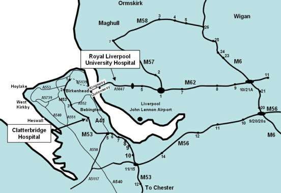
Proton beam radiotherapy, using vitreous cutter, („vacuum cleaner‟)
special equipment, located at or through a trapdoor in the eye;
Clatterbridge Centre for Oncology, Computerised tomography, for
on the Wirral Peninsula; producing x-ray images of the eye;
Trans-scleral local resection, Magnetic resonance imaging,
removing the tumour through a producing fine images of the eye;
trap-door in the wall of the eye; and
Trans-retinal local resection, Cytogenetic studies of melanoma,
„hoovering‟ the tumour through a for detecting DNA abnormalities
hole in the retina; in the tumour, which give an
Trans-pupillary thermotherapy; indication of prognosis.
sterilizing the tumour with a beam
of infra-red laser; We have instituted several protocols to
Photodynamic therapy, injecting a help patients understand their condition
light sensitizer into a vein in the and its treatment. These include:
arm and then shining low-energy Giving patients a series of guides
laser onto the back of the eye to and information leaflets;
activate the sensitizer as it passes Giving all new patients a CD-
through the tumour, ROM or tape cassette with an
Topical chemotherapy, using audio-recording of their first
eyedrops to treat tumours on the consultation;
surface of the eye, and Mailing patients a copy of the
Enucleation, removing the eye, if correspondence we send to the GP
other methods are unlikely to and ophthalmologist;
conserve the eye with useful Creating a website; and
vision. Providing a telephone helpline, run
by our Specialist Nurses.
These treatments are useful both for
benign and malignant tumours. The wide Our clinics include:
choice of treatment enables us to design New patient clinics on Mondays;
the optimal strategy for each patient, if Follow-up clinics on Thursdays
necessary combining different methods to and on Friday afternoons;
achieve the best possible results. Conjunctival tumour clinics on
Thursday afternoon, once a month
Special investigations include: Nurse Oncology Clinics on
Colour photography using a range Thursday afternoons.
of cameras, for documenting
tumour appearances; To maintain high standards we also:
Fluorescein angiography, for Conduct multidisciplinary team
investigating tumour circulation; meetings;
Ocular ultrasonography; for Prepare staff guidelines;
measuring tumour dimensions and Conduct patient satisfaction
tissue consistency; surveys and quality of life studies;
Optical coherence tomography for Measure satisfaction of referring
assessing the layers of the retina ophthalmologists;
and detecting fluid under the
Conduct research;
retina;
Encourage complaints; and
Tumour biopsy, performed either
Perform continuous outcomes
using a fine needle, or a 25-gauge
assessments (ie, Audit).
5
Your Care Pathway

                    6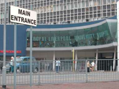
2____________________________________
The Referral Process
You have been referred because your eye If you live too far from Liverpool to travel
specialist has identified a tumour (lump) on the same day, our secretary will
in or on your eye. In many cases this will arrange accommodation for you. At
be benign but there is a chance that it may present, we pay the hotel for the room
be malignant. We aim to see all patients (single or double) so you will only need to
within two weeks of receipt of your pay for breakfast and any extras
referral from your specialist. (telephone, parking, etc). The
accommodation will either be at a hotel or
Eye specialists refer patients to us by in an apartment across the street from the
sending a letter or fax or by making a hospital.
telephone call. As soon as we receive your
referral we will send you:
details of your appointment time and
place;
this guide;
a questionnaire, to confirm that details
regarding your name, date of birth,
address and general practitioner are all
correct;
a request for permission to send a copy
of a report on your condition to your
optometrist (i.e., optician) after your
first visit to our centre. It is important Royal Chambers
to give optometrists this feedback, for
educational purposes, but your consent The hotel selected is a short taxi ride from
is needed as optometrists are not the hospital. There are, of course, several
doctors; and other hotels around Liverpool and if you
a request for permission to use your do not mind travelling a greater distance to
data and any images for research, the hospital or contributing to the cost, if
teaching and quality control studies. the hotel is more expensive, then our
secretary would be happy to find
alternative accommodation. If you travel
to our centre by car, please aim to arrive
early in case you are delayed.
The Ocular Oncology Office If you have any special requirements
please let us know by mail or phone and
Please return the completed forms to the we will do everything we can to meet your
ocular oncology secretary as soon as you needs.
can.
7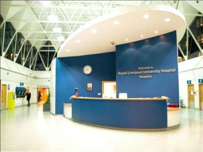
3____________________________________
Your first visit
Registration loss or theft, so please keep any valuable
When you arrive at the eye outpatient items with you.
clinic make your way to the reception
desk. You will be asked for your Examination by Surgical Trainee or
appointment card and registered on the Fellow
computer system. You also be given a After seeing the sift nurse, you will be
questionnaire about your health. seated in the waiting area again until you
are called to see the specialist registrar,
Examination by Nurse who will:
Once you have registered, you will be Introduce himself or herself;
asked to take a seat in the waiting area Confirm your name and age;
until you are called by the sifting nurse, Ask you about how and when you
who will: first became aware of your ocular
Measure your vision with the condition;
letters chart; Ask about the sequence of events
Measure the intraocular pressure leading to your referral to our
(i.e. tonometry); centre;
Test the pupillary reflexes with a Ask about your general health and
torch; previous illnesses;
Ask about your general health, as Review your health questionnaire
well as any medications and with you, if you have already
allergies; and completed it;
Instill drops into both your eyes to Examine the front of your eyes
enable examination of each fundus with a slit-lamp; and
(i.e., back of the eye) by Examine the back of your eyes
ophthalmoscopy. with an ophthalmoscope.
These drops will probably blur your vision We are interested in how your tumour was
so that you may be unable to read without detected and how your condition was
appropriate spectacles for up to four hours. managed prior to your referral to our
Remember also that you should not drive a centre. This is because we are conducting
car until you are once again able to read a a study into the detection of ocular
number plate from the legal distance. tumours in the community. We hope this
investigation will in future result in earlier
If your tumour is located on the iris, the diagnosis and treatment.
pupils will not be dilated until the
consultant has examined you. Photography
After seeing the specialist registrar, you
Outpatient Clinic will be asked to wait outside the
The nurse can store your luggage in a consulting room until you are called for
cupboard, if you wish. However, the photography. The photographer will check
hospital cannot be held responsible for any your name and age before asking you to
position yourself at the camera. Please try
8to keep your eye wide open while the perform ultrasonography, which is
photographs are being taken. useful for diagnosis and for
measuring tumour size;
With your consent, your portrait may be perform wide-angle photography
taken. This is only so that we can see this of the back of your eye, if this is
on our computer screen whenever we are likely to be useful;
speaking to you by telephone. explain to you as much as possible
about your condition; and
plan your future care with you.
If any close relatives or friends have come
with you to the hospital they are welcome
to accompany you during your
consultation.
Fundus photography in progress
Your ocular photographs will be used:
To compare tumour appearances
with any future photographs so as
to be able to recognize growth or
regression; and Counselling by consultant
For teaching in lectures at
scientific meetings and A plastic model eye or 3-D photographs
publications. will be used to help you understand the
structure of the eye.
In any publications, your anonymity will
be respected. Your permission for You are of course encouraged to ask
publication will be requested using a questions, although these are best left to
consent form. A special section of the the end of the examination.
form will need to be signed if you may be
recognised from the photographs. An audio-recording of your consultation
will be given to you on CD-ROM or tape
After the photography you will be asked to cassette to help you remember what was
sit in the waiting area until you are seen by said. Most patients seem to find this very
the consultant ocular oncologist. useful and are quite surprised by the
amount of information they missed the
Examination by Consultant first time.
We now have three consultants
specialising in ocular oncology and you Discharge from Clinic
will be under the care of one of these. The If on the basis of size and appearance your
consultant will: tumour is considered to be benign and if it
introduce himself; does not require treatment, you will be
review your referral letter and the allowed home and discharged from our
case notes; clinic.
examine your eyes;
9A letter will be written to the consultant
ophthalmologist at your home hospital
describing the clinical findings, stating the
diagnosis and advising on future care.
Copies of the letter will be sent to you,
your GP and, with your consent, your
optometrist. If there is anything you do not
understand please phone our nurse.
If you need to return to our centre, we will
give you an appointment sheet. This
should be taken to the reception desk, Multidisciplinary meeting
where a specific date for your next
appointment will be selected. Our Counselling by Nurse
receptionist will give you an appointment After your consultation you will be taken
card. to a quiet room, where a nurse will go
over what was said, answering any more
Treatment Selection questions that might come to mind. You
If you need treatment, then all the will receive an information prescription
therapeutic options will be discussed, (www.nhs.uk/ips) specifically tailored for
together with treatment schedules, your health condition providing
possible side effects, and likely outcomes. information on a several topics ranging
from cancer charities to local information
You will be helped to choose the best about cancer.
treatment for your particular condition. If
possible, a decision is made by the end of If you would like to speak to another
your visit, but you would still be able to patient who has previously received the
change your mind afterwards. same treatment as yourself then the nurse
would be able to arrange for you to speak
If you are to receive radiotherapy for an to this volunteer by telephone.
intraocular melanoma, the risks and
benefits of tumour biopsy will be A computerised kiosk in the waiting area
discussed with you. This involves is available so that you can find out more
obtaining a tiny tumour sample just before about your condition and its treatment.
or after your radiotherapy. Lab tests will
show whether or not the tumour is life-
threatening.
Your care will be discussed at a
multidisciplinary meeting immediately
after the clinic. This is attended by
doctors, nurses, and administrative staff. It
is possible that your treatment plans might
be altered following these discussions, in
which case we will speak to you about any
revisions without delay.
Kiosk in waiting area with information
If you need more time to reach a decision, for patients and relatives
this is quite possible, of course.
104____________________________________
Ocular investigations
This chapter describes the various Most tumours can be diagnosed by their
examinations and tests that are in common appearance on ophthalmoscopy or slit-
use at our clinic. You may find it useful to lamp examination. It may be necessary to
refer to the diagram of the eye in the monitor a lesion over several months or
Glossary. years to detect growth, thereby confirming
the diagnosis.
Slit-lamp examination
The slit-lamp allows a highly-magnified Difficulties can arise if the tumour is not
view of the eye, with well-controlled visible because of haemorrhage or
illumination providing a clear view of the cataract. These can be overcome by
tumour. The source of light can either be treating the cataract, waiting for the
diffuse or slit-like (hence the name of the haemorrhage to clear spontaneously, or
instrument). It is possible to adjust the perhaps removing the haemorrhage
length of the slit, which can therefore be surgically.
used to measure the size of a tumour.
Colour photography
Ophthalmoscopy Colour photography is useful for
The back of the eye is examined with a documenting the appearances of the
variety of ophthalmoscopes, which give a tumour so that any change over time is
stereoscopic and panoramic view of the readily detected. Standard cameras can
tumour and its surroundings. only image tumours extending far back in
the eye, near the fovea and optic disc. We
now have wide-angle cameras, which can
photograph tumours that are beyond the
reach of standard cameras. The Panoret
camera uses a contact lens whereas the
Optos is a non-contact device. Both
cameras are sponsored entirely with
donations and legacies from our patients.
Binocular indirect ophthalmoscopy
Wide-angle photography in progress
11The injected fluorescein dye tends to
cause yellowing of the skin and urine for a
few hours and about one in ten patients
experience transient nausea, although
vomiting is rare. About one in 2000
patients develops an allergic reaction,
which very rarely is fatal (i.e., in about
one in two hundred thousand patients).
Optical Coherence Tomography
This camera optically produces an image
Optos photograph showing a panoramic showing a „slice‟ of the retina, with the
view of the back of the right eye. (This various layers having different colours. It
shows a choroidal melanoma after is useful for detecting abnormal fluid at
successful plaque radiotherapy.) the back of the eye.
Contrast the wide view with that of the
photograph below, taken with a
conventional camera.
Angiography
Angiography is performed by injecting a
dye into a vein in the arm and then taking
a series of photographs of the back of the
eye. OCT Scan of macula
The dye is fluorescent; that is, it has the
property of changing light from one colour
to another. The photographs are taken
using a flash and a filter of the appropriate
colours. The dye can therefore be seen
shining brightly as it passes through the
arteries and veins and as it leaks through
any abnormal areas. There are two kinds
of angiography: fluorescein angiography OCT scan showing cystoid macular
and indocyanine green angiography, oedema
which use blue and red light respectively.
Ultrasonography
With ultrasonography, high-frequency,
inaudible sound waves are emitted into the
eye. These waves bounce off any tissue
surface back towards the probe, which
measures the „loudness‟ of the reflected
sound and the time taken for the sound to
travel into the eye and back again. The
intensity of the reflected signal gives an
idea of the „hardness‟ of the reflecting
tissue. The time taken for the reflected
Fluorescein angiogram signal to be received gives an indication of
the distance travelled by the sound.
12A-scan ultrasonography produces a linear Ultrasound probe in position
signal, with a series of waves, which
reveal the consistency of the tumour. With The front of the eye is assessed with a
B-scan ultrasonography, the beam sweeps special high-frequency probe, which
the eye from side to side, producing a requires the use of a small eye-bath or a
visual slice of the eye and a good idea of sheath filled a clear jelly-like fluid.
the size and shape of any tumour in the
eye. Magnetic Resonance Imaging
MRI is performed by emitting pulses of
magnetism through the body so that all the
atoms spin in the same direction thereby
giving rise to electrical fields, which are
measured and converted into images. This
method produces very clear pictures of the
eye, with different tissues showing
different degrees of brightness.
Computerized tomography
CT scans are obtained by passing very fine
x-rays through the head from different
Ultrasonogram, with tumour at back of directions and then reconstructing the
eye, next to nerve results to create an image „slice‟ of the
eye.
Ultrasonography has several applications
in assessing an eye with a tumour: MRI and CT scans do not usually provide
If the media are opaque, it enables more information than ultrasonography,
the tumour to be detected. which is more convenient and less
Ultrasonography can also reveal expensive.
tumour extension outside the eye.
By demonstrating a mushroom Biopsy
shape, ultrasonography can help Biopsy is useful for:
establish the diagnosis. Making the diagnosis, when this
With calipers, it is possible to remains uncertain after clinical
measure tumour thickness and examination and ultrasonography;
basal diameter. These Confirming a suspected diagnosis
measurements are useful when of intraocular metastasis (i.e.,
selecting treatment and measuring tumour travelling to the eye from
how a tumour is growing or elsewhere), also indicating the
regressing over time. most likely site of origin, if this is
not known; and
Determining whether an
intraocular melanoma has the
capacity to metastasize to the liver
and other parts of the body.
Trans-vitreal biopsy
This is performed by passing either a fine
25-gauge needle or a 25-gauge vitreous
cutter (like a vacuum cleaner) through the
eye into the middle of the tumour and
13taking small samples for analysis. The diagnosis and a cure. It is mostly
vitreous cutter gives a better yield so that performed if local resection would be the
it is more reliable. treatment of choice in any case. In
exceptional situations, if the eye is blind
and painful the most practical solution is
to remove the eye and to establish the
diagnosis by pathological examination.
Biopsy of intraocular tumours is
associated with a number of risks, which
include:
An inconclusive result, either
because the sample taken was too
small or because technical
problems in the laboratory. An
inadequate sample is more likely
with very small tumours.
Seeding of intraocular tumour onto
the surface of the eye. This is very
rare.
Choroidal tumour biopsy with 25-gauge
vitreous cutter Intraocular haemorrhage, which is
common but mild, usually
Trans-scleral fine needle aspiration biopsy resolving spontaneously over a few
A fine, sharp needle is passed through the weeks.
wall over the eye directly into the tumour Other rare ocular complications,
to obtain a tiny sample. such as retinal detachment and
intraocular infection.
Incisional biopsy
Incisional biopsy is performed by The benefits of biopsy include:
removing a small sample with scissors or a Making an immediate diagnosis so
scalpel. With intraocular tumours, this is a that treatment can be given without
more difficult procedure than trans-ocular delay, also avoiding unnecessary
biopsy and is usually performed under scans.
general anaesthesia, with moderate Providing reassurance when an
lowering of the blood pressure. With intraocular melanoma is most
conjunctival tumours, this is a small unlikely to affect general health
operation, performed under local Enabling special investigations to
anaesthesia on an outpatient basis. be performed and other care to be
provided when an intraocular
Excisional biopsy melanoma is life-threatening.
Excisional biopsy involves total removal
of the tumour, thereby providing both a
145____________________________________
Treatment of ocular tumours
This chapter gives an overview of the wall of the eye directly over the
methods available for treating ocular tumour and held in place with
tumours, benign and malignant. More sutures. If possible, a biopsy is
detailed information will be given to you performed immediately before the
once the most appropriate form of radioactive plaque is sutured to the
treatment for your particular tumour has eye.
been selected. A second, 25-minute operation
under general or local
Plaque radiotherapy anaeasthesia. The plaque is
This treatment is indicated for selected removed between one and seven
choroidal melanomas, some malignant days later, once the appropriate
conjunctival tumours and some tumours dose of radiation has been
travelling to the eye from elsewhere (i.e. delivered.
metastases). It enables a high dose of
radiation to be focused onto a small area We can select between ruthenium plaques,
and has the advantage of being completed which are suitable for tumours up to
in a few days. approximately 5 mm thick, and iodine
plaques, which can treat tumours up to 9
mm thick (albeit giving a higher dose of
radiation to normal ocular structures).
Ruthenium plaques are available within a
day, whereas iodine plaques need to be
constructed for each patient and this takes
up to six weeks.
This radiation does not travel beyond the
eye so there is no risk of hair loss or other
general problems. There is no radiation
once the plaque is removed.
We have developed techniques for
Ruthenium plaque sutured to the eye positioning a ruthenium plaque
wall directly over the tumour eccentrically in relation to the tumour so
that we can increase the dose of radiation
The radiotherapy is administered by to the tumour without a corresponding
means of a saucer-shaped plaque, which increase in the radiation delivered to optic
has an inner, concave radioactive surface nerve and fovea.
and an outer, convex protective shield.
The treatment involves:
A 45-minute operation under
general anaesthesia, during which
the plaque is placed against the
15 An examination at your local eye
hospital about four weeks after the
radiotherapy.
The radiotherapy at CCO is painless.
Good result after plaque radiotherapy,
with conservation of 6/6 vision ten years
after treatment. The black area is dead
tumour (i.e., „pile of soot‟) and the white
area corresponds to the plaque‟s Plan for proton beam radiotherapy
location, which was eccentric in relation
to the tumour. Stereotactic radiotherapy
This treatment involves directing radiation
Proton beam radiotherapy at the tumour from several directions so as
This treatment is selected when the to maximise the dose of radiation within
tumour is not suitable for the more the tumour while minimising the radiation
convenient plaque radiotherapy, that is, delivered to surrounding healthy tissues.
small intraocular melanomas near the This treatment is not performed at our
optic nerve, some choroidal hospital because we prefer other methods.
haemangiomas, iris melanomas, and some
large intraocular melanomas. Trans-scleral local resection
Trans-scleral local resection involves
The treatment involves: removing the tumour through a large
A 45-minute operation under trapdoor in the wall of the eye. To prevent
general anaesthesia, to suture four bleeding, the intraocular pressure is
tiny tantalum markers to the wall lowered, using hypotensive anaesthesia,
of the eye, at known distances monitoring the brain and heart using
from the tumour margins; special equipment. Each trapdoor
Treatment planning for half a day operation takes about two or three hours.
at Clatterbridge Centre for This is difficult surgery and mostly
Oncology (CCO), a few weeks reserved for large tumours that tend to
after the marker insertion; become toxic to the eye after radiotherapy.
If local resection is performed in the first
A few weeks later, a course of
instance, a radioactive plaque is usually
radiotherapy at CCO, with one 60-
placed over the treated area to reduce the
minute session on five consecutive
chances of tumour recurrence, and this is
days;
done either at the end of the operation or a
Tumour biopsy after completion of
month later.
the radiotherapy; and
16.
Right eye showing choroidal melanoma
before local resection (a)
Endoresection involves (a) tumour
removal, (b) temporarily filling the eye
with air, (c) laser treatment, and
(d) filling the eye with silicone
Conjunctival excision
Discrete tumour nodules on the surface of
Post-operative view showing (a) inner the eyeball can be removed surgically, if
surface of white sclera, (b) retinal vein, not too extensive. This can be done under
and (c) small residual haemorrhage. local or general anaesthesia.
Trans-retinal endoresection Transpupillary thermotherapy
The tumour is cut into fragments, which Laser treatment involves heating the
are sucked up a fine, metal tube that is tumour for about one minute, using an
passed through a hole in the retina. Laser infrared laser beam. The treatment lasts
treatment is administered during the about half an hour and is delivered under
operation to „weld‟ the retina in place. The local anaesthesia on an outpatient basis.
eye is filled with silicone oil to hold the
retina in place until scarring has welded This treatment is suitable for small
the retina in position. This operation is tumours, when there is uncertainty as to
usually performed to remove moderately whether the lesion is a benign mole or a
sized tumours after radiotherapy if they malignant melanoma. It is also useful for
become toxic to the eye. In rare cases melanomas that are leaking excessive
when we perform endoresection as the amounts of fluid after previous
first procedure, we administer laser radiotherapy.
treatment and perhaps radiotherapy to
prevent tumour recurrence. Cryotherapy
Very thin tumours on the surface of the
The treatment involves: eyeball can be given „freezing treatment‟,
a two-hour operation under general using either a spray of liquid nitrogen or a
anaesthesia, and special pencil-like device. This treatment
after approximately 12 weeks, a can be administered under local or general
30-minute operation under local anaesthesia.
anaesthesia to remove the silicone
oil, often with cataract surgery at
the same time.
17External beam radiotherapy angiogenic factors (e.g. Avastin), which
External beam radiotherapy is indicated shrink blood vessels. Avastin was
for some tumours travelling to the eye developed for intravenous administration
from another part of the body (i.e., for treatment of cancer spreading to the
metastases). These tumours respond to liver from the large bowel. It is not
doses of radiation that are usually low licensed for intraocular administration but
enough to be well tolerated by the whole has nevertheless been found to be safe and
eye. The equipment required for this effective for conditions such as diabetic
treatment is available at most hospitals. To retinopathy and age-related macular
reduce complications, the treatment is degeneration. We have had some very
given in small doses over days or weeks. good results in patients with irradiated
intraocular melanoma and with a variety
Photodynamic therapy of other tumours. Ocular complications
With this treatment, a dye called such as haemorrhage and infection are
Verteporfin is injected into a vein in the rare. Care has to be taken with patients
arm, so that it circulates around the body having high blood pressure, angina and
and through the tumour. After a few kidney disease. The risks and benefits are
minutes, an infra-red laser is directed at discussed in detail and are listed in an
the tumour for about 83 seconds and this information sheet given to all patients
activates the dye so that it kills the tumour. undergoing this treatment.
This treatment is painless. It is necessary
to stay out of bright sunlight for about two Enucleation
days afterwards. Removal of the eye is indicated when the
chances of conserving a useful eye are not
Photodynamic therapy is very effective for good enough to justify the risks involved.
choroidal haemangiomas and other
tumours arising from blood vessels. There The operation is usually performed under
is some evidence that this treatment may general anaesthesia. A long-acting
also be useful for some other tumours, anaesthetic injection is given to minimize
such as melanoma and vasoproliferative pain during the initial post-operative
tumour. period. The enucleated eye is replaced by
a ball implant. The eye muscles are
Topical chemotherapy sutured to this implant so that the artificial
If too extensive for surgical removal, very eye will move with the fellow eye. At the
thin conjunctival tumours on the surface end of the operation a transparent
of the eye can be treated with special „conformer‟, similar to a rigid contact
drops („weedkiller‟), consisting of lens, is placed in the socket. About ten
Mitomycin C or 5-FU. These drops are weeks after surgery, this is replaced by a
administered on an outpatient basis, with „tailor-made‟ permanent artificial eye, at
each course lasting between four and the patient‟s referring hospital. The
seven days. It is usually necessary to artificial eye is like a coloured contact
repeat the course of treatment every two or lens, painted to match the fellow eye. This
four weeks, about three or four times. usually gives a good cosmetic result.
Intraocular injections To avoid any risk of Creuzfeld Jacob
Intraocular injections, which are quite Disease (CJD) (i.e., a human form of
painless, are given under local anaesthesic. „mad-cow disease‟) the operation is
These include steroids for inflammation, performed with disposable instruments
chemotherapeutic agents such as and tissue transplants are not used.
methotrexate for lymphoma, and anti-
18A district nurse will visit you daily to help before proton beam radiotherapy to
you with the conformer. If any problems agree therapeutic strategy;
arise, you can phone the Specialist Ocular after biopsy, local resection or
Oncology Nurse (i.e. Key Worker) at any enucleation, to discuss findings of
time (0151 706 3976). microscopic tissue examination (i.e.,
pathology);
Exenteration after any adverse radiotherapy events,
In very rare instances it is necessary to to determine whether complications
remove not only the eye but also the might be avoided;
surrounding tissues and the eyelids. A once a year, with all members of the
special cosmetic prosthesis is made after team present, to announce new
the operation. developments and review policy.
Multidisciplinary Meetings Operational meetings are also held
Meetings with staff from different regularly to discuss organizational matters,
disciplines (e.g. ophthalmologists, nurses, equipment, and working protocols.
pathologists, radiotherapists, oncologists,
etc) are held:
at the end of each new patient clinic,
to discuss diagnosis and treatment
plans;
196____________________________________
Your hospital admission
Treatment schedule anaesthesia. Scans for tumour in other
We try to perform treatment the day after parts of the body are performed selectively
your arrival at our centre. This is to reduce (e.g., if an intraocular melanoma is large).
the amount of travel between your home
and the hospital, if you live far away from You will be asked to sign a consent form,
Liverpool. We also assume that you would which specifies the nature of the operation
like to get your treatment over and done and the eye to be treated. If the operation
with as quickly as possible so that you do involves removal of the tumour or the eye,
not worry with anticipation and so that then you may be asked for permission to
you can quickly return to normal life. If use a small tumour specimen and blood
you do not wish to start treatment samples for research purposes. You are of
immediately, we can easily postpone your course under no obligation to participate
operation. in any studies.
If we receive more patients than we can Please do not leave the clinic until you
treat on a single day we need to prioritise have had all investigations and completed
patients according to the size of the the necessary forms.
tumour and the distance that they would
need to travel, should surgery be delayed. Admission to Ward
Tumours rarely change much from week You will be admitted to our inpatient ward
to week so a delay of a few days or weeks (9Y) or the daycase unit on the ground
should not worsen the outcome. floor. This will happen immediately after
your clinic visit or on the day of your
If your operation is scheduled for the operation. A nurse will welcome you and
afternoon as a day case, we will still show you to your bed. You will be asked
require you to arrive at our unit in the more questions about your general health,
morning and this is in case there are any medications, allergies, and other matters.
cancellations or changes to the theatre list.
A named ophthalmic nurse will be
Pre-operative Investigations allocated to your care, to look after your
If you are to be admitted to hospital, you individual needs. All nurses and doctors
will first have a number of investigations. wear name badges at all times.
These include:
Haematological studies (ie, “full The ward is divided into several rooms,
blood count”), to exclude anaemia; with each multi-bed room housing patients
Serum biochemistry (ie, “U‟s and of the same gender. There are a few single
E‟s and LFT‟s”), to check liver and rooms for patients requiring isolation.
kidney function; and
Electrocardiography (ie, “ECG”), You will be visited by our specialist ocular
to check your heart. oncology nurse. She will see how you are
settling into the hospital and will answer
These tests are performed mainly to any questions you may have.
ensure that you are fit for general
20The Surgical Trainee (ST) will perform a If you are to have a general anaesthetic,
full clinical examination and will prescribe you will need to fast for at least six hours
any of your usual medications that you beforehand, that is, from midnight if your
need to continue taking during your stay in operation is scheduled in the morning and
hospital. from about 7 am if you are due to have
your operation in the afternoon. While
This is another opportunity for you to ask fasting, you must have nothing at all to eat
any more questions that come to mind. At or drink.
the appropriate time, you will be shown
how to instill eye drops. If the tumour is at the back of the eye, we
will dilate the pupil of the eye to be
As soon as possible, a doctor or nurse will operated upon.
obtain the results of all investigations,
ensuring that these are adequately The anaesthetic room
documented in your casesheet. When it is time for your operation, you
will be moved to the anaesthetic room and
The anaesthetist will visit you before your transferred from your bed to the operating
operation. The aims of this visit are to table.
ensure that you are fit for anaesthesia and
to explain to you the details of your The operating department assistant (ODA)
anaesthetic. The results of your ECG and will attach electrical leads to your chest
any other relevant investigations will be and arms to monitor your heart. If local
reviewed. You will be asked about any resection is to be performed, additional
previous anaesthetics you have received. leads will be attached to your head for
Please feel free to ask any questions. brain monitoring. All leads are attached to
the skin with adhesive tape.
Once you have seen the anaesthetist you
can leave the hospital for a few hours, if The anaesthetist will put you to sleep by
you wish. Before you do so, please check giving you an injection on the back of
whether you are to be seen by the your hand.
anaesthetist as your operation may need to
be postponed if you miss this assessment. Once you are anaesthetised, the
Also be sure to inform the nursing staff of anaesthetist will maintain your airway by
your plans and to agree upon a time when placing either a laryngeal tube or mask at
you should return. Please also give them the back of your throat. This is to ensure
your mobile number if you have one. that you have no difficulty breathing
during the operation.
Visiting times are 2.00-4.00 pm and 6.00-
8.00 pm, although special arrangements If the eye is to be removed, a mixture of
can be made at the discretion of the nurse local anaesthetic with adrenaline solution
in charge. A limit of two visitors per is injected behind the eye to reduce
bedside should be observed. Children are bleeding and post-operative pain.
allowed only if visiting a close family
member and are the responsibility of the The operating theatre
accompanying adult. The electrical leads and a breathing tube
will be linked up to the anaesthetic
Pre-operative Ward Round machine so that your pulse, blood
On the day of your operation, you will be pressure, oxygen level and your heart will
visited by an ophthalmologist to check any be monitored continuously.
details and to ensure that all is well.
21If you are to receive hypotensive
anaesthesia, which is necessary for local
resection of the tumour (ie, „trapdoor
operation‟), then your brain activity will
be monitored continuously. All these
precautions ensure that there is a healthy
exchange of fresh air through your lungs
and a good circulation of blood throughout Surgery in progress
your body while you are under A scrub nurse, also wearing sterile gloves
anaesthesia. and gown, looks after the instrument
trolley, passing instruments as necessary
The anaesthetist will stay with you to the surgeon and the assistant.
throughout the operation.
Your identity will be confirmed by a
nurse, by checking your wrist band and
the signed consent form, which will also
be used to check that the correct type of
operation is to be performed.
If you are to have a local resection, the
lashes will be trimmed (These re-grow
back to normal in six weeks).
The skin around the eye to be operated is Scrub nurses at work
cleaned with an antiseptic solution called
Betadine, which contains iodine (so please A „runner‟ is also present in the theatre in
be sure to let us know if you are allergic to case any items need to be transferred to
iodine). and from the instrument trolley, to collect
any tumour specimens and to ensure that
The eye not to be operated on is taped everything runs smoothly.
shut, so that it is well protected. A sterile
drape is placed over your head and upper The ODA is at hand to help with the
chest. This has a small window, which is operating microscope, operating lights,
sealed with an adhesive, transparent film. video recorder and any other equipment.
This film is cut with scissors to expose the
eye, which is held open with a speculum. There may occasionally be visitors, such
as ophthalmologists from other hospitals
At this point (and not before), the eye to or medical students.
be operated upon is examined by the
surgeon by ophthalmoscopy so that the A video of the operation may be taken for
tumour is located. This makes it absolutely teaching purposes, with your consent. This
impossible to operate on the wrong eye. will not be shown without your consent if
there is any chance that you might be
During the operation, the surgeon is recognised.
assisted either by another ophthalmologist
or by the specialist ocular oncology nurse. The Recovery Room
At the end of your operation, you will be
transferred back to your own bed and
taken to the recovery room, where you
22will be monitored by your own nurse (i.e., Placing you in a single room;
one nurse per patient). The anaesthetist Displaying a hazard notice on the
will also be present in the operating suite door of your room;
until you regain consciousness. Covering your eye with two
shields, to block any stray
The Ward radiation from escaping.
Once you have recovered from the
anaesthetic and are comfortable, two Do not be alarmed by all these
porters and a nurse will take you to your precautions. The amount of radiation is
ward. quite minimal.
In the ward, a nurse will look after you You do not need to wear a cover over your
and will give you any medications that eye if there is no one else in the room.
you require for the relief of pain or nausea.
We recognise that the period following
After some types of surgery (e.g., diagnosis and treatment can be difficult. It
endoresection and local resection), it may is quite normal for people to feel a range
be necessary for you to remain postured of emotions at this time. A health
for the first day, for example, lying on psychologist or specialist nurse will
your left side with your left cheek against therefore visit you on the ward. This is to
the pillow. This is so that any retinal discuss any worries so that these can be
haemorrhage will gravitate away from the addressed without delay. If the need for
fovea and not damage your vision. more support is identified, arrangements
will be made for you to be telephoned
The Post-operative Ward Round after you leave the hospital or to receive
Once a day, and more often if necessary, further care close to home.
an ophthalmologist will examine your eye.
This is mostly to ensure that there is no Discharge from Hospital
infection and to check that the intraocular You can expect to return home one or two
pressure is normal. days after your operation.
Routinely, all patients are examined first When the time comes for you to be
by a surgical trainee between 8.15 am and discharged from hospital, you will be
8.45 am, and then by the consultant given a supply of any drops and oral
between 8.45 am and 9.00 am. Please be medications that are required. These will
sure to stay by your bed between these need to be obtained from the hospital
times. pharmacy. We order these medications a
few days in advance so that you will not
Drops will be given to you to keep your be kept waiting when it is time for you to
pupil dilated. This is to prevent discomfort return home.
and to enable the back of your eye to be
examined. Antibiotic and steroid drops You will also be given a note to take to
will also be given to prevent infection and your general practitioner, an information
inflammation. leaflet and a satisfaction questionnaire (to
be completed anonymously). Please return
If you have a radioactive plaque in place, the questionnaire, even if there is anything
special precautions are necessary to you are not satisfied with.
prevent persons around you from being
exposed to unnecessary radiation. These Please ask the ward nurse for a „Sick
include: Note‟ if you need one. If an urgent
23appointment is needed at your hospital, terms with things, taking advantage of
this will be arranged and you will be whatever help is available from family,
informed of the date and time of the friends and various care workers.
appointment before your discharge from
the ward. You can wash your hair and take a shower
or bath at any time as long as you are
The consultant will send your careful to avoid water splashing into your
ophthalmologist a discharge letter with all eye, at least for the first four or five days.
relevant information about your condition. It should be safe to go for a walk and to
Any follow-up arrangements will be resume mild exercise after one or two
mentioned in the letter so that your days, but more strenuous activities such as
hospital can send you an appointment if running may need to be postponed for one
necessary. Copies of this letter will be sent or two weeks, depending on the operation.
to your general practitioner and to you.
With your consent, a copy of the letter is Your operated eye will probably be quite
also sent to your optometrist. red and tender for the first week or two,
but should settle down, especially if you
As soon as the results of any pathology remember to take your medications as
studies are received, these are sent to your instructed. Until healing occurs, your eye
ophthalmologist and general practitioner will be particularly sensitive to irritants
together with a covering letter explaining such as soapy water, chlorinated water in
the significance of the results and swimming pools, and smoke.
suggesting further management as
appropriate. We do not send patients Within two weeks of your return home,
copies of these reports, which we feel you will receive a telephone call from our
should be discussed in person. You will specialist ocular oncology nurse in case
receive a letter with appropriate you have any problems. If you have any
instructions. We usually need to wait a questions, however, you can ring her up at
few weeks for the laboratory results to any time (Tel: 0151 706 3976). Similarly,
become available to us. if you have any worries or concerns about
how you are coping, our specialist nurse
Convalescence at Home can get you in touch with our health
When you return home, you may feel psychologist.
surprisingly tired and you may even feel a
little „low‟ for a few days. These feelings If the specialist ocular oncology nurse or
are quite normal, bearing in mind the the health psychologist is not available
stress of the previous few weeks, the when you phone, please leave a message
inactivity in hospital, and having to cope on the answer-phone and you will receive
with one or two general anaesthetics. a reply as soon as possible
Your convalescence is a time for peace,
good food, gentle exercise and coming to
247____________________________________
Follow up
Follow-up at home hospital Initially, arrangements are made for these
You should be examined by an examinations to alternate between your
ophthalmologist about one week after local hospital and our centre. This system
local resection or endoresection and within of alternating examinations is designed to:
a month of any other procedures (i.e., ensure that the ophthalmologists at
plaque radiotherapy, enucleation). your hospital keep in touch with
your progress and become familiar
Unless you live very close to Liverpool, with your condition;
this examination will be performed at your make it easier for the
own hospital. ophthalmologists at your hospital
to take over full responsibility for
Some general ophthalmologists are your care when the time comes for
anxious about having to look after a you to stop attending our centre;
patient who has undergone treatment for a reduce the need for you to travel
tumour, because, quite understandably, long distances between your home
they may not be very familiar with the and our hospital, if you live far
surgical techniques involved. from Liverpool; and
Nevertheless, all your ophthalmologist reduce the number of patients
needs to do at this early stage is to ensure attending our follow-up clinics,
that there is no infection, no raised making it possible for a consultant
intraocular pressure and, indeed, no sign to see every patient at every clinic
of any other complication that would be visit.
common to any eye operation.
Follow-up at the Oncology Centre
After radiotherapy or phototherapy, the Our follow-up clinics are held on
tumour does not usually begin to regress Thursdays (morning and afternoon) and on
for several weeks or months. This is why Friday afternoons. We also have a Nurse-
follow-up assessments at our centre are run clinic for patients who do not need to
delayed for six or nine months after such be examined by an ophthalmologist. The
treatments. nurses are fully trained and can obtain
immediate advice from a doctor if the
If it is likely that treatment is required need arises.
before this time, for example, after local
resection or endoresection, then The procedure at our follow-up clinic is
arrangements will be made for you to similar to that of your first consultation.
attend our centre as necessary.
You will be registered at the reception
Ocular examination is recommended desk. Next, the sift nurse will check your
every six months for the first six years vision and, if necessary, dilate your pupils.
then once a year for several years and
eventually once every 18 months to two A surgical trainee or fellow will first ask
years for the rest of your life. you a number of questions, using a
25structured questionnaire. These questions The consultant will review the case notes
are designed to cover all possible with you, discuss any symptoms or
symptoms and to get some idea of how concerns you might have, and examine
worried you might be about tumour your eye, if necessary also performing
recurrence and spread. These questions are ultrasonography. If appropriate, he will
asked so that we can alleviate symptoms compare the clinical findings with
and discuss any of your worries. This is so previous photographs. Next, he will
that appropriate reassurance can be given discuss your results with you, answer any
if such fears are groundless (as they often questions, dictate a letter to your
are) or so that advice can be given if there ophthalmologist with a copy to your
is genuine cause for concern. general practitioner and give you an
appointment sheet to hand to the clerk at
You will next have your photographs the reception desk.
taken. We will ask you to sign a consent
form for these images for teaching and The specialist nurse can speak to you in a
research. quiet room if there are any matters you
wish to discuss with her. You may also
After your photography, you will be asked ask to meet our health psychologist.
to wait until a consultant sees you.
Unfortunately, this can be quite a long You, your GP and any other practitioner
wait because the consultants routinely see involved in your care will receive a copy
all patients at every clinic, which means of your report, but it is not our current
that the morning clinic rarely finishes policy to send copies of letters from
before 1.30 to 2.00 pm. We have tried to follow-up clinics to optometrists.
reduce waiting times by spreading
appointment times more evenly across the Discharge from our Centre
clinic session but this tended to result in You will be discharged from our centre
slack periods in the early part of the clinic when we feel it is safe for you to stop
and a rush to catch up later on. It only attending.
needs one or two patients to arrive late,
because of a train delay or congestion on
the motorways, to disrupt the whole clinic.
268____________________________________
After your discharge from our centre
We would like to continue to be involved selective intrahepatic radiotherapy
in your care even after you have stopped using Yttrium beads.
coming to our follow-up clinics.
Please ask us for information about
Screening for metastatic disease screening for metastasis and treatment of
If you have been treated for an intraocular metastatic disease from uveal melanoma if
melanoma you will be informed of your these ever become relevant to your care.
prognosis in an appropriate manner,
depending on how good or bad the news We hope it will one day be possible to
is. Your GP and ophthalmologist will also administer adjuvant therapy that can delay
be informed of the results. or prevent the onset of symptomatic
metastatic disease.
Depending on your prognosis, plans will
be made for your further care, which will How we are informed of your progress
be discussed with you. These may involve We ask your general ophthalmologist to
six- or twelve-monthly blood tests and send us a copy of any letter to your GP.
liver scans. There are different types of All relevant information is computerised
scan, which include: in our database. This information is used
ultrasound scans; to perform long-term studies, which are so
magnetic resonanance imaging important in evaluating our care.
(MRI), which can be performed
with or without contrast and How you can get in touch with us
diffusion weighting; and We would all like patients to feel that they
positron-emission tomography can get in touch no matter how many
(PET), mostly after liver tumours years have elapsed since treatment.
are found, to look for tumours
elsewhere. We routinely send a quality-of-life
questionnaire to all patients on the
Compared to US, MRI is more sensitive, anniversary of their first ocular treatment.
especially with contrast, but also more This helps us evaluate our care, from the
expensive. patient‟s point of view, so that future
patients can be given appropriate advice
Treatment of metastatic disease from when selecting treatment. Please make a
uveal melanoma special effort to complete and return this
A variety of treatments are available for questionnaire, even if there seem to be an
metastatic disease from uveal melanoma awful lot of difficult and strange
and these include: questions.
systemic chemotherapy;
intra-hepatic chemotherapy, How we keep you informed of progress
delivered into the liver; We have set up a website on the internet
surgical removal of liver tumours; (www.eyetumour.com). This contains a
radiofrequency ablation; copy of this guide and other information.
There are also links to sites you might find
ipilimumab immunotherapy;
useful.
27You can also read