Transgenic mice Cre-dependently expressing mutant polymerase-gamma: novel test-system for pharmacological study of mitoprotective drugs
←
→
Page content transcription
If your browser does not render page correctly, please read the page content below
Research Results in Pharmacology 7(3): 33–39
                                                                                                                                  UDC 577.212.2
                                                                                                             DOI 10.3897/rrpharmacology.7.72784

                                                                 Research Article

Transgenic mice Cre-dependently expressing
mutant polymerase-gamma: novel test-system
for pharmacological study of mitoprotective drugs
Marina V. Kubekina1,2, Yulia Yu. Silaeva2, Alexandra V. Bruter1,2, Diana S. Korshunova1,
Leonid A. Ilchuk1,2, Yulia D. Okulova1,2, Mariya O. Soldatova3, Evgeniya Seryogina4,
Inga M. Kolesnik5, Polina A. Ukolova4, Mikhail V. Korokin5, Alexey V. Deykin1
1 Center for Precision Genome Editing and Genetic Technologies for Biomedicine, Institute of Gene Biology, Russian Academy of Sciences, 34/5
  Vavilova St., Moscow 119334, Russia
2 Core Facility Centre, Institute of Gene Biology (IGB), Russian Academy of Sciences, 34/5 Vavilova St., Moscow 119334, Russia
3 Department of Biology, Medical Genetics and Ecology, Kursk State Medical University, 3 Karl Marx St., Kursk 305041, Russia
4 Cell Physiology and Pathology Laboratory, Orel State University, 95, Komsomolskaya, St., Orel 302026, Russia
5 Department of Pharmacology, Belgorod State National Research University, 85 Pobedy St., Belgorod 308015, Russia

Corresponding author: Marina V. Kubekina (kubekina@genebiology.ru)

Academic editor: Oleg Gudyrev ♦ Received 2 June 2021  ♦  Accepted 9 August 2021  ♦  Published 17 September 2021

Citation: Kubekina MV, Silaeva YuYu, Bruter AV, Korshunova DS, Ilchuk LA, Okulova YuD, Soldatova MO, Seryogina E, Kolesnik
IM, Ukolova PA, Korokin MV, Deykin AV (2021) Transgenic mice Cre-dependently expressing mutant polymerase-gamma:
novel test-system for pharmacological study of mitoprotective drugs. Research Results in Pharmacology 7(3): 33–39. https://doi.
org/10.3897/rrpharmacology.7.72784

       Abstract
       Introduction: PolG-alpha is a nuclear-encoded enzyme which provides replication and repair of mitochondrial DNA.
       D257A mutation of PolG-alpha leads to change in the N-terminal ”proofreading” domain, which deprives the enzyme
       of 3′-5′ exonuclease activity, resulting in accumulation of mutations in the mitochondrial genome.
       Materials and methods: Murine zygotes were microinjected with transgene construction carrying mutant murine Polg
       coding sequence and GFP coding sequence by a loxP-flanked STOP-cassette. Two Cre-activator strains, CMV-Cre
       (systemic activation) and Tie2-Cre (endothelial activation), were used for activation of the transgene. To confirm the
       insertion and Cre-dependent activation of the transgene, genotyping and qPCR copy number measurement of mutant
       Polg were performed, and GFP fluorescence was assessed.
       Results: Two primary transgenic animals were used as the founders for two lines with copy numbers of transgene ~7
       and ~5. After systemic activation, the number of the transgene copies decreases to ~1.0 while endothelial specific acti-
       vation does not affect the number of transgene copies in tail tissue.
       Discussion: A murine model with spatial control of mutant Polg expression has been developed. To our knowledge, this
       is the first transgenic model of tissue-specific mitochondrial dysfunction.
       Conclusion: Transgenic mice Cre-dependent expressing mutant polymerase-gamma are a novel test-system for study-
       ing mitochondrial biology and efficacy of mitoprotective drugs.

       Keywords
       Cre-LoxP system, genome editing, IVF, mitochondrial polymerase-gamma, transgene copy number determination,
       transgenic mice

Copyright Kubekina MV et al. This is an open access article distributed under the terms of the Creative Commons Attribution License (CC-BY 4.0), which permits
unrestricted use, distribution, and reproduction in any medium, provided the original author and source are credited.
34 Kubekina MV et al.: A strain of mice with conditional expression of mut. Polg was created
Introduction created, but their viability and fertility are critically re-
duced, which greatly complicates the experimental work
Mitochondrial dysfunction (MD) represents an inade- (Trifunovic et al. 2004).
quate number of mitochondria, or an inability to provide
necessary substrates to mitochondria, or a dysfunction
in their electron transport and ATP-synthesis machinery Materials and methods
(Nicolson 2014). Besides a large group of monogenic
mitochondrial diseases (MD) is a key link in the patho- Design and manufacture of genetic construct
biology of a wide spectrum of disorders, including car-
diovascular (Zakirov et al. 2020), neurodegenerative (An- The genetic construct was created based on the pKB1
gelova et al. 2021) and metabolic diseases (Prasun 2020). vector and the mouse Polg gene ORF. The pKB1 vector
The mitochondrial genome is characterized by a high was designed for Cre-dependent expression of the genes
density of nucleotide substitutions and significant instabil- of interest. It contained an ampicillin resistance gene, as
ity (10–17 times higher than the mutation rate of nuclear well as insulators and terminators to protect the transge-
genes) (Wallace and Chalkia 2013; Aryaman et al. 2019). ne from the position effect (Bruter et al. 2021), a tran-
In addition, the mitochondrial genome is characterized by scriptional unit under the control of the CAG promoter, a
heteroplasmy, that is, copies can differ from each other in STOP-cassette flanked by LoxP sites (Deykin et al. 2019),
length, a set of genes, and the presence of mutations (Su multiple cloning site for transgene cloning, an encephalo-
et al. 2018). These features are due to rapid mutation pro- myocarditis virus IRES element in the one reading frame
cesses (Fontana and Gahlon 2020), lack of recombination with GFP gene and an SV40polyA signal. It is assumed
(El-Hattab et al. 2017), and cytoplasmic gene inheritance that transgene expression in pKB1 is activated only after
(Wei and Chinnery 2020) in the mitochondrial genome. The site-specific recombination at LoxP sites and subsequent
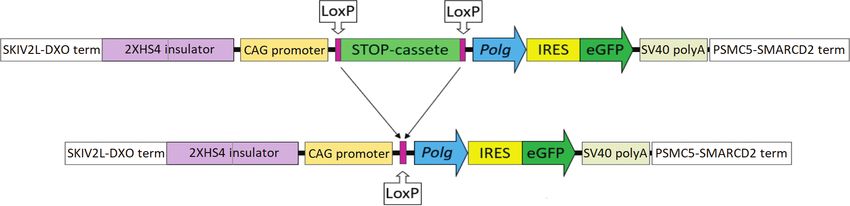
main reason for such mitochondrial genome instability lies STOP-cassette excision by Cre recombinase (Fig. 1).
in the absence of histones, the peculiarity of repair mecha- Total RNA for Polg cloning was extracted from mouse
nisms, and the presence of free oxygen radicals, which are liver biopsy. The tissue was homogenized using the Pre-
by-products of aerobic respiration (Woo et al. 2012; Nis- cellys 24 homogenizer, and RNA was extracted using a
sanka and Moraes 2018). Many pathologies are associated QIAGEN RNeasy Mini Kit. cDNA was synthesized us-
with mitochondrial mutations, such as Leber hereditary op- ing RevertAid Reverse Transcriptase (Thermo Scientific)
tic neuropathy, chronic fatigue syndrome, encephalomyo- and specific primer P1 (all primer sequences are present-
pathy, etc. (Orekhov et al. 2015; Hsu et al. 2016). ed in Table 1) for mouse Polg gene 3′-UTR. Polg gene
It is obvious that the improvement of approaches to (3653 bp) was amplified with Platinum SuperFi polymer-
treating mitochondrial pathologies requires adequate test ase (Invitrogen), using primers P2 and P3. The amplified
systems. Herein, we describe an approach for creating a
Table 1. Primer sequences used in the study
murine model that accumulates mutations in the mito-
chondrial genome of somatic and germ cells due to the Code Sequence 5′→3′
presence of Polg gene mutants. P1 ATGGCTGCCTTTTGCAAAAAAGC
P2 ATTAACGCGTATGAGCCGCCTGCTCTGGAA
PolG-alpha is an enzyme that mediates mitochondri- P3 TAATACGCGTCTAGGGTCCAGGCTGGCTTCG
al DNA replication and repair (Copeland and Longley P4 CTCGAGCCCATATCAGGGAACAG
2003). PolG-alpha is encoded by the nuclear genome and P5 CAAAGGAAACATTGTGCCCCAC
its mutant form PolG-alpha D257A leads to a change in P6 GTTAGATCTGCTGCCACCGT
the N-terminal “proofreading” domain, which deprives P7 AGGTGGCAAGTGGTATTCCG
P8 GCGGTCTGGCAGTAAAAACTATC
the enzyme of 3′-5′ exonuclease activity. The mutant form P9 GTGAAACAGCATTGCTGTCACTT
of the protein is unable to correct polymerization errors, P10 CTAGGCCACAGAATTGAAAGATCT
which leads to the accumulation of mutations in the mi- P11 GTAGGTGGAAATTCTAGCATCATCC
tochondrial genome. Previously, transgenic animals with P12 GTGCATGTTTGCCTATAAGCTGG
constitutive ubiquitous mutant Polg expression have been P13 GGTAAATATCCAGTGCTTCACCCT
Figure 1. Linearized pKB1-Polg genetic construct design.Research Results in Pharmacology 7(3): 33–39 35
fragment was subjected to site-directed mutagenesis after cultivation. ”Spermoprep” medium was used for sperm
cloning into the intermediate T-vector pTZ57R/T (Ad- culture (Paneco, Russia), and IVF was carried out in the
dgene). Site-directed mutagenesis was carried out using G-IVF PLUS medium (Vitrolife Sweden AB, Sweden).
primers P4 and P5, due to which it was possible to in-
troduce the D257A mutation into the native mouse Polg Transgenic animals creation
gene ORF, which deprives the enzyme of 3′-5′-exonucle-
ase “proofreading” activity. This mutated Polg gene was Primary F0 transgenic animals were obtained through a
sequenced, amplified using primers P2 and P3, treated microinjection of a gene construct into zygote pronucleus.
with the restriction enzyme MluI (Thermo Fisher Scien- The method for obtaining primary transgenic animals was
tific) with subsequent insertion into the pKB1 vector, en- described in detail earlier (Zvezdova et al. 2010; Silaeva
zyme treatment and dephosphorylation FastAP, Thermo et al. 2018).
Fisher Scientific).
Then the construction was prepared for microinjec- In vitro fertilization
tions: circular plasmid DNA was treated with the re-
striction enzyme PvuI (Thermo Fisher Scientific); the The method of intravital surgical sperm collection was
resulting fragments were separated by electrophoresis in used for IVF according to the protocol (Val and Robleda-
agarose gel. A linear fragment 14832 bp, devoid of bacte- no 2013) to obtain F1 offspring from the primary transge-
rial sequences, was extracted using a Monarch kit for iso- ne #1683. Two-blastomeric embryos obtained after IVF
lation of DNA fragments from gels and reaction mixtures were transferred to recipients.
(New England Biolabs) and dissolved in TE buffer at a
concentration of 1 ng/μl (Zvartsev et al. 2019). Genotyping
Animals and keeping conditions Mouse DNA is derived from tail tissue. Tissue samples
were lysed in alkaline lysis (25 mM NaOH, 0.2 mM
Several mice strains were used in the work. 6–8-week-old EDTA) and purified by the phenol-chloroform method.
males of hybrid F1 (CBA X C57BL/6) (Stolbovaya Nur- The mice were genotyped using the PCR method and the
sery) were used as breeders; immature females (12–13 g) following reagents: HS Taq-polymerase, Red buffer, and
CBA X C57BL/6 were used as donors; 8–10-week-old dNTP (Evrogen), and the STOP-cassette specific primers
females CD1 were used as recipients. During the expe- – P6 and P7 (292 bp fragment amplification, Fig. 2).
rimental work, the mice were kept in the vivarium of the Genotyping of Cre mice was carried out in accordance
Institute of Gene Biology of the Russian Academy of Sci- with the Jackson Laboratory recommendations using
ences. The mice were kept in conditions of free access primers P8, P9, P10, and P11.
to water and food, a 12/12 light cycle, air temperature
23±1℃, and humidity of 42±5%. Copy number determination
Two activator mice of the Cre strain obtained from
Jackson Laboratory were used to activate transgene ex- Transgene copy number was measured by real-time PCR
pression, namely B6.C-Tg(CMV-cre)1Cgn/J Stock No: as described earlier (Bruter et al. 2021). We used primers
006054|CMV-Cre (systemic activation) and B6.Cg- P12 and P13 to measure Polg transgene copy number.
Tg(Tek-cre)1Ywa/J Stock No: 008863|Tie2-Cre (en-
dothelial activation). Ethical approval
Solutions and media All the animal experiments were approved and controlled
by the IGB RAS Bioethics Commission (the conclusion
The M2 medium (Sigma-Aldrich, USA) was used to wash of the Bioethical Commission dated 30/04/2021). The
out the oocytes, the ”Droblenie” medium (Paneco, Russia) animal experiments neither posed any biological hazards,
and mineral oil (Sigma, USA) were used for the oocytes nor required any biosafety facility.
Figure 2. Polg F0 mice genotyping. Samples by numbers #1251, #1254, #1683 and #1752 are transgenes.36 Kubekina MV et al.: A strain of mice with conditional expression of mut. Polg was created
Table 2. Polg transgene copy number in the strains of genetical-
Results ly modified mice
Primary transgenic mice creation with mutant Polg Line Mouse number Generation Copy number
gene inducible expression Polg 1251 F0 0.35
Polg 1254 F0 0.30
Polg 1683 F0 7.37
In the process of creating the model, 1081 microinjec-
Polg 1752 F0 0.1
ted embryos were obtained; they were transplanted to Polg 5479 F1 6.71
104 recipients. As a result, 29 mice were born from 17 Polg 6981 F1 5.44
recipients, of which 4 turned out to be the primary Polg Polg 7513 F2 6.90
transgenes. These were mice #1251♂, #1254♂, #1683♂, Polg 7515 F2 7.36
Polg 7538 F2 6.39
#1752♂. The results of genotyping are shown in Fig. 2.
Polg 7539 F2 6.46
Transgene copy number was determined for all pri- Polg*Tie2 893 F3 6.59
mary transgenes. Primary transgenic mice often have a Polg*Tie2 898 F3 6.34
mosaic pattern of transgene construct insertion into the Polg*Tie2 903 F3 5.39
genome. The results of the copy number measurement are Polg*Tie2 904 F3 5.29
Polg*Tie2 905 F3 5.66
presented in Table 2. For mouse #1251, the copy number Polg*CMV 889 F3 1.77
was 0.35 transgene copies per genome, for mouse #1752 Polg*CMV 1503 F3 1.61
– 0.1 transgene copies per genome, for mouse #1254 – Polg*CMV 8429 F2 1.20
0.30 transgene copies per genome, and for mouse #1683,
the copy number was ~7 transgene copies per genome. (the method is described above) to obtain F1 offspring.
Two-blastomeric embryos resulting from IVF were trans-
Obtaining two independent animal lines with induci- ferred to the recipients. As a result, one transgenic mouse
ble expression of the mutant Polg gene F1 was obtained with number 6981♀, with a ~5 copy
number. Many F2 offspring with a ~7 copy number were
All primary transgenes were put into crosses to obtain F1 obtained from F1 5479♂ (Table 2).
generation of transgenic animals. Mice #1251 and #1752 As a result, we obtained two independent lines of mice
died, leaving no offspring. One F1 transgenic mouse (of with a transgene ~7 and ~5 copy number for the inducible
90 offspring) 5479♂, with a ~7 copy number, was obtain- expression of the mutant form of Polg.
ed from transgenic mouse #1254 (Table 2). Transgenic
animal #1683♂ with the highest copy number turned out Transgene copies in three Polg strain variants
to be viable, but not capable of independent reproducti-
on. So, we performed a surgical collection of sperm from As a result of Polg transgenic mice crossing of the strain
the epididymis and used the obtained material for IVF with mice expressing Cre-recombinase systemically and
Figure 3. The results of copy number determination in different Polg strains. Transgene copy number of the Polg*CMV is sig-
nificantly reduced compared to Polg and Polg*Tie2. Copy numbers of Polg and Polg*CMV significantly differ (p < 0.01). Copy
numbers of Polg and Polg*Tie insignificantly differ (p~0.05).Research Results in Pharmacology 7(3): 33–39 37
in the vascular endothelium, we obtained double trans- in somatic cells is associated with a decrease in life ex-
genes Polg*Tie2 and Polg*CMV. The copy number was pectancy and premature onset of aging. In the mice of this
determined in all samples from double transgenic mice strain, the following was observed: weight loss, decreased
(Table 2, Fig. 3). subcutaneous fat content, hair loss, kyphosis, osteoporo-
From the chart above, with systemic activation, the sis, anemia, decreased fertility, and enlarged heart (Tri-
transgene copy number decreases to ~1, while with en- funovic et al. 2004). This phenotype negatively affects
dothelial activation, the transgene copy number remains at the viability of transgenes and significantly complicates
the same level in the tail tissue. This is the expected result, the maintenance of the animal strain. Our model is free
since systemic activation presupposes widespread recombi- from these drawbacks since it assumes inducible and tis-
nation of the STOP-cassette from the entire genomic DNA sue-specific expression of the mutant form of the protein.
of the organism, leaving only one copy of the transgene per To obtain a universal and viable model, we used a genet-
genome. This allows us to evaluate the efficiency of the ic construct based on the Cre-LoxP system, due to which
Cre-LoxP system. In transgenic Polg*Tie2 mice, similar the inducible tissue-specific expression of the mutant Polg
recombination occurs only in vascular endothelial cells. gene is possible. Due to the presence of a STOP-cassette, the
expression of the protein mutant form and the subsequent
GFP fluorescence of Polg*CMV pup phenotypic manifestation are possible only after crossing
the obtained model animals with mice with tissue-specific
We detected GFP fluorescence in transgenic Polg*CMV Cre-recombinase activation (Bruter et al. 2021).
mouse skin via a UV lamp in just one day after birth For the first time, we were able to develop a model
(Fig. 4). This means that mutant PolG-alpha and GFP of tissue-specific rather than systemic mitochondrial dys-
coexpression is activated during embryogenesis. function. We believe that mutant PolG-alpha tissue-spe-
cific expression will lead to an increase in the number of
mitochondrial genome mutations in certain tissues (de-
pending on the activator used), for example, in the vascu-
lar endothelium. These animals can be further used in ba-
sic research and preclinical trials of mitoprotective drugs.
Separately, it is worth mentioning that our murine model
with spatial control of mutant Polg expression has been de-
veloped. This means that we can obtain mice with different
Figure 4. Transgenic pup (left) skin in the UV chamber com- degrees of heteroplasmy, and at the same time mitochondri-
pared to two non-transgenic siblings (right). al dysfunction. An increase in a degree of heteroplasmy is
achieved by crossing Polg females of the same line in each
generation with males of any lines. Thanks to this, we can
We only see GFP fluorescence in one pup out of three, assess the effect of a degree of heteroplasmy on the pheno-
the other two are not transgenic, which is confirmed by type and assess a phenotypic effect of mitotherapeutic drugs.
genotyping results. This strain will make it possible to assess an increased
mutational load effect of the mitochondrial genome of a
certain tissue on the vital activity of the whole organism.
Discussion The model of mitochondrial dysfunction proposed by us
is more viable and adequate for studying this patholo-
Mutations of several genes involved in the maintenance gy in specific tissues. It provides wide opportunities for
of vital functions of mtDNA and in mitochondrial protein the study of mitochondrial dysfunction in neurons, im-
synthesis have been described. One of these genes is poly- mune, muscle, endothelial cells, which is of considerable
merase-gamma, which replicates and repairs mitochond- scientific interest. In this work, we did not confirm the
rial DNA. PolG-alpha is an attractive target for the creati- mitochondrial dysfunction phenotype in transgenic mice,
on of model animals, since this enzyme is encoded by the which is the subject of further research.
nuclear genome, within which it is much easier to make
genetic engineering modifications. Moreover, a decrease
in PolG-alpha function leads to spontaneous mutations Conclusion
accumulation, resulting in heteroplasmic mutant mtDNA,
which represents genuine MD caused by oxidative and Here we describe the creation of the first mouse strain with
nitrosative stress, inflammation, xenobiotics exposure, conditional expression of the mutant polymerase-gamma.
etc. (Payne and Chinnery 2015). As a result of microinjection and embryo transfer of ge-
Trifunovic et al. (2004) carried out an experiment to netically modified embryos, primary transgenic animals
create mice with a mutant form of this enzyme, name- were born and bred. After breeding with Cre activators,
ly, they deprived it of its 3′-5′-exonuclease activity. Thus, the transgene expression was confirmed by GFP fluores-
these mice did not repair the errors created by PolG-alpha cence and a decrease in the copy number of the mutant
in the mitochondrial DNA of all mouse organism cells. Polg transgene. Thus, transgenic mice Cre-dependently
That study showed that an increase in mtDNA mutations expressing mutant polymerase-gamma is a novel test-sys-38 Kubekina MV et al.: A strain of mice with conditional expression of mut. Polg was created
tem for studying mitochondrial biology and efficacy of Funding
mitoprotective drugs.
The reported study was funded by the Russian Foundati-
Conflict of interest on for Basic Research, project number 19-34-90073 and
by grant from the President of the Russian Federation No.
The authors have declared that no competing interests exist. MD-757.2020.7.
References
Angelova PR, Esteras N, Abramov AY (2021) Mitochondria and Biophysica Acta 1847(11): 1347–1353. https://doi.org/10.1016/j.
lipid peroxidation in the mechanism of neurodegeneration: Finding bbabio.2015.05.022 [PubMed] [PMC]
ways for prevention. Medicinal Research Reviews 41(2): 770–784. Prasun P (2020) Mitochondrial dysfunction in metabolic syndrome.
https://doi.org/10.1002/med.21712 [PubMed] Biochimica et Biophysica Acta – Molecular Basis of Disease 1866(10):
Aryaman J, Johnston IG, Jones NS (2019) Mitochondrial het- 165838. https://doi.org/10.1016/j.bbadis.2020.165838 [PubMed]
erogeneity. Frontiers in Genetics 9: 718. https://doi.org/10.3389/ Silaeva YY, Kirikovich YK, Skuratovskaya LN, Deikin AV (2018)
fgene.2018.00718 [PubMed] [PMC] Optimal number of embryos for transplantation in obtaining genet-
Bruter AV, Korshunova DS, Kubekina MV, Sergiev PV, Kalinina ic-modified mice and goats. Russian Journal of Developmental Bi-
AA, Ilchuk AA, Silaeva YY, Korshunov EN, Soldatov VO, Deykin ology 49(6): 356–361. https://doi.org/10.1134/s106236041806005x
AV (2021) Novel transgenic mice with Cre-dependent co-expression [in Russian]
of GFP and human ACE2: a safe tool for study of COVID-19 patho- Su T, Turnbull DM, Greaves LC (2018) Roles of mitochondrial DNA
genesis. Transgenic Research 30: 289–301. https://doi.org/10.1007/ mutations in stem cell ageing. Genes (Basel) 9(4): 182. https://doi.
s11248-021-00249-8 [PubMed] [PMC] org/10.3390/genes9040182 [PubMed] [PMC]
Copeland WC, Longley MJ (2003) DNA polymerase gamma in mi- Trifunovic A, Wredenberg A, Falkenberg M, Spelbrink JN, Rovio AT,
tochondrial DNA replication and repair. The Scientific World Journal Bruder CE, Bohlooly-Y M, Gidlöf S, Oldfors A, Wibom R, Törnell
3: 34–44. https://doi.org/10.1100/tsw.2003.09 [PubMed] [PMC] J, Jacobs HT, Larsson NG (2004) Premature ageing in mice express-
Del Val GM, Robledano PM (2013) In vivo serial sampling, of epi- ing defective mitochondrial DNA polymerase. Nature 429(6990):
didymal sperm in mice. Laboratory Animals 47(3): 168–174. https:// 417–423. https://doi.org/10.1038/nature02517 [PubMed]
doi.org/10.1177/0023677213478411 [PubMed] Wallace DC, Chalkia D (2013) Mitochondrial DNA genetics and the
Deykin A, Tikhonov M, Kalmykov V, Korobko I, Georgiev P, Mak- heteroplasmy conundrum in evolution and disease. Cold Spring Har-
simenko O (2019) Transcription termination sequences support the bor Perspectives in Biology 5(11): a021220. https://doi.org/10.1101/
expression of transgene product secreted with milk. Transgenic cshperspect.a021220 [PubMed] [PMC]
Research 28(3–4): 401–410. https://doi.org/10.1007/s11248-019- Wei W, Chinnery PF (2020) Inheritance of mitochondrial DNA
00122-9 [PubMed] in humans: implications for rare and common diseases. Journal
El-Hattab AW, Craigen WJ, Scaglia F (2017) Mitochondrial DNA of Internal Medicine 287(6): 634–644. https://doi.org/10.1111/
maintenance defects. Biochimica et Biophysica Acta – Molecular joim.13047 [PubMed]
Basis of Disease 1863(6): 1539–1555. https://doi.org/10.1016/j.bba- Woo DK, Green PD, Santos JH, D’Souza AD, Walther Z, Martin
dis.2017.02.017 [PubMed] WD, Christian BE, Chandel NS, Shadel GS (2012) Mitochondrial
Fontana GA, Gahlon HL (2020) Mechanisms of replication and repair in genome instability and ROS enhance intestinal tumorigenesis in AP-
mitochondrial DNA deletion formation. Nucleic Acids Research 48(20): C(Min/+) mice. The American Journal of Pathology 180(1): 24–31.
11244–11258. https://doi.org/10.1093/nar/gkaa804 [PubMed] [PMC] https://doi.org/10.1016/j.ajpath.2011.10.003 [PubMed] [PMC]
Hsu YR, Yogasundaram H, Parajuli N, Valtuille L, Sergi C, Oudit GY Zakirov FH, Zhang D, Grechko AV, Wu WK, Poznyak AV, Orekhov
(2016) MELAS syndrome and cardiomyopathy: linking mitochondri- AN (2020) Lipid-based gene delivery to macrophage mitochondria
al function to heart failure pathogenesis. Heart Failure Reviews 21(1): for atherosclerosis therapy. Pharmacology Research & Perspectives
103–116. https://doi.org/10.1007/s10741-015-9524-5 [PubMed] 8(2): e00584. https://doi.org/10.1002/prp2.584 [PubMed] [PMC]
Nicolson GL (2014) Mitochondrial dysfunction and chronic disease: Zvartsev RV, Korshunova DS, Gorshkova EA, Nosenko MA,
Treatment with natural supplements. Integrative Medicine (Encini- Korneev KV, Maksimenko OG, Korobko IV, Kuprash DV, Druts-
tas, Calif.) 13(4): 35–43. [PubMed] kaya MS, Nedospasov SA, Deikin AV (2019) Neonatal lethality
Nissanka N, Moraes CT (2018) Mitochondrial DNA damage and reac- and inflammatory phenotype of the new transgenic mice with over-
tive oxygen species in neurodegenerative disease. FEBS Letters 592(5): expression of human interleukin-6 in myeloid cells. Doklady Bio-
728–742. https://doi.org/10.1002/1873-3468.12956 [PubMed] [PMC] chemistry and Biophysics 483(1): 344–347. https://doi.org/10.1134/
Orekhov AN, Zhelankin AV, Kolmychkova KI, Mitrofanov KY, S1607672918060157 [PubMed] [in Russian]
Kubekina MV, Ivanova EA, Sobenin IA (2015) Susceptibility of Zvezdova ES, Silaeva YY, Vagida MS, Maryukhnich EV, Deikin
monocytes to activation correlates with atherogenic mitochondrial AV, Ermolkevich TG, Kadulin SG, Sadchikova ER, Goldman IL,
DNA mutations. Experimental and Molecular Pathology 99(3): 672– Kazansky DB (2010) Generation of transgenic animals express-
676. https://doi.org/10.1016/j.yexmp.2015.11.006 [PubMed] ing the alpha- and beta-chains of the autoreactive T-cell recep-
Payne BA, Chinnery PF (2015) Mitochondrial dysfunction in ag- tor. Molecular Biology 44(2): 311–322. https://doi.org/10.1134/
ing: Much progress but many unresolved questions. Biochimica et S0026893310020135 [PubMed] [in Russian]Research Results in Pharmacology 7(3): 33–39 39 Author contributions Marina V. Kubekina, postgraduate student, junior researcher at the Institute of Gene Biology, Russian Academy of Sciences, e-mail: kubekina@genebiology.ru, ORCID ID https://orcid.org/0000-0002-8834-1111. Writing the article, molecular cloning, analyzing transgenic mice PCR, and measuring copy number. Yulia Yu. Silaeva, Ph.D, research fellow at the Institute of Gene Biology, Russian Academy of Sciences, e-mail: yulya.silaeva@gmail.com, ORCID ID https://orcid.org/0000-0003-2070-9001. Writing the article and developing a research design. Alexandra V. Bruter Ph.D, research fellow at the Institute of Gene Biology, Russian Academy of Sciences, e-mail: aleabruter@gmail.com, ORCID ID https://orcid.org/0000-0002-2090-2488. Developing a research design. Diana S. Korshunova, senior technician at the Institute of Gene Biology, Russian Academy of Sciences, e-mail: korshunova@genebiology.ru, ORCID ID https://orcid.org/0000-0002-0259-7045. Coducting microinjections and working with the animals in all stages. Leonid A. Ilchuk, senior technician at the Institute of Gene Biology, Russian Academy of Sciences, e-mail: le- chuk12@gmail.com, ORCID ID https://orcid.org/0000-0002-0157-2102. Measuring copy number and working with graphic material. Yulia D. Okulova, senior technician at the Institute of Gene Biology, Russian Academy of Sciences, e-mail: oku- lova@genebiology.ru. Analyzing transgenic mice PCR analysis. Mariya O. Soldatova, 2nd year student of Biology, Medical Genetics and Ecology Department, Kursk State Medi- cal University, e-mail: mar.sold46@gmail.com, ORCID ID https://orcid.org/0000-0001-6637-1654. Genotyping Evgeniya Seryogina, research assistant of Cell Physiology and Pathology Laboratory, Orel State University, e-mail: e.s.seryogina@gmail.com, ORCID ID https://orcid.org/0000-0002-2796-4040. Writing the article. Inga M. Kolesnik, PhD in Medical Sciences, Associate Professor, Department of Pharmacology and Clinical Pharmacology, e-mail: kolesnik_i@bsu.edu.ru. The author was engaged in collection, analysis and interpretation of the data for publication. Polina A. Ukolova, research assistant of Cell Physiology and Pathology Laboratory, Orel State University, e-mail: polya.ukolova@yandex.ru, ORCID ID https://orcid.org/0000-0002-9636-513X. Writing the article. Mikhail V. Korokin, Doctor Habil. of Medical Sciences, Professor, Department of Pharmacology and Clinical Pharmacology, e-mail: mkorokin@mail.ru, ORCID ID https://orcid.org/0000-0001-5402-0697. Conceiving the idea and planning research. Alexey V. Deykin, Ph.D, senior scientist at the Institute of Gene Biology, Russian Academy of Sciences, e-mail: alexei@deikin.ru, ORCID ID https://orcid.org/0000-0001-9960-0863. Conceiving the idea and planning research.
You can also read