Watt W. Webb: His measurements of the seemingly inaccessible broadened the horizons of biophysics - PNAS
←
→
Page content transcription
If your browser does not render page correctly, please read the page content below
RETROSPECTIVE
RETROSPECTIVE
Watt W. Webb: His measurements of the
seemingly inaccessible broadened the horizons
of biophysics
Elliot Elsona,1
Watt W. Webb’s scientific career was a series of triumphs
over challenges to make difficult measurements of im-
portant physical and biological phenomena: “impos-
sible problems of experimental physiology,” as he
described them (1). A common theme was his use of
light microscopy in new ways to reveal equilibrium
and dynamic properties in biomolecular systems and
in organisms. Webb’s deep understanding of physics
directly contributed to new instruments and methods
for detecting and interpreting the hitherto undetect-
able and undecipherable. Webb and his collaborators
opened approaches adopted far and wide to investi-
gate important questions in areas of physics, biology,
biochemistry, and biophysics.
Webb was born on August 27, 1927 in Kansas City,
Missouri. He entered the Massachusetts Institute of
Technology at 16, where he majored in business and
engineering administration. He then worked as an
industrial engineer at Union Carbide. During that period,
he resumed studies at the Massachusetts Institute of
Technology and by 1955 had completed a Doctorate of
Science in materials science physics and mathematics.
Early Engineering Physics
Beginning in the late 1950s and continuing through
the early 1970s, Webb contributed to a wide variety of
physics topics, including crystal growth and disloca-
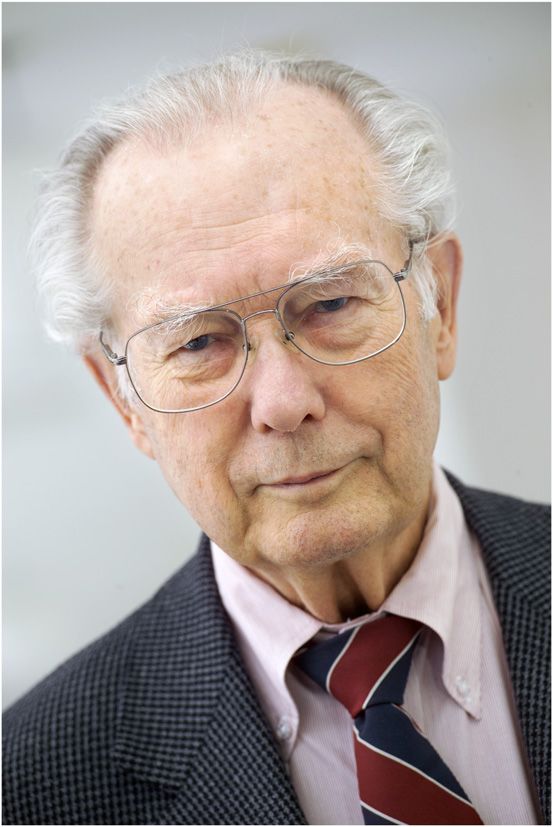
tions, magnetization, continuous transitions at critical Watt W. Webb. Image credit: Cornell University,
liquid–vapor interfaces, and fluctuations in supercon- licensed under CC BY-NC-ND.
ductors. Even at this early stage in his career, Webb
showed an interest in statistical fluctuations, albeit in
quantum systems, statistical noise, and phase trans- from equilibrium. Even in equilibrium the concentra-
formations that prefigured his later work on more tions of chemical reactants fluctuate spontaneously
biologically oriented areas. about their equilibrium values. The reaction kinetics
could be determined from the time courses of these
Entering Biophysics—Fluorescence Correlation tiny fluctuations. Webb’s interest in the problem may
Spectroscopy have reflected his earlier work on fluctuations in
Webb’s entry into biophysics in the early 1970s was quantum systems (2). These later studies were origi-
driven by a tantalizing challenge: How to measure the nally motivated by questions about the kinetic mech-
kinetics of chemical reactions without displacing them anism of the helix to random coil transition of DNA
a
Department of Biochemistry and Molecular Biophysics, Washington University School of Medicine in St. Louis, St. Louis, MO 63110-1093
Author contributions: E.E. wrote the paper.
The author declares no competing interest.
Downloaded by guest on November 20, 2021
This open access article is distributed under Creative Commons Attribution-NonCommercial-NoDerivatives License 4.0 (CC BY-NC-ND).
1
Email: elson@wustl.edu.
Published March 10, 2021.
PNAS 2021 Vol. 118 No. 12 e2101879118 https://doi.org/10.1073/pnas.2101879118 | 1 of 4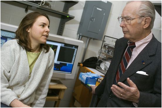
Measurement of the diffusion rates and molecular in-
teractions of lipids and proteins on and within cell
membranes. The fluid mosaic model of membrane
proteins embedded in a fluid lipid bilayer (15) and
theory (16) suggested that the diffusion rates of the
proteins should be comparable to that of the mem-
brane lipids. FRAP and FCS measurements found that
protein diffusion was much slower than expected (17,
18). Since then, many studies using FRAP, single-
particle tracking, and other methods have been fo-
cused on this subject.
Hearkening back to his early work on phase be-
havior of nonbiological fluid mixtures, Webb and his
Watt W. Webb in his laboratory. Image credit: Cornell
colleagues studied the phase equilibria of lipid bilayer
University, licensed under CC BY-NC-ND.
membranes (19–21). Lipid molecules with diverse
molecules (3). A promising simpler reaction system polar head groups, and both saturated and unsatu-
with which to begin, however, was the binding of rated hydrocarbon chains of varying lengths, yielded
ethidium to DNA, which causes a large enhancement membranes with subtle differences in phase behavior.
of the ethidium fluorescence (4). Work on this system Experiments were carried out on giant unilamellar
led Webb and coworkers to develop fluorescence vesicles, single lipid bilayer “bubbles” 10 μm or more
in diameter. Fluorescent lipid molecules that parti-
correlation spectroscopy (FCS) (5–7). FCS at that time
tioned selectively into (quasi-solid) gel and fluid phase
was difficult and was never suitable to measure fluc-
domains were essential for this work (20, 21). These
tuations of DNA helicity. Nevertheless, FCS is now
studies revealed informative relationships between
routine in many laboratories throughout the world and
the shapes of domains, their lipid compositions, the
is being applied to a wide range of subjects (8).
interfacial tension of domain boundaries, and mem-
Although the main motivation for developing FCS
brane curvature. Moreover, they demonstrated that
was to measure chemical reaction kinetics via fluctua-
the compositions of the two phases could approach
tions, an easier application, which is still of great interest,
one another until, at a critical temperature, the dis-
is to measure translational diffusion via the fluctuations
tinguishable phases disappear in a continuous phase
of fluorescence that arise as fluorescent molecules dif-
transition. Measurement of thermal shape fluctuations
fuse across the laser beam that measures the sample
of the giant unilamellar vesicles led to evaluation of
fluorescence. The small size of the laser-illuminated spot
their curvature elastic modulus (22).
makes FCS particularly suited to measuring diffusion
in small systems (e.g., within or on the surfaces of bio-
Multiphoton Microscopy
logical cells). One of most elegant aspects of FCS is Confocal fluorescence microscopy makes possible the
its extraction of macroscopic rate parameters from mi- detection of a small number of molecules and there-
croscopic fluctuations. Related work on fluorescence fore was essential to the development of FCS and
fluctuations due to molecular rotational motion was FRAP (11). Moreover, laser-scanning confocal micros-
also proceeding at this time (9). It is sometimes easier, copy provided an important and widely used tool for
however, to measure the response to displacing the visualizing cells beginning in the 1980s. The Webb
system from equilibrium macroscopically by photo- laboratory pioneered the next major advance in fluo-
bleaching a faction of the fluorophore within the focal rescence microscopy, multiphoton microscopy (MPM)
spot and then measuring the reequilibration of the (23–26). MPM provided new capabilities both for sci-
fluorescence due to diffusion back into the bleached entific investigation and for diagnostic and surgical
region. This is the basis of fluorescence photobleaching uses. Returning to engineering practical contributions,
recovery, now called fluorescence recovery after photo- Webb devoted considerable effort to develop endo-
bleaching (FRAP) (10, 11). This approach was also de- scopic uses of MPM. A Google Scholar search on
veloped with technical variations independently around “Watt Webb, endoscopy” returns many papers de-
this time by other laboratories (12–14). voted to endoscopic instrumentation (27) and to the
This work demonstrates Webb’s strength in the evaluation of diagnostic studies, such as the areas of
design of measurement approaches and his experience human bladder cancer (28) and lung cancer (29).
in the interpretation of microscopic properties of physical MPM consists of combining two or more photons
systems. These were crucial to the development of FCS absorbed essentially simultaneously to excite fluores-
and were hallmarks of his many and important later cence. The sum of the energies of the absorbed
contributions to molecular and cellular biophysics. photons must match the total energy required for one-
photon excitation of the fluorescence. Hence, the
Diffusion of Cell Membrane Components and wavelength of each of the combined photons is
Membrane Lipid Phase Behavior greater than the wavelength required for conventional
The availability of FCS and FRAP allowed Webb and one-photon excitation. Mode-locked lasers are
Downloaded by guest on November 20, 2021
his coworkers to address a major set of subjects still needed to produce short but very intense light pulses
currently of interest to biophysicists and biochemists: needed for the very high photon fluxes required for
2 of 4 | PNAS Elson
https://doi.org/10.1073/pnas.2101879118 Watt W. Webb: His measurements of the seemingly inaccessible broadened the
horizons of biophysicsthe simultaneous absorption of two or more photons. lipid vesicles (35). Related work included studies of the
Substantial advantages accrue from the use of a “giga-seal” high-resistance adherence of the channel-
longer wavelength of exciting light. The background containing membrane to a micropipette, essential for
fluorescence excited by the light of longer wavelength these measurements (36), and also the effect of mem-
as it travels through the medium toward and from the brane tension on conductance by alamethicin channels
focal point is much less than would arise from one- (37). More directly, biological studies addressed audi-
photon excitation at a shorter wavelength. In addition, tory mechanisms in crickets (38, 39) and in the saccular
photobleaching of the fluorophore would be confined hair cells of the frog inner ear (18, 26, 40–42).
to the focal volume rather than over the entire path of
the excitation light. Furthermore, because excitation Concluding Thoughts
occurs only in the focal volume, even scattered fluo- Watt W. Webb made many important and diverse
rescent light can be used to construct the image. As a contributions to biophysics, including the develop-
result, a major advantage of multiphoton technology is ment of methods for measuring difficult phenomena,
its suitability for deep tissue imaging (30). Webb’s long such as the kinetics of spontaneous concentration
and diverse experience with lasers was especially fluctuations in chemical reaction systems, MPM,
beneficial to the development of MPM. membrane phase equilibria, and auditory transduction
Another version of MPM uses second harmonic mechanisms, and the interpretation of these mea-
generation (SHG) in which two photons of the same surements to provide mechanistic understanding of
frequency of light are combined by a nonlinear ma- important biochemical and biophysical phenomena.
terial to generate a photon of twice the frequency His work provides a roadmap to some of the most
(energy). Early applications of this phenomenon were interesting and significant work in these areas to occur
carried out by Webb and his coworkers, including over the last half century. This accounting would be
studies of collagen fibrils, which generate a strong incomplete, however, if I failed to mention Webb’s
SHG signal (31) and are useful to analyze how cells great personal influence as a teacher and mentor to a
remodel collagen matrices in engineered tissue con- large cadre of contemporary biophysical scientists. To
structs (see, e.g., ref. 32). SHG is also useful to mea- his students and collaborating scientists, Webb was
sure fast action potentials (33) and uniformly polarized always a supportive friend and source of sound guid-
microtubules in brain tissue (34). ance. Those of us who had the pleasure of visiting or
working in his laboratory in the basement of Clark Hall
Electrophysiology at Cornell University will always remember and trea-
Even a brief account of Webb’s contributions should sure the pleasure of seeing a difficult measurement
not omit his work on electrophysiology, including an yield a long-sought result and the lively discussions of
early use of patch clamping to record conductance by science and many other topics during the 4 o’clock
chloride channels from electric eels in reconstituted coffee hour.
1 W. W. Webb, Commentary on the pleasures of solving impossible problems of experimental physiology. Annu. Rev. Physiol. 68, 1–28
(2006).
2 W. W. Webb, R. J. Warburton, Intrinsic quantum fluctuations in uniform filamentary superconductors. Phys. Rev. Lett. 20, 461
(1968).
3 E. L. Elson, Fluorescence correlation spectroscopy: Past, present, future. Biophys. J. 101, 2855–2870 (2011).
4 J. L. Bresloff, D. M. Crothers, DNA-ethidium reaction kinetics: Demonstration of direct ligand transfer between DNA binding sites.
J. Mol. Biol. 95, 103–123 (1975).
5 E. Elson, D. Magde, Fluorescence correlation spectroscopy. I. Conceptual basis and theory. Biopolymers 13, 1–27 (1974).
6 D. Magde, E. L. Elson, W. W. Webb, Thermodynamic fluctuations in a reacting system—Measurement by fluorescence correlation
spectroscopy. Phys. Rev. Lett. 29, 705–708 (1972).
7 D. Magde, E. L. Elson, W. W. Webb, Fluorescence correlation spectroscopy. II. An experimental realization. Biopolymers 13, 29–61
(1974).
8 M. A. Digman, E. Gratton, Lessons in fluctuation correlation spectroscopy. Annu. Rev. Phys. Chem. 62, 645–668 (2011).
9 M. Ehrenberg, R. Rigler, Rotational Brownian-motion and fluorescence intensity fluctuations. Chem. Phys. 4, 390–401 (1974).
10 D. Axelrod, D. E. Koppel, J. Schlessinger, E. Elson, W. W. Webb, Mobility measurement by analysis of fluorescence photobleaching
recovery kinetics. Biophys. J. 16, 1055–1069 (1976).
11 D. E. Koppel, D. Axelrod, J. Schlessinger, E. L. Elson, W. W. Webb, Dynamics of fluorescence marker concentration as a probe of
mobility. Biophys. J. 16, 1315–1329 (1976).
12 K. Jacobson, A. Ishihara, R. Inman, Lateral diffusion of proteins in membranes. Annu. Rev. Physiol. 49, 163–175 (1987).
13 R. Peters, J. Peters, K. H. Tews, W. Bähr, A microfluorimetric study of translational diffusion in erythrocyte membranes. Biochim.
Biophys. Acta 367, 282–294 (1974).
14 M. Poo, R. A. Cone, Lateral diffusion of rhodopsin in the photoreceptor membrane. Nature 247, 438–441 (1974).
15 S. J. Singer, G. L. Nicolson, The fluid mosaic model of the structure of cell membranes. Science 175, 720–731 (1972).
16 P. G. Saffman, M. Delbrück, Brownian motion in biological membranes. Proc. Natl. Acad. Sci. U.S.A. 72, 3111–3113 (1975).
17 J. Schlessinger et al., Lateral transport on cell membranes: Mobility of concanavalin A receptors on myoblasts. Proc. Natl. Acad. Sci.
U.S.A. 73, 2409–2413 (1976).
18 J. L. Thomas, T. J. Feder, W. W. Webb, Effects of protein concentration on IgE receptor mobility in rat basophilic leukemia cell plasma
membranes. Biophys. J. 61, 1402–1412 (1992).
Downloaded by guest on November 20, 2021
19 J. S. Huang, W. W. Webb, Diffuse interface in a critical fluid mixture. J. Chem. Phys. 50, 3677 (1969).
20 T. Baumgart, S. T. Hess, W. W. Webb, Imaging coexisting fluid domains in biomembrane models coupling curvature and line tension.
Nature 425, 821–824 (2003).
Elson PNAS | 3 of 4
Watt W. Webb: His measurements of the seemingly inaccessible broadened the https://doi.org/10.1073/pnas.2101879118
horizons of biophysics21 T. Baumgart, G. Hunt, E. R. Farkas, W. W. Webb, G. W. Feigenson, Fluorescence probe partitioning between Lo/Ld phases in lipid
membranes. Biochim. Biophys. Acta 1768, 2182–2194 (2007).
22 M. B. Schneider, J. T. Jenkins, W. W. Webb, Thermal fluctuations of large cylindrical phospholipid vesicles. Biophys. J. 45, 891–899
(1984).
23 W. Denk, J. H. Strickler, W. W. Webb, Two-photon laser scanning fluorescence microscopy. Science 248, 73–76 (1990).
24 A. Ustione, D. W. Piston, A simple introduction to multiphoton microscopy. J. Microsc. 243, 221–226 (2011).
25 W. R. Zipfel, R. M. Williams, W. W. Webb, Nonlinear magic: Multiphoton microscopy in the biosciences. Nat. Biotechnol. 21,
1369–1377 (2003).
26 S. Maiti, J. B. Shear, R. M. Williams, W. R. Zipfel, W. W. Webb, Measuring serotonin distribution in live cells with three-photon
excitation. Science 275, 530–532 (1997).
27 D. R. Rivera, C. M. Brown, D. G. Ouzounov, W. W. Webb, C. Xu, Multifocal multiphoton endoscope. Opt. Lett. 37, 1349–1351 (2012).
28 S. Mukherjee et al., Human bladder cancer diagnosis using multiphoton microscopy. Proc. SPIE Int. Soc. Opt. Eng. 7161, 716117
(2009).
29 M. Jain et al., Multiphoton microscopy: A potential “optical biopsy” tool for real-time evaluation of lung tumors without the need for
exogenous contrast agents. Arch. Pathol. Lab. Med. 138, 1037–1047 (2014).
30 D. R. Miller, J. W. Jarrett, A. M. Hassan, A. K. Dunn, Deep tissue imaging with multiphoton fluorescence microscopy. Curr. Opin.
Biomed. Eng. 4, 32–39 (2017).
31 R. M. Williams, W. R. Zipfel, W. W. Webb, Interpreting second-harmonic generation images of collagen I fibrils. Biophys. J. 88,
1377–1386 (2005).
32 F. Toki et al., Second harmonic generation reveals collagen fibril remodeling in fibroblast-populated collagen gels. Cell Struct. Funct.
38, 227–236 (2013).
33 D. A. Dombeck, M. Blanchard-Desce, W. W. Webb, Optical recording of action potentials with second-harmonic generation
microscopy. J. Neurosci. 24, 999–1003 (2004).
34 D. A. Dombeck et al., Uniform polarity microtubule assemblies imaged in native brain tissue by second-harmonic generation
microscopy. Proc. Natl. Acad. Sci. U.S.A. 100, 7081–7086 (2003).
35 D. W. Tank, C. Miller, W. W. Webb, Isolated-patch recording from liposomes containing functionally reconstituted chloride channels
from Torpedo electroplax. Proc. Natl. Acad. Sci. U.S.A. 79, 7749–7753 (1982).
36 L. R. Opsahl, W. W. Webb, Lipid-glass adhesion in giga-sealed patch-clamped membranes. Biophys. J. 66, 75–79 (1994).
37 L. R. Opsahl, W. W. Webb, Transduction of membrane tension by the ion channel alamethicin. Biophys. J. 66, 71–74 (1994).
38 P. R. Dragsten, W. W. Webb, J. A. Paton, R. R. Capranica, Auditory membrane vibrations: Measurements at sub-angstrom levels by
optical heterodyne spectroscopy. Science 185, 55–57 (1974).
39 P. R. Dragsten, W. W. Webb, J. A. Paton, R. R. Capranica, Light-scattering heterodyne interferometer for vibration measurements in
auditory organs. J. Acoust. Soc. Am. 60, 665–671 (1976).
40 W. Denk, R. M. Keolian, W. W. Webb, Mechanical response of frog saccular hair bundles to the aminoglycoside block of
mechanoelectrical transduction. J. Neurophysiol. 68, 927–932 (1992).
41 W. Denk, W. W. Webb, Thermal-noise-limited transduction observed in mechanosensory receptors of the inner ear. Phys. Rev. Lett.
63, 207–210 (1989).
42 W. Denk, W. W. Webb, Forward and reverse transduction at the limit of sensitivity studied by correlating electrical and mechanical
fluctuations in frog saccular hair cells. Hear. Res. 60,89–102 (1992).
Downloaded by guest on November 20, 2021
4 of 4 | PNAS Elson
https://doi.org/10.1073/pnas.2101879118 Watt W. Webb: His measurements of the seemingly inaccessible broadened the
horizons of biophysicsYou can also read