Whitish genital lesions - RACGP
←
→
Page content transcription
If your browser does not render page correctly, please read the page content below
Clinical

Whitish genital lesions

                                                                                                             QUESTION 3
Tim Aung, Devita Surjana, Manisha Singh                        •   lichen simplex chronicus
                                                               •   lichen planus                             What is lichen sclerosus and what are the
                                                               •   post-inflammatory hypopigmentation        clinical features of LSV?
CASE                                                           •   morphoea
A woman aged 40 years presented for a                          •   extramammary Paget disease                QUESTION 4

cervical screening test (CST). She was a                       •   vulvar intraepithelial neoplasia (VIN).   What are the aetiology and epidemiology
mother of three and a recently arrived                         The pale-white and ivory-white                of LSV?
refugee. She had never had a Pap smear                         discolouration of the vulva and clitoris
or CST in the past because she had been                        with some anatomical distortion make          ANSWER 3

living in a remote area for decades. The                       LSV the most likely diagnosis in this case.   Lichen sclerosus is a chronic inflammatory
purpose of the test was fully explained                        If there is a prominent erythematosus         dermatosis commonly affecting the
to the patient. When asked before the                          with scales, then psoriasis and contact       anogenital region. The condition is
procedure about routine symptomatology                         dermatitis can also both be considered as     characterised by white sclerotic patches
including menstruation, discharge and                          differential diagnoses. Table 1 compares      that subsequently coalesce, becoming
itchiness, the patient had no complaints.                      and contrasts the features of LSV with        shiny porcelain-white or ivory-white
   During the procedure, in the presence                       other similar conditions.                     colour. When it affects the vulva, it is
of a chaperone female practice nurse,
whitish discolouration of the vulva,                           ANSWER 2

involving both labia majora and minora,                        A diagnosis of LSV can be made
and clitoris with distorted anatomy was                        clinically without a mandatory
noted (Figure 1). On further enquiry, the                      biopsy. However, a punch biopsy
patient stated she had experienced mild                        from the white sclerotic area is highly
pruritus for several years, and she thought                    recommended to confirm the diagnosis
it was due to friction from walking.                           and exclude alternative diagnoses
                                                               including squamous cell carcinoma
                                                               (SCC). The histopathology usually
QUESTION 1                                                     illustrates atrophic or hyperkeratotic
What is the differential diagnosis based                       epidermis with lichenoid infiltrate in
on this presentation, and what is the most                     the dermal–epidermal junction and
likely diagnosis?                                              the homogenisation of collagen in the
                                                               upper dermis.2–4
QUESTION 2

How will you definitively diagnose this
condition?                                                     CASE CONTINUED

                                                               The patient was informed about
ANSWER 1                                                       the unusual discolouration and the
Given whitish discolouration of the vulva                      possibility of a genital skin disorder.
in a woman aged 40 years, the differential                     She was scheduled for a biopsy to guide
                                                                                                             Figure 1. Lichen sclerosus of vulva with some
diagnosis for this particular case can                         management and rule out SCC. The
                                                                                                             anatomical distortion (A = Buried clitoris,
include:1,2                                                    biopsy confirmed lichen sclerosus with        B = Involvement of clitoris hood, C = Distorted
• lichen sclerosus of the vulva (LSV)                          no evidence of neoplasia (Figure 2).          labia minora, D = Extending to perineum)
• vitiligo

© The Royal Australian College of General Practitioners 2021                                                   Reprinted from AJGP Vol. 50, No. 1–2, Jan–Feb 2021   55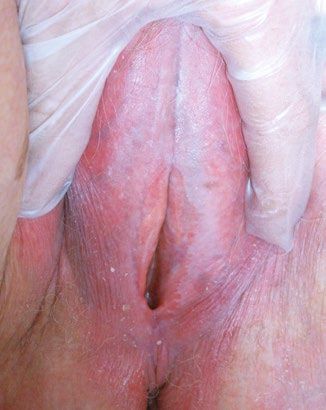
Clinical Whitish genital lesions
Table 1. Differential diagnoses of lichen sclerosus of the vulva9–13
Primary lesion Main Genital Extragenital Possible
features symptom involvement involvement dermoscopic features Histology/microscopy
Lichen sclerosus White patches with Prominent Very common Rare White structureless Hyalinisation of
of the vulva (LSV) sclerotic skin texture, itch (+++) (80%) (15–20%) areas with linear upper dermis, with a
fissures, erosions vessels, ice slivers, band of lymphocytes
comedo-like openings below; atrophic or
(hair-bearing area) hyperkeratosis epidermis
Vitiligo Well-defined No itch Less* More common* Homogenous white Total absence of
depigmentation with structureless areas functioning melanocytes,
no alteration of skin with absent or reduced with the inflammatory
texture pigment network lymphocyte on the edges
of the lesions
Morphea Thick, hard skin No itch Rare More common* White-yellowish Atrophic epidermis with
(sclerotic/fibrosis) structureless areas, increased collagen in
linear vessels within the dermis and loss of
the lilac ring appendageal structures
Lichen planus Hypertrophic erosions Pain is Less* More common* White crossing streaks Hyperkeratosis and
of the vaginal introitus greater (Wickham striae) acanthosis; saw-toothing
(LSV does not affect than itch of rete pegs, band-like
vaginal mucosa) chronic inflammatory
infiltrate obscuring
the dermo-epidermal
junction
Lichen simplex Lichenification Itch (+++); Less* More common* Scales, exaggerated Marked hyperkeratosis
chronicus with scaling scratching is skin markings associated with foci of
pleasurable parakeratosis, prominent
granular cell layer,
papillary dermal fibrosis
Dermatitis Erythematosus with Itch (++) Common, Common, Red dots in a patchy Extensive spongiosis;
(atopic/contact) or without scaling depending on depending on distribution and yellow initially acute spongiotic
and lichenification triggers triggers scales dermatitis, evolving into
subacute or chronic
spongiotic dermatitis
Psoriasis Erythematosus with Itch (+) Less* More common* Scales and dotted Regular acanthosis,
or without scaling vessels; under confluent parakeratosis,
and fissures high-power imaging, supra-papillary plate
dilated, elongated and thinning
convoluted capillaries
are visible
Cicatricial Blisters (bullae) or Pain Rare Mouth NA Immunofluorescence
pemphigoid erosions analysis – linear
deposition of
immunoglobulin (Ig) G
or IgA, and complement
(C3) at the basement
membrane zone
Vulvovaginal Red, inflamed mucosa Itch (+++) Common NA NA Confirmation of
candidiasis with white-curd candidiasis using vaginal
discharge swab for microscopy,
culture and sensitivities
Note: Itch is classified as mild (+), moderate (++) or severe (+++)
*When compared with LSV
NA, not applicable
56 Reprinted from AJGP Vol. 50, No. 1–2, Jan–Feb 2021 © The Royal Australian College of General Practitioners 2021
Whitish genital lesions Clinical
called vulvar lichen sclerosus or LSV. It neck, thigh, buttock and breast, is seen in sclerosus is underreported because of a
was previously known as lichen sclerosus 15–20% of cases.2 Figure 3 shows various number of factors: lack of awareness of the
et atrophicus, kraurosis vulvae, leukoplakic morphologies of genital lichen sclerosus. patient and practitioner; embarrassment
vulvitis and lichen albus. When it affects Dermoscopically, patchy white and reluctance to disclose symptoms; and
the penis, the term balanitis xerotica structureless areas, ice slivers, presentation at different practitioners
obliterans has been used historically.1,5 comedo-like openings (hair bearing area of general practice, sexual health,
Occasionally LSV can be asymptomatic only), purpuric globules and dotted or gynaecology, urology and dermatology.1,3,6
and discovered incidentally during CST.3,6 sparse thin linear vessels can be seen As a result, delays in diagnosis and
The possible clinical manifestations of LSV (Figure 2).3 undertreatment are not uncommon.
include: Lichen sclerosus can be associated with Although earlier literature reported that
• pruritus (often intractable), pain and autoimmune-related diseases such as lichen sclerosus and LSV generally affect
bleeding from fissuring and erosion thyroid disease, vitiligo, alopecia areata individuals of Caucasian descent, the
• dyspareunia and other sexual and pernicious anaemia.2,7 condition can be seen in patients of any
dysfunction ethnicity.1
• constipation and painful defecation if ANSWER 4
perianal skin is involved The exact aetiology of lichen sclerosus
• atrophy and distortion of anatomical remains speculative. Several theories CASE CONTINUED
structures including burying of the such as autoimmune (approximately The patient underwent a blood test for
clitoris, fusion or loss of labia minora, 20% association), genetics (12% positive autoantibody screening, and the results
stenosis of the introitus, and distortion family history), hormonal factors and showed no associated autoimmune
of urethral orifice resulting in urinary chronic trauma/irritation have been disorders; such investigation is not
problems. proposed.1,7,8 LSV commonly affects routinely required for every case. She had
Morphologically, LSV results in an atrophic individuals from the fifth decade onwards no other skin disorders. She was referred
or hyperkeratotic surface of the vulva with but can be seen at any age including to a dermatology clinic at public hospital.
white sclerotic papules, patches or plaques, prepuberty. Approximately 30–50% While waiting to be seen by a specialist,
which may extend to the perineum and of affected women develop symptoms the patient was treated with potent topical
perianal area. Areas of purpura, fissures prior to menopause.6,9 The prevalence corticosteroid (TCS; mometasone furoate
and erosion can sometimes be seen. of LSV is estimated to be one in 30 older 0.1% ointment) with general advice as
Extragenital lichen sclerosus, which women and one in 900 prepubertal girls.1 per current guidelines. She had some
predominantly affects the shoulder, Most scholars suggest that genital lichen improvement in symptoms and skin
texture within six weeks.
QUESTION 5
What are the complications of LSV?
QUESTION 6
What are the management options for LSV?
ANSWER 5
The complications of LSV are:
• anatomical distortion/alteration – as
described in the clinical manifestations,
resulting in sexual dysfunction plus
urinary and bowel opening problems
• psychological – LSV significantly affects
the individual’s sexual function and
quality of life, resulting in psychological
distress and low self-esteem
A B
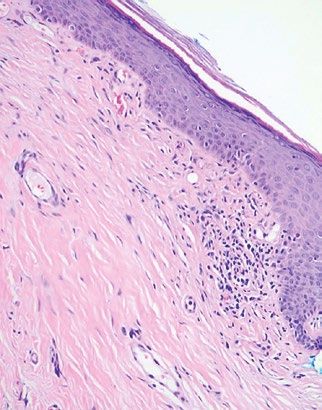
• cancer – there is an increased risk of
vulvar SCC of approximately 5%.1,2
Figure 2. Histology and dermoscopy of lichen sclerosus of the vulva (LSV)
ANSWER 6
a. Histology of LSV; b. Dermoscopy of LSV (A = Structureless white patches [sclerosus], B = Ice
sliver, C = Scattered linear vessels mixed with the whitish structure) Goals of treatment are to: 1) alleviate
symptoms of pruritus, fissuring and pain,
© The Royal Australian College of General Practitioners 2021 Reprinted from AJGP Vol. 50, No. 1–2, Jan–Feb 2021 57
Clinical                                                                                                                                         Whitish genital lesions

2) improve sexual function and quality                    and inexpensive to use, and it is effective         irritants may include hygiene products,
of life and 3) reduce scarring (structural                in 90% of patients.10                               creams, lubricants, contraceptives and
distortion) and risk of cancer.8 The                         It is advisable to review the patient in         procedures (eg improper hair removal).
detailed management for LSV is outlined                   4–6 weeks and three months from the                 Tight underwear and any activities
as follows.                                               start of TCS therapy. A 6–12-monthly                that can rub onto the sensitive mucosa
   Ultra-potent or potent TCSs are the                    follow-up is recommended during                     (eg riding a bicycle or horse) should also
first line of treatment. They provide                     maintenance treatment. Treatment failure            be avoided.10,11
both symptomatic relief and clinical                      at any stage indicates the need to consider      • Advise patients to become familiar with
improvement, reducing complications of                    an incorrect diagnosis, noncompliance               the appearance of their genital area,
scarring and malignant change.3,4,9 TCSs                  issue, development of VIN or SCC, or                as lifelong monitoring is required to
(one fingertip unit = 0.5 g) are applied                  superimposed factors such as allergic               detect, diagnose and treat new lesions
twice daily until symptoms (itchiness,                    reaction to specific medications, infection         or scarring.
soreness) are relieved (1–2 weeks),                       (candida, virus [herpes simplex virus],          Consider multidisciplinary care involving
then reduced to daily application until                   bacteria [Staphylococcus spp., Gardnerella       a local vulvar clinic (if available) or
the return of normal texture of the                       spp.]), and irritation from excess sweat         dermatologist or gynaecologist with an
skin (usually 1–2 months), and later on                   or urinary and faecal incontinence.              interest in lichen sclerosus. For example,
alternative days, totalling approximately                 There is no role for topical testosterone        surgery for correction of anatomical
three months from the start of treatment.                 and oestrogen.                                   distortion or treatment of early carcinoma
Afterward, maintenance treatment using                       General measures include the following:       may be needed.
lower potency (mid-strength) TCSs such                    • Counsel patients about the nature of
as betamethasone valerate (0.02%),                           disease, course, treatment and need for
triamcinolone acetonide (0.02%) or                           follow-up. Some individuals may need          Discussion and conclusion
methylprednisolone aceponate (0.1%)                          reassurance that lichen sclerosus is not a    General practitioners (GPs) have an
twice weekly is generally recommended.                       sexually transmissible infection.             opportunity to detect genital skin lesions
Frequency of TCS application can                          • Advise patients to avoid scratching and        while performing a CST. It is prudent
be individualised depending on                               irritants to the genital area by using soap   for GPs to be familiar with characteristic
hyperkeratosis. Ointment-based TCSs are                      substitutes for cleaning and applying         features of genital skin disorders to be
commonly preferred to cream in genital                       a protective barrier (eg soft paraffin or     effectively able to carry out further actions.
areas because of better absorption as well                   emollient) to minimise the contact with       Early detection and treatment with timely
as barrier function.6,9 TCS therapy is safe                  sweat, urine and faeces. Other vulvar         referral for genital skin disorders such as
                                                                                                           LSV will reduce the impact on the patient’s
                                                                                                           morbidity both physically and mentally.
                                                                                                           Management of vulvar skin disorders
                                                                                                           spans dermatology, gynaecology and
                                                                                                           sexual health, and referral to a pertinent
                                                                                                           specialist is recommended depending
                                                                                                           on the severity and complication of the
                                                                                                           disease. The prognosis of LSV is usually
                                                                                                           favourable if diagnosed and treated in the
                                                                                                           early nonscarring stages.

                                                                                                           Key points
                                                                                                           •   Genital skin disorders are generally
                                                                                                               underreported because of various
                                                                                                               factors.
                                                                                                           •   GPs have an opportunity to detect
                                                                                                               genital skin lesions while performing a
                                                                                                               CST or investigating a patient’s direct
                                                                                                               concerns.
 A                                                        B
                                                                                                           •   Early detection and treatment
Figure 3. Various morphology of lichen sclerosus of the vulva (LSV)                                            for genital skin disorders such as
a. LSV with typical shiny porcelain-white vulva (image courtesy of DermNet NZ); b. LSV with                    LSV will reduce the impact on the
erosion and fissures (image courtesy of DermNet NZ)                                                            patient’s morbidity physically and
                                                                                                               psychologically.

58   Reprinted from AJGP Vol. 50, No. 1–2, Jan–Feb 2021                                                         © The Royal Australian College of General Practitioners 2021
Whitish genital lesions Clinical
Authors 3. Lee A, Fischer G. Diagnosis and treatment Int 2016;113(19):337–43. doi: 10.3238/
Tim Aung FRACGP, FRNZCGP, ProfDip (Skin of vulvar lichen sclerosus: An update arztebl.2016.0337.
Cancer Surg), ProfDip (Gen Derm), Primary Care for dermatologists. Am J Clin Dermatol 10. Welsh BM, Berzins KN, Cook KA, Fairley CK.
Practitioner, Qld 2018;19(5):695–706. doi: 10.1007/s40257-018- Management of common vulval conditions. Med
0364-7. J Aust 2003;178(8):391–95. doi: 10.5694/j.1326-
Devita Surjana MBBS, PhD, FACD, Dermatologist,
Cutis Medical, Northern Dermatology and Sunnybank 4. Cyrus N, Jacobe HT. Morphea and lichen 5377.2003.tb05257.x
Dermatology, Qld sclerosus. In: Kang S, Amagai M, Bruckner AH, 11. Drummond C. Common vulval dermatoses. Aust
et al, editors. Fitzpatrick’s Dermatology. 9th edn. Fam Physician 2011;40(7):490–96.
Manisha Singh MD, FRCPA, Dermatopathologist,
New York, NY: McGraw-Hill Education, 2019; p.
Infinity Pathology, Qld 12. Errichetti E, Stinco G. Dermoscopy in general
1116–122.
Competing interests: None. dermatology: A practical overview. Dermatol Ther
5. Oakley A. Lichen sclerosus. Hamilton, NZ: (Heidelb) 2016;6(4):471–507. doi: 10.1007/s13555-
Funding: None. DermNet NZ, 2016. Available at https:// 016-0141-6.
Provenance and peer review: Not commissioned, dermnetnz.org/topics/lichen-sclerosus [Accessed
13. Lallas A. Dermoscopy in general dermatology/
externally peer reviewed. 17 June 2020].
inflammoscopy. Graz: Dermoscopedia, 2019.
Correspondence to: 6. Fisher G. Vulval lichen sclerosus – Diagnosis and Available at https://dermoscopedia.org/w/index.
timmynz2006@gmail.com treatment. Med Today 2019;20(1): 21–29. php?title=Inflammoscopy&oldid=16575 [Accessed
7. Fistarol SK, Itin PH. Diagnosis and treatment of 17 June 2020].
lichen sclerosus: An update. Am J Clin Dermatol
References
2013;14(1):27–47. doi: 10.1007/s40257-012-0006-4.
1. Marfatia Y, Surani A, Baxi R. Genital lichen
8. Lewis F. Dermotoses of the female genitalia. In:
sclerosus et atrophicus in females: An update.
Griffiths CEM, Baraker J, Bleiker T, Chalmers R,
Indian J Sex Transm Dis AIDS 2019;40(1):6–12.
Creamer D, editors. Rook’s textbook of dermatology.
doi: 10.4103/ijstd.IJSTD_23_19.
9th edn. Oxford, UK: John Wiley & Sons, 2016.
2. Nair PA. Vulvar lichen sclerosus et atrophicus.
9. Kirtschig G. Lichen sclerosus-presentation,
J Midlife Health 2017;8(2):55–62. doi: 10.4103/jmh.
diagnosis and management. Dtsch Arztebl
JMH_13_17. correspondence ajgp@racgp.org.au
GLW0001_RACGP ad_210x125_art.indd 1 11/1/21 4:06 pm
© The Royal Australian College of General Practitioners 2021 Reprinted from AJGP Vol. 50, No. 1–2, Jan–Feb 2021 59You can also read