A case of metastatic primary cardiac angiosarcoma
←
→
Page content transcription
If your browser does not render page correctly, please read the page content below
J R Coll Physicians Edinb 2021; 51: 156–9 | doi: 10.4997/JRCPE.2021.213                                                CASE REPORT

Clinical
A case of metastatic primary cardiac
angiosarcoma
C Intrator1, TA Muhammad2, S Gu3, B Lambourne4, AC Thompson5

                  We report a 49-year-old Southeast Asian woman diagnosed with metastatic               Correspondence to:
                  primary right atrial angiosarcoma (PCA) and the difficulties encountered              Christina Intrator
                                                                                                        34 Northumberland
 Abstract in         this diagnosis and its subsequent management. Diagnosis of PCA is
                  often delayed due to non-specific clinical presentation of patients. These            Gardens
                  tumours often present once metastatic spread has occurred, restricting                Newcastle upon Tyne
                  treatment options and leading to very poor prognosis. Patients undergo a              NE2 1HA
multidisciplinary team (MDT) approach involving chemotherapy, radiotherapy and, if eligible,            UK
surgery, but evidence-based treatment guidelines have yet to be established due to the rarity
of the tumour.                                                                                          Email:
                                                                                                        christina.intrator@nhs.net
Keywords: angiosarcoma, right atrial mass, pericardial mass, cardiac tumour

Informed consent: Written informed consent was obtained from the patient for publication
of this case report and any accompanying images.

Introduction                                                         Case presentation

Angiosarcomas are rare aggressive malignancies formed from           A 49-year-old woman from China presented with new pleuritic
the inner lining of blood vessels. Primary malignant tumours         chest pain, one month of exertional dyspnoea and three
of the heart are extremely rare, of which angiosarcomas              months of cough, poor appetite and night sweats. Prior
are the most common in adults.1 Diagnosis of PCA is often            to that, she was fit and well. On examination the patient
delayed due to non-specific clinical presentation, and can           was tachypnoeic and tachycardic, and there were bi-basal
sometimes be missed altogether, only being diagnosed at              crackles and a pericardial rub on the chest and precordial
autopsy.2 At the time of diagnosis, up to 75% of patients            auscultation. Blood tests showed normocytic anaemia
have systemic metastases, restricting treatment options and          (haemoglobin 92 g/l), raised C-reactive protein (199 mg/l) and
leading to very poor prognosis with a median survival of three       alkaline phosphatase (291 u/l). Troponin-T was marginally
to six months.3 Therefore a high degree of clinical suspicion        elevated (12 ng/l). An electrocardiogram demonstrated sinus
and sensitive investigations are crucial for diagnosis.4             tachycardia and a chest X-ray showed a globular heart. A
                                                                     transthoracic echocardiogram showed a small volume
Current management of patients with PCA is an MDT approach           pericardial effusion and echodense mass adjacent to the
involving a combination of chemotherapy, radiotherapy                right atrium (RA) and right ventricle (RV) free wall (Figure 1).
and, if the disease is localised, radical surgery. However,          Left ventricular function was normal with an ejection fraction
specific evidence-based treatment guidelines have yet to be          over 55%. The suspected diagnosis was pericarditis, treated
established due to the rarity of the tumour.4                        with ibuprofen, although its cause was still undetermined.

In this paper we report the case of a 49-year-old Southeast          Due to patient symptoms, previous travel to China and
Asian woman with a delayed diagnosis of metastatic primary           pericardial mass, it was felt the patient was at high risk
right atrial angiosarcoma and the difficulties encountered           of pericardial tuberculosis and she was admitted to the
in this diagnosis and its subsequent management. Patient             infectious diseases ward. Sputum for acid-fast bacilli and
survival from the initial presentation to death was seven            a QuantiFERON-TB Gold blood test were sent, which were
months. Literature surrounding PCAs is then critically               both negative. A computerised tomography (CT) scan of
reviewed.                                                            the chest, abdomen and pelvis showed extensive bilateral
                                                                     miliary pulmonary lesions, a large right renal mass, multiple
                                                                     liver lesions and pericardial enhancement. A hypervascular

1,2
   Senior House Officer, Newcastle upon Tyne NHS Foundation Trust, Newcastle upon Tyne, UK; 3Clinical Research Fellow, Division of
Cardiovascular Medicine, Addenbrooke’s Hospital, Cambridge, UK; 4Consultant Medical Oncologist, Northern Centre for Cancer Care,
Freeman Hospital, Newcastle upon Tyne NHS Foundation Trust, Newcastle upon Tyne, UK; 5Consultant Cardiologist, Department of
Cardiology, Royal Victoria Infirmary, Newcastle upon Tyne NHS Foundation Trust, Newcastle upon Tyne, UK

156    JOURNAL OF THE ROYAL COLLEGE OF PHYSICIANS OF EDINBURGH VOLUME 51 ISSUE 2 JUNE 2021        50TH ANNIVERSARY YEAR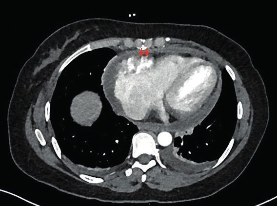
Primary cardiac angiosarcoma
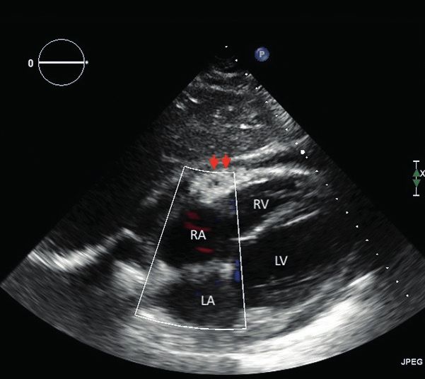
Figure 1 Transthoracic echocardiography, subcostal view. Figure 2 Computerised tomography of chest abdomen pelvis with
Subcostal view of conventional two-dimensional transthoracic contrast. Transverse section of chest tomography at initial
echocardiogram at initial presentation shows a small volume presentation demonstrates a hypervascular pericardial mass (red
pericardial effusion (1.3 cm) and an echodense cardiac mass arrows) arising from the right atrium and right atrioventricular
adjacent to the right atrium/right ventricle free wall. RA: right groove measuring 55 mm × 25 mm with associated pericardial
atrium, RV: right ventricle, LA: left atrium, LV: left ventricle, red effusion. There are also extensive bilateral miliary pulmonary
arrows: pericardial mass lesions and small bilateral pleural effusions. No mediastinal or
axillary lymphadenopathy was seen. The abdominal images not
viewed here showed multiple indeterminate liver lesions and a
large right lower pole renal mass consistent with an incidental
renal cell carcinoma radiologically (43 × 38 mm). Reported as
consistent with extensive tuberculosis and incidental renal cell
carcinoma. Red arrows: pericardial mass
pericardial mass of 55 × 25 mm was noted in the right
atrioventricular groove (Figure 2). Due to the CT findings,
an initial diagnosis of extensive miliary tuberculosis with Treatment for tuberculosis was discontinued. Needle
an incidental renal cell carcinoma was made. The patient pericardiocentesis was attempted to avoid impending cardiac
was started on quadruple antituberculous therapy and high- tamponade, but the fluid was too thick to drain.
dose steroids.
The sarcoma MDT concluded that, given the extent of disease
Due to the pericardial thickening and risk of constrictive and speed of progression, the patient’s life expectancy was
pericarditis she also underwent a surgical pericardial biopsy unlikely to exceed one year. Palliative chemotherapy was the
and pericardial window formation. The pericardial biopsy was management of choice as pericardial stripping was felt to
sent for microbiology culture and sensitivity, and histology. A offer little long-term benefit. The patient was started on a six-
thick gelatinous pericardial collection was also found at the cycle chemotherapy regime with doxorubicin and ifosfamide.
time of the operation and sent for microbiology. Neither sample Follow-up imaging showed a mixed response to treatment
grew mycobacteria. It was later noted that the pericardial with some response of lung, liver and pericardial lesions,
biopsy was exposed to formalin, making the isolation of but further progression of skeletal metastases. Disease
mycobacteria suboptimal. Histological examination of the progression remained rapid and five months after diagnosis
biopsy showed features of fibrinous pericarditis and chronic the pericardial tumour had increased significantly in size,
inflammation of uncertain cause with no granulomas. The distorting the RA and RV and encasing the aortic root and
patient was discharged on antituberculous treatment with pulmonary artery. The patient went on to develop bilateral
outpatient follow-up. Her symptoms failed to improve and pleural effusions thrombocytopenia and a haemorrhagic
she re-presented to hospital with chest pain and palpitations stroke. Seven months after the initial presentation, the
two weeks later. patient was transferred to a hospice and subsequently died.
Interval CT imaging showed an increase in the number and Discussion
size of lung nodules and liver lesions, worsening pericardial
nodularity and an unchanged pericardial mass with an Primary cardiac tumours are very rare and only 25% are
increased pericardial effusion compressing the RV. An urgent malignant, of which sarcomas are the most common. 5
ultrasound-guided biopsy of a liver lesion was performed. Angiosarcomas are the most common type of cardiac
Immunohistochemistry detected tumour cells strongly and sarcoma with an incidence of 0.0001% and tend to affect
diffusely positive for CD31 (endothelial marker) and ERG younger patients, with a male preponderence.5 While most
(nuclear vascular marker). Histological appearances were types of sarcoma arise from the left side of the heart, the
consistent with a high-grade angiosarcoma. A new diagnosis majority of angiosarcomas arise from the lateral wall of the
of metastatic PCA was made with lung and liver metastases. right atrium, sparing the septum and often infiltrating the
50TH ANNIVERSARY YEAR JUNE 2021 VOLUME 51 ISSUE 2 JOURNAL OF THE ROYAL COLLEGE OF PHYSICIANS OF EDINBURGH 157C Intrator, TA Muhammad, S Gu et al.
pericardium.6 PCAs are aggressive and 75% of patients have Due to the rarity, complexity and rapidly progressing nature of
metastases at diagnosis, mostly affecting the lungs, and PCAs, an MDT approach is required. Specific evidence-based
occasionally the lymph nodes, bone and liver.5,6 algorithms for PCAs are not yet available due to the rarity of
cases so management decisions are often extrapolated from
Late diagnosis of PCAs can occur due to the rarity of disease extracardiac soft-tissue sarcomas. Surgical resection with
and the non-specific nature of its presenting symptoms. adjuvant chemotherapy is possible in local, non-metastatic
Patients tend to present abruptly and symptoms will depend disease; however, complete resection is difficult since
on location of tumour, size and presence or absence of PCAs are large and often involve vital cardiac structures.
metastases. The most common symptoms include exertional Some studies show that even incomplete resection may
dyspnoea, chest pain, haemoptysis, fatigue and fever.7 Many help with tumour control and a prolonged symptom-free
patients will re-present to health services several times period.12 Radiotherapy is a recognised treatment in localised
before being diagnosed. Localising symptoms of PCAs disease, but it can cause myocardial injury. In advanced
occur later on when there is extensive local infiltration of metastatic PCAs, anthracycline-ifosfamide regimens have
the tumour. This may cause haemodynamic effects due to been used providing better overall survival than surgery or
cavity obstruction or vessel occlusion leading to right-sided radiotherapy.13,14 Doxorubicin-based regimens are currently
heart failure, superior vena cava obstruction, arrhythmias, gold standard chemotherapy, but they have cardiotoxic
recurrent pericardial effusions and cardiac tamponade.7 effects, which can limit their use.9,13,14 Recent studies suggest
Diagnosis of PCAs thus requires a high degree of clinical that taxanes such as paclitaxel may be of more benefit,
suspicion, especially in patients with recurrent pericardial with less cardiotoxic effects.4 However, due to the poor
effusions, a right-sided cardiac mass and pulmonary lesions. condition of these patients at presentation, chemotherapy
may have limited use and newer therapies still in trials may
Early diagnosis may allow more time for optimal management prove promising, such as tyrosine kinase inhibitors (TKIs)
and the mainstay investigation for diagnosing PCAs is of angiogenesis.4,13 Approaches offering a combination of
imaging. Transthoracic echocardiography (TTE) is non-invasive surgery, radiotherapy and chemotherapy, or targeted TKIs,
and most commonly used to detect a right atrial mass or may offer hope for increased survival in certain patients.
pericardial effusion, raising suspicion of PCA. Further imaging
techniques such as CT or cardiac magnetic resonance Conclusion
imaging (cMRI) help to better differentiate tumours, the extent
of their local infiltration and metastatic spread.8,9 Chest X-rays We presented the case of a 49-year-old Southeast Asian
usually show cardiomegaly, a non-specific sign. woman of metastatic PCA. It highlights the non-specific
clinical presentation of the disease, its diagnostic difficulties
Tissue diagnosis is also crucial, especially in cases with and the choice of diagnostic modalities on offer to aid early
diagnostic uncertainty such as ours. PCAs are defined diagnosis. It also highlights the rapid and aggressive course
histopathologically as malignant tumour cells that of PCAs. Although doxorubicin-based chemotherapy is
display endothelial differentiation with mitotic figures and currently the mainstay of treatment for metastatic PCAs, more
areas of necrosis.1,10 Microscopically they form irregular studies are required to develop evidence-based algorithms
vascular spaces and are immunohistochemically positive for optimal management, including the use of newer more
for endothelial markers such as CD31.10 Cytology from targeted therapies.
pericardiocentesis, however, rarely yields a conclusive
diagnosis, and tissue specimens obtained from myocardial Acknowledgement
biopsy via thoracoscopy can be non-diagnostic, with a high Dr Petra Dildey, Consultant Cellular Pathologist, RVI for the
risk of cardiac rupture.11 Therefore tissue diagnosis is not review of histopathology slides.
always possible, but if metastases are present, these can
sometimes be biopsied too.
References
1 Burke A, Virmani R, Rosai J et al. Atlas of Tumor Pathology: 5 Shanmugam G. Primary cardiac sarcoma. Eur J Cardiothorac
Tumors of the Heart and Great Vessels. 3rd series. Surg 2006; 29: 925–32.
Washington DC: American Registry of Pathology; 1995. 6 Virmani R. Tumors and tumor-like conditions of the heart. In:
2 Lahat G, Dhuka AR, Lahat S, et al. Outcome of locally recurrent Silver MD, Gotlieb AI, Schoen FJ. Cardiovascular Pathology.
and metastatic angiosarcoma. Ann Surg Oncol 2009; 16: 3rd ed. Churchill Livingstone; 2001; 583–605.
2502–9. 7 Wang H, Shi J, Liu H et al. Clinical and diagnostic features of
3 Lee CH, Chan GS, Chan WM. Unexplained recurrent pericardial angiosarcoma with pulmonary metastases: a retrospective
effusion: a lethal warning? Heart 2003; 89: e11. observational study. Medicine (Baltimore) 2017; 96: e8033.
4 Penel N, Bui BN, Bay J-O et al. Phase II trial of weekly 8 Yang HS, Sengupta S, Umland MM et al. Primary cardiac
paclitaxel for unresectable angiosarcoma: the ANGIOTAX angiosarcoma evaluated with contrast two-dimensional
Study. J Clin Oncol 2008; 26: 5269–74. and real-time three-dimensional echocardiography. Eur J
Echocardiogr 2008; 9: 733–8.
158 JOURNAL OF THE ROYAL COLLEGE OF PHYSICIANS OF EDINBURGH VOLUME 51 ISSUE 2 JUNE 2021 50TH ANNIVERSARY YEARPrimary cardiac angiosarcoma
9 Gaballah AH, Jensen CT, Palmquist S et al. Angiosarcoma: 13 Cioffi A, Reichert S, Antonescu CR et al. Angiosarcomas and
clinical and imaging features from head to toe. Br J Radiol other sarcomas of endothelial origin. Hematol Oncol Clin
2017; 90:20170039. North Am 2013; 27: 975–88.
10 Nayar S, Nayar PG, Cherian KM. Angiosarcoma presenting as 14 Pervaiz N, Colterjohn N, Farrokhyar F et al. A systematic
syncope. Asian Cardiovasc Thorac Ann 2008; 16: 154–6. meta-analysis of randomized controlled trials of adjuvant
11 Randall MB, Geisinger KR. Angiosarcoma of the heart: chemotherapy for localized resectable soft-tissue sarcoma.
pericardial fluid cytology. Diagn Cytopathol 1990; 6: 58–62. Cancer 2008; 113: 573–81.
12 Bouma W, Lexis CP, Willems TP et al. Successful surgical
excision of primary right atrial angiosarcoma. J Cardiothorac
Surg 2011; 6: 1–6.
50TH ANNIVERSARY YEAR JUNE 2021 VOLUME 51 ISSUE 2 JOURNAL OF THE ROYAL COLLEGE OF PHYSICIANS OF EDINBURGH 159You can also read