A Case Report of Multiple Capillary Hemangioma in a Chronic Myeloid Leukemia Patient Taking Tyrosine Kinase Inhibitors
←
→
Page content transcription
If your browser does not render page correctly, please read the page content below
HJ Byun, et al
                                                                                              pISSN 1013-9087ㆍeISSN 2005-3894
   Ann Dermatol Vol. 33, No. 3, 2021                                                       https://doi.org/10.5021/ad.2021.33.3.278

   CASE REPORT

A Case Report of Multiple Capillary Hemangioma in a
Chronic Myeloid Leukemia Patient Taking Tyrosine
Kinase Inhibitors
Hyun Jeong Byun, Donghwi Jang, Jongeun Lee, Se Jin Oh, Youngkyoung Lim, Ji-Hye Park,
Jong Hee Lee, Dong-Youn Lee

Department of Dermatology, Samsung Medical Center, Sungkyunkwan University School of Medicine, Seoul, Korea

                                                                                                                              1
A capillary hemangioma is a vascular tumor with small capil-                 that shows proliferation of the endothelial cells . Depending
lary sized vascular channel. Multiple capillary hemangioma                   on the size of the vascular channel, a tumor with small ca-
in relation with drugs have been rarely reported. Here in, we                pillary sized vascular channel is classified as a capillary
report a case of multiple capillary hemangioma in patient di-                hemangioma1. A capillary hemangioma is primarily pap-
agnosed with chronic myeloid leukemia who received ty-                       ular or nodular in shape, and multiple capillary hemangio-
                                                                                                                                      2
rosine kinase inhibitors (TKIs). Histopathological findings                  ma in relation with drugs have rarely been reported . In
have shown capillary proliferation in the upper dermis,                      the present case, we report multiple capillary hemangio-
which is consistent with capillary hemangioma. Since TKIs                    ma developed after taking bcr-abl tyrosine kinase in-
can paradoxically activate the MEK/ERK pathway which is re-                  hibitors (TKIs).
quired for angiogenesis, we presumed that the lesions as the
cutaneous side effects of TKIs. (Ann Dermatol 33(3) 278∼280,                 CASE REPORT
2021)
                                                                             A 57-year-old male presented with multiple erythematous
-Keywords-                                                                   papules and plaques on the trunk, which developed two
Capillary hemangioma, Chronic myeloid leukemia, Dasatinib,                   months ago. Ten months ago, he was diagnosed with
Imatinib, Nilotinib                                                          bcr-abl positive chronic myeloid leukemia (CML) and was
                                                                             treated with nilotinib (300 mg twice daily for 7 weeks), a
                                                                             bcr-abl TKI. Seven weeks later, nilotinib was changed to
INTRODUCTION                                                                 dasatinib, an inhibitor of bcr-abl kinase and SrC family
                                                                             kinase, due to the exfoliative skin rash. Dasatinib was ad-
Hemangioma is a benign blood containing vascular tumor                       ministered 50 mg once daily for 10 weeks. Dasatinib treat-
                                                                             ment was then interrupted because of neutropenia for a
Received November 22, 2019, Revised January 7, 2020, Accepted for publi-     month, and then treatment was restarted with imatinib
cation February 1, 2020                                                      mesylate, which binds to an ATP-binding site on bcr-abl,
Corresponding author: Ji-Hye Park, Department of Dermatology, Samsung        KIT, and platelet-derived growth factor receptors3. Imatinib
Medical Center, Sungkyunkwan University School of Medicine, 81
Irwon-ro, Gangnam-gu, Seoul 06351, Korea. Tel: 82-2-3410-6578, Fax:          mesylate was administered 100 mg once daily for two
82-2-3410-3869, E-mail: jh1204.park@samsung.com                              weeks. Multiple erythematous papules and plaques were
ORCID: https://orcid.org/0000-0002-6699-5202                                 found by the time around the start of imatinib treatment.
This is an Open Access article distributed under the terms of the Creative   Physical examination revealed approximately 75 eryth-
Commons Attribution Non-Commercial License (http://creativecommons.
org/licenses/by-nc/4.0) which permits unrestricted non-commercial use,       ematous to violaceous round or rod-shaped papules or
distribution, and reproduction in any medium, provided the original work     plaques mainly on the anterior and lateral trunk (Fig. 1).
is properly cited.                                                           We received the patient’s consent form about publishing
Copyright © The Korean Dermatological Association and The Korean             all photographic materials. According to the patient, the
Society for Investigative Dermatology
                                                                             lesions grew bigger over time, with no specific symptoms.

278 Ann Dermatol
Capillary Hemagioma Developed after Taking TKIs
Fig. 1. (A, B) Multiple round or rod-
shaped erythematous papules and
plaques on the trunk.
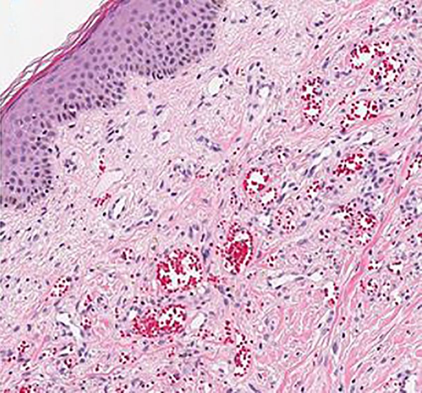
Fig. 2. (A) Diffuse capillary prolifer-
ation in the upper dermis, and
vascular dilatation involving mid-
dermis (H&E, ×40). (B) Capillary
proliferation involving the upper
dermis (H&E, ×200).
The lesions were not in the typical nodular shape, but case, multiple capillary hemangioma, which had never
rather rod like, and that made us suspect the lesions as occurred before, developed after taking TKIs. As the le-
scars. However, the fact that multiple lesions developed sions began to develop after taking TKIs, cutaneous side
without trauma was inconsistent with the clinical manifes- effects of the drugs were suspected. We suspect that the
tations of scars. Therefore, biopsy was performed for accu- TKIs caused paradoxical angiogenesis. Nilotinib, dasati-
rate diagnosis. Skin biopsy was done for the erythematous nib, and imatinib are all drugs with anti-angiogenic effect,
plaque on the chest. The biopsy revealed capillary pro- which are usually used as a treatment for angiogenic CML
7,8
liferation in the upper dermis, which is consistent with ca- cells . They are known to reduce angiogenic factors
7
pillary hemangioma (Fig. 2). No other capillary hemangio- such as vascular endothelial growth factor in CML patients .
ma were found in the abdomen and pelvic computed to- However, it is reported that TKIs can paradoxically acti-
mography scan. The cutaneous lesions were gradually im- vate the MEK/ERK pathway, which is required for angio-
proved without any special treatment. geneseis9. In an experiment that studied whether various
protein kinase inhibitors affected MEK/ERK pathways, the
DISCUSSION authors found that imatinib, nilotinib, dasatinib paradoxi-
cally stimulated MEK/ERK phosphorylation10. Since Raf-MEK-
The cutaneous side effects of TKIs include superficial ede- ERK signal transduction pathway is required for angio-
11
ma, maculopapular eruptions, and pigmentary changes genesis , we could consider the possibility that such
etc4. Also, few cases of capillary proliferative lesions mechanism have induced paradoxic angiogenesis, caus-
caused by these drugs have been reported. A case of scro- ing multiple capillary hemangioma. As a similar case, pre-
5
tal hemangioma developed after taking sunitinib , and a vious literature has reported an occurrence of Kaposi sar-
case of periungual pyogenic granuloma following im- coma following an imatinib mesylate administration in a
atinib administration6 had been reported. In the present CML patient12. The mechanism underlying the develop-
Vol. 33, No. 3, 2021 279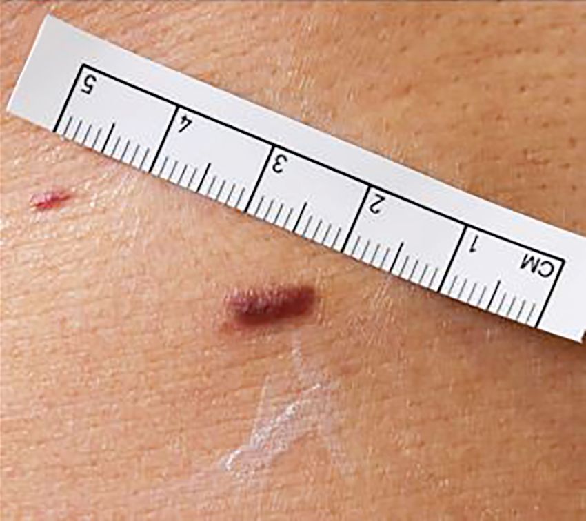
HJ Byun, et al

ment of Kaposi sarcoma was not clear. However, accord-        REFERENCES
ing to the adverse drug reaction probability scale, the
score estimated that the development of Kaposi sarcoma         1. George A, Mani V, Noufal A. Update on the classification
was probably associated with the imatinib treatment12. In         of hemangioma. J Oral Maxillofac Pathol 2014;18(Suppl 1):
the present case, Kaposi sarcoma could be excluded, be-           S117-S120.
                                                               2. Usui S, Kogame T, Shibuya M, Okamoto N, Toichi E. Case
cause only the capillary proliferation in the upper dermis
                                                                  of multiple disseminated cutaneous lobular capillary hemangioma
was observed in the biopsy, and no tissue findings that           that developed while taking oral contraceptive pills. J
would suspect Kaposi sarcoma such as slit like vascular           Dermatol 2019;46:e202-e203.
space, and proliferating spindle cells were found. In addi-    3. Ciarcia R, Damiano S, Puzio MV, Montagnaro S, Pagnini F,
tion to TKIs, factors that may have caused hemangioma in          Pacilio C, et al. Comparison of dasatinib, nilotinib, and
this case include history of exfoliative dermatitis and           imatinib in the treatment of chronic myeloid leukemia. J
leukemia. We cannot exclude these factors, because there          Cell Physiol 2016;231:680-687.
                                                               4. Lee WJ, Lee JH, Won CH, Chang SE, Choi JH, Moon KC, et
was a case report of multiple hemangioma in relation
                                                                  al. Clinical and histopathologic analysis of 46 cases of
with exfoliative dermatitis, and another report with under-
                                                                  cutaneous adverse reactions to imatinib. Int J Dermatol
lying leukemia13,14. These reports however, did not clarify       2016;55:e268-e274.
the mechanism of development of hemangioma by the              5. Tonini G, Intagliata S, Cagli B, Segreto F, Perrone G, Onetti
underlying diseases. The lesions developed after the ad-          Muda A, et al. Recurrent scrotal hemangiomas during
ministration of TKIs, and they gradually improved after           treatment with sunitinib. J Clin Oncol 2010;28:e737-e738.
drug discontinuation. Considering this temporal relation-      6. Dika E, Barisani A, Vaccari S, Fanti PA, Ismaili A, Patrizi A.
ship, it is reasonable to consider the possibility that the       Periungual pyogenic granuloma following imatinib therapy
                                                                  in a patient with chronic myelogenous leukemia. J Drugs
secondary neoplasms were caused by TKIs. To the best of
                                                                  Dermatol 2013;12:512-513.
our knowledge, this is the first case to report of multiple
                                                               7. Yıldırım R, Sincan G, Pala Ç, Düdükcü M, Kaynar L, Urlu
hemangioma occurred after the use of TKIs. It is mean-            SM, et al. Effects of Imatinib, Nilotinib, Dasatinib on VEGF
ingful that we added possible cutaneous side effects of           and VEGFR-1 levels in patients with chronic myelogenous
TKIs.                                                             leukemia. Eur J Gen Med 2016;13:111-115.
                                                               8. Pandey N, Yadav G, Kushwaha R, Verma SP, Singh US,
                                                                  Kumar A, et al. Effect of imatinib on bone marrow morphology
CONFLICTS OF INTEREST
                                                                  and angiogenesis in chronic myeloid leukemia. Adv
                                                                  Hematol 2019;2019:1835091.
The authors have nothing to disclose.
                                                               9. Greuber EK, Smith-Pearson P, Wang J, Pendergast AM. Role
                                                                  of ABL family kinases in cancer: from leukaemia to solid
FUNDING SOURCE                                                    tumours. Nat Rev Cancer 2013;13:559-571.
                                                              10. Packer LM, Rana S, Hayward R, O'Hare T, Eide CA,
None.                                                             Rebocho A, et al. Nilotinib and MEK inhibitors induce
                                                                  synthetic lethality through paradoxical activation of RAF in
                                                                  drug-resistant chronic myeloid leukemia. Cancer Cell 2011;
DATA SHARING STATEMENT
                                                                  20:715-727.
                                                              11. Murphy DA, Makonnen S, Lassoued W, Feldman MD,
Research data are not shared.
                                                                  Carter C, Lee WM. Inhibition of tumor endothelial ERK
                                                                  activation, angiogenesis, and tumor growth by sorafenib
ORCID                                                             (BAY43-9006). Am J Pathol 2006;169:1875-1885.
                                                              12. Campione E, Diluvio L, Paternò EJ, Di Marcantonio D,
Hyun Jeong Byun, https://orcid.org/0000-0002-4354-5655            Francesconi A, Terrinoni A, et al. Kaposi's sarcoma in a
Donghwi Jang, https://orcid.org/0000-0002-3495-4772               patient treated with imatinib mesylate for chronic myeloid
Jongeun Lee, https://orcid.org/0000-0002-1999-9948                leukemia. Clin Ther 2009;31:2565-2569.
                                                              13. Torres JE, Sánchez JL. Disseminated pyogenic granuloma
Se Jin Oh, https://orcid.org/0000-0001-7525-4740
                                                                  developing after an exfoliative dermatitis. J Am Acad
Youngkyoung Lim, https://orcid.org/0000-0002-6409-2704
                                                                  Dermatol 1995;32(2 Pt 1):280-282.
Ji-Hye Park, https://orcid.org/0000-0002-6699-5202            14. Pembroke AC, Grice K, Levantine AV, Warin AP. Eruptive
Jong Hee Lee, https://orcid.org/0000-0001-8536-1179               angiomata in malignant disease. Clin Exp Dermatol 1978;3:
Dong-Youn Lee, https://orcid.org/0000-0003-0765-9812              147-156.

280 Ann Dermatol
You can also read