A New Species of Spongilla (Porifera, Demospongiae) from a Karst Lake in Ha Long Bay (Vietnam) - MDPI
←
→
Page content transcription
If your browser does not render page correctly, please read the page content below

Journal of
           Marine Science
           and Engineering

Article
A New Species of Spongilla (Porifera, Demospongiae)
from a Karst Lake in Ha Long Bay (Vietnam)
Barbara Calcinai 1, * , Carlo Cerrano 1,2 , Laura Núñez-Pons 2 , Maurizio Pansini 3 ,
Do Cong Thung 4 and Marco Bertolino 3
 1    Department of Life and Environmental Sciences, Università Politecnica delle Marche, 60131 Ancona, Italy;
      c.cerrano@univpm.it
 2    Department of Integrated Marine Ecology (EMI), Stazione Zoologica Anton Dohrn (SZN), 80121 Napoli,
      Italy; laura.nunezpons@szn.it
 3    Department of Earth, Environmental and Life Sciences, University of Genova, 16132 Genova, Italy;
      pansisml@gmail.com (M.P.); marco.bertolino@edu.unige.it (M.B.)
 4    Institute of Marine Environment and Resources (IMER), Hai Phong City, Vietnam; thungdocong@gmail.com
 *    Correspondence: b.calcinai@univpm.it; Tel.: +39-071-2204283

 Received: 26 October 2020; Accepted: 5 December 2020; Published: 9 December 2020

 Abstract: Cahong in Ha Long Bay (Vietnam) is a small lake with a reduced, invisible connection
 with the open sea. The water column conditions locally experience notable fluctuations across the
 year, mostly driven by biannual monsoon seasons. Salinity, temperature, and pH often reach extreme
 values, unsustainable for the majority of the marine fauna. Therefore, the biodiversity of the benthic
 macrofauna in this peculiar habitat is remarkably low. In particular, a single sponge species new
 to science was found solely populating this characteristic brackish lake during our last survey in
 August 2018. Spongilla manconiae sp. nov. is a new Porifera species described here. It belongs to an
 exclusively freshwater taxon and seems to have acquired adaptive traits to tolerate extreme peaks
 of temperature and salinity. The mitochondrial Cytochrome C Oxidase subunit 1 (COI) and the
 nuclear Internal Transcribed Spacers 1 and 2 (ITSs) gene markers were used for barcoding tagging and
 phylogenetic analyses. The new species revealed large genetic distances and separate clustering in
 the tree topology, with respect to other reference spongillid sequences from various geographic areas.
 The study provides evidence for an urgency to protect these unique marine lake systems because
 they represent rare, fluctuant, fragile habitats that may speed up speciation processes.

 Keywords: Porifera; freshwater sponge; new species; karstification; Cat Ba Archipelago

1. Introduction
     Cat Ba Archipelago—in Ha Long Bay (North-Eastern Vietnam)—is characterized by thousands
of karst towers and islands dating back to the Tertiary Period (about 35 mya). Karst processes have
induced the formation of numerous sea lakes [1]. Besides Vietnam, in the Indo-Pacific area, analogous
marine lakes are found in Indonesia and Palau. Recent reports from Vermeulen et al. [2] registered
138 sea lakes in Ha Long Bay, which correspond to about 1/3 of the 400 estimated saltwater lakes
existing all over the world.
     Marine lakes are unique habitats characterized by brackish to marine water, according to
their degree of connection with the open sea, which can be secluded or visible [3]. In Vietnam,
the physical–chemical characteristics of the more cloistered marine lakes are for the most part extreme
and regulated by intense fluctuations of temperature and rainfall. During the monsoon season
occurring in late spring and summer, high temperatures and heavy rains cause severe stratification in
the water column. Under such extreme conditions, a large portion of the marine fauna in the lakes

J. Mar. Sci. Eng. 2020, 8, 1008; doi:10.3390/jmse8121008                            www.mdpi.com/journal/jmse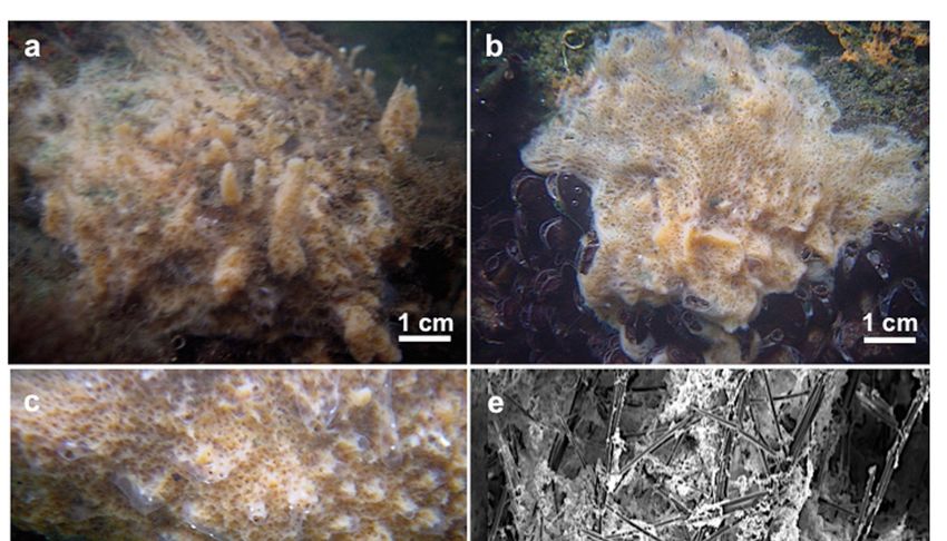
J. Mar. Sci. Eng. 2020, 8, 1008                                                                   2 of 14

partly degenerates or totally disappear [3]. Highly isolated lakes, such as the large marine Kakaban
lake in Indonesia, are known to host a high number of endemic species of several marine taxa [4] and
are consequently optimal scenarios for rapid evolution and speciation processes [5].
     Sponges are elemental components of the marine benthic fauna from tropical, temperate to polar
regions [6–9], acting as ecosystem engineers and performing key ecological roles [10,11]. The sponge
fauna of Vietnam has been moderately studied, with 299 demosponges (201 identified at species level)
reported up to 2013 [12,13]. In the frame of international projects with Vietnam, some of the present
authors recorded during several expeditions 46 species in nine surveyed marine karst lakes of Ha Long
Bay [12,14]; 23 of these are exclusive of such lake environment. Recently, Cerrano and co-workers [15]
noticed three new sponge species never recorded before in Vietnam and updated the inventory to
302. However, according to the last monitoring surveys, the sponge diversity in the area appears
to be drastically decreasing since more than half the species reported until 2007 were not recorded
again [15]. This paper provides the description of a sponge species new to science coming from an
enclosed marine karst lake, with the additional aim to increase awareness toward the singularity of
these rare, fragile, and threatened habitats, and encourage their protection.

2. Materials and Methods

2.1. Sampling Site and Sponge Collection
      The survey was conducted in August 2018 in Cahong lake (Ha Long Bay) in the frame of
the cooperative project “Studying biodiversity of limestone islands in Vietnam’s waters; proposing
solutions and models to use, conserve, and sustainably develop” (codes KC09.11/16-20), involving the
Polytechnic University of Marche, the University of Genoa, and the Institute of Marine Environment
and Resources (IMER) of Hai Phong. Cahong lake is an enclosed lake with no evident connection with
the open water. It is one of the numerous karstic saltwater lakes of Ha Long Bay, whose formation
goes back to the last glaciation period (c. 18 000 yr BP) [16]. This isolated lake is located inside the
small island of Congdo (20◦ 520 46” N–107◦ 120 12” E) (Figure 1). This lake has been identified after
interviewing local fishermen, exploiting a Local Ecological Knowledge (LEK) approach. Fishermen
told us that the lake is among those frequently visited and occasionally exploited as a natural fishing
pond for promoting fish farming. The physical–chemical characteristics of the superficial layer of the
lake were measured in December 2017 and August 2019 by the staff of the Hai Phong Institute using a
multifunctional environment meter (model WQC-24).
      Sponge collection was qualitative. Seven samples were collected by SCUBA diving, following
visually oriented transects, spaced about 5 m from one another to cover a significant area of the lake.
The maximum depth in the lake was 6 m; however, our sampling went down to 3 m, where the limit
of sponge life seemed to be established. Following reference [17], below this depth, the temperature
recorded by the dive computer (Ratio, iDive Easy) was very high, around 38 ◦ C and there were no
signs of living organisms. The only sponge species found in the lake was the one reported here. This
peculiar vertical distribution of environmental parameters reminds of solar ponds, rarely reported as
natural systems. A solar pond is a body of a certain liquid, relatively shallow and generally artificial
that maintains a downward positive density gradient and thereby entraps heat in bottom layers. Such
a density gradient counteracts the change of density induced by the absorption of solar radiation,
creating denser and warmer waters, which challenge the development of life toward the deeper layers.
The sponges were photographed in situ and fresh on board. Specimens were divided into subsamples
(about 3 cm3 ) and fixed in ethanol 70% for morphological observations and in 4% formaldehyde
solution for histological analyses; smaller portions (~1.5 cm) were preserved in absolute ethanol at
4 ◦ C for DNA barcoding.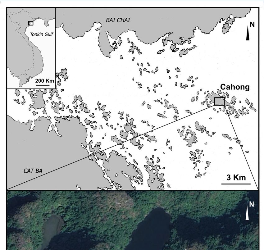
J. Mar. Sci. Eng. 2020, 8, 1008                                                                                 3 of 14
      J. Mar. Sci. Eng. 2020, 8, x FOR PEER REVIEW                                                        3 of 17

                       Figure 1. Map of the sampling site. The image below illustrates the Cahong lake.
                  Figure 1. Map of the sampling site. The image below illustrates the Cahong lake.
     2.2. Morphological
2.2. Morphological StudyStudy
          The material
     The material   for for
                        thethe  study
                             study  ofof spiculemorphometry
                                       spicule   morphometry andandthe
                                                                     theskeletal organization
                                                                          skeletal            waswas
                                                                                   organization  prepared
                                                                                                     prepared
     following Rützler [18] and observed under light microscopy. Dissociated spicules were transferred
following Rützler [18] and observed under light microscopy. Dissociated spicules were transferred
     onto stubs and coated with gold for SEM analyses (Philips XL 20 and Vega3_TESCAN Microscope
onto stubs and coated with gold for SEM analyses (Philips XL 20 and Vega3_TESCAN Microscope
     type LMU). The skeletal architecture was studied by examining hand-cut sections of sponge
type LMU).   The
     portions, by skeletal
                  SEM [19].architecture was studied by examining hand-cut sections of sponge portions,
by SEM [19].
          Spicule dimensions (obtained from 30 spicules) are given as the smallest length-(mean ±
     Spicule dimensions
     SD)-largest            (obtained
                 length x smallest    from 30 spicules)
                                    width-(mean          are deviation)-largest
                                                 ± standard  given as the smallest  length-(mean ± SD)-largest
                                                                                width.
length x smallest  width-(mean
          Type material            ± standard
                         was deposited          deviation)-largest
                                         at the Museo                width.
                                                      di Storia Naturale  di Genova (MSNG), Italy.
     Type material was deposited at the Museo di Storia Naturale di Genova (MSNG), Italy.
      2.3. Genotyping
2.3. Genotyping
          Genomic DNA of sponge subsamples was purified using HiPura Soil DNA Purification Kit
    (HiMedia DNA
    Genomic   Laboratories, Mumbai,
                     of sponge       India) following
                                 subsamples            manufacturer’s
                                              was purified              protocols,
                                                              using HiPura    Soil and
                                                                                   DNA was   stored at Kit
                                                                                         Purification
    −20°C. Two gene marker regions were employed for barcoding. The mitochondrial Cytochrome C ◦
(HiMedia Laboratories, Mumbai, India) following manufacturer’s protocols, and was stored at −20 C.
    Oxidase subunit 1 (COI) was amplified using two pairs of primers. The standard COI partition (~640
Two gene marker regions were employed for barcoding. The mitochondrial Cytochrome C Oxidase
subunit 1 (COI) was amplified using two pairs of primers. The standard COI partition (~640 bp) was
amplified with the universal [20] dgHCO2198 (50 -GGTCAACAAATCATAAAGAYATYGG-30 ) and
dgLCO1490 (TAAACTTCAGGGTGACCAAARAAYCA-30 ) primers. Additionally, the Erpenbeck’s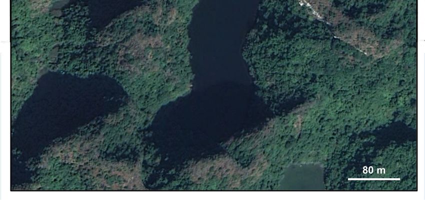
J. Mar. Sci. Eng. 2020, 8, 1008                                                                       4 of 14

‘I3-M11’extension of ~560 bp, which overlaps ~60 bp with Folmer’s 30 COI partition was amplified
using the sponge specific primers PorCOI2fwd (50 -AATATGNGGGCNCCNGGNATNAC-30 ) and
PorCOI2rev (50 -ACTGCCCCCATNGATAAAACAT-30 ) [21]. These two fragments were then
concatenated with bioinformatic approaches (see below) to obtain a fragment of ~1100 bp, which
seems to improve taxonomic resolution in certain groups of Porifera and other diploblasts [22].
The nuclear ITSs fragment that included both Internal Transcribed Spacers 1 and 2, and covering
18S-ITS1-5.8S-ITS2-28S (~850 bp) was amplified with the specific primer RA2 (50 -ACTGCCCC
CATNGATAAAACAT-30 ) priming on the 30 terminus of the 18S small ribosomal subunit, and the
ITS2.2 primer (50 -CCTGGTTAGTTTCTTTTCCTCCGC-30 ) targeting the 50 terminus of the 28S large
ribosomal subunit [23]. A third fragment targeting the 28S rDNA C-Region, recently included among
the recommended taxonomic markers for Porifera [24], continuously failed in amplification trials and
could not be sequenced for the present study. PCR amplifications were performed in 25 µL volume
reactions containing 2.5 µL (2 mM) of Buffer, 1 µL Deoxynucleotide (dNTP) solution mix (2 mM),
0.8 µL (20 µg/µL) of bovine serum albumin (BSA), 0.3 µL (5 U/µL) of Roche Taq DNA Polymerase,
0.8 µL (10 mM) of each primer, and 1 µL of template DNA, and following a thermocycling profile of
5 min at 95 ◦ C; followed by 35 cycles of 30 s at 94 ◦ C, 45 s at 57–50 ◦ C, and 90 s at 72 ◦ C; and a final
extension for 10 min at 72 ◦ C. The amplified PCR products were checked in 1% agarose gel under UV
light and quantified on NanoDrop® . Sanger sequencing was performed bi-directionally at in-house
facilities (Servizio di Biologia Molecolare RIMAR, SZN) on a Thermo Fisher Scientific 48 capillary
ABI 3730xl DNA Analyzer, using 4.5 micromolar of each of the corresponding PCR primer pairs, and
15 femtomoles/uL of DNA template. Chromatograms of forward and reverse reads were visualized,
assembled, and corrected on Geneious Prime® 2020.1.1 http://www.geneious.com [25], and poriferan
origin was checked by BLAST against NCBI GenBank (http://www.ncbi.nlm.nih.gov/BLAST/ [26].
All the sequences in this study were retrieved from a single specimen and have been deposited, along
with the morphological descriptions, in GenBank (accession numbers: MT176773 and MT177208) and
Sponge Barcoding Project databases for public access.

2.4. Genetic Analysis
     Reference sequences for the two markers, COI and ITSs, from sponges in the Family Spongillidae
(including all the available entries for Genus Spongilla), along with two outgroup demosponges
Echinospongilla brichardi (Brien, 1974) and Oncosclera sp. in the Order Spongillida, were downloaded
from Genbank for phylogenetic approaches (Table S1 in the Supplementary Materials for accession
numbers of reference sequences). The resulting data set was aligned against our sequences with
MAFFT (Multiple Alignment using Fast Fourier Transform) [27] under default settings in Geneious
Prime® 2020.1.1 [25]. COI alignments were checked by amino acid translation. GBlocks 0.91b
was applied to identify and exclude poorly aligned blocks with large gap positions variability
in the ITSs alignment. The program was run with stringent settings via the web interface (http:
//phylogeny.lirmm.fr/phylo_cgi/one_task.cgi?task_type=gblocks) [28]. Curated ITS alignments from
GBlocks were then used for downstream analyses. For each marker separately, and for the concatenated
alignment, COI-ITSs Maximum Likelihood (ML) trees were built on IQTree with 1500 ultrafast bootstrap
replicates [29] under the most fitting genetic models by partitions according to ModelFinder [30], i.e.,
TPM3u+F+I for COI and TPM2+F+G4 for ITSs. Concatenated data returned the most informative
outcomes and, hence, was further used for Bayesian analysis (BA) and tree construction on MrBayes [31]
with GTR GAMMA model and Markov Chain Monte Carlo (MCMC) set for 1,100,000 generations,
4 chains, 200 generations sampling frequency, and 500 burnin values. All the phylogenetic analytic
tools were run on the CIPRES web portal (http://www.phylo.org/) [32].
     To estimate species delimitation, two methods were applied: the Automatic Barcode Gap Discovery
(ABGD) based on barcode gap [33] and the Poisson-Tree-Processes (PTP), which relies on branch
lengths representing the number of substitutions in a phylogenetic tree [34]. The ABGD analysis was
performed on our alignments through the server (www.abi.snv.jussieu.fr/public/abgd/) with parameters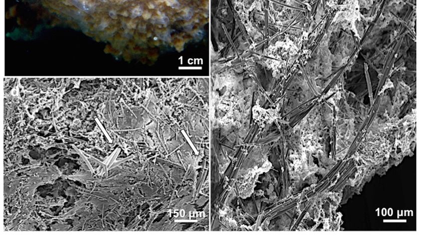
J. Mar. Sci. Eng. 2020, 8, 1008                                                                     5 of 14

set to genetic model Kimura (K80) TS/TV 2.0 and 1.5 relative gap width. For bPTP method, the above
ML trees from IQTree (with their corresponding substitution models by partition) were used as inputs
on the webserver (https://species.h-its.org/ptp/).

3. Results and Discussion
     Marine lakes of this area are not monitored with continuity and historical time series are not
available. Information regarding human exploitation of the lakes has been collected through Local
Ecological Knowledge (LEK), interviewing some fishermen encountered in the bay. The environmental
characteristics of the lake are shown in Table 1. The salinity reflected remarkably low values, especially
during the summer, when monsoon rains are frequent. The temperature showed wide oscillations,
escalating in the hot season by over 13 ◦ C and reaching particular high values on the lake bottom (~6 m
depth). Such increased temperatures in the lower layers of the lake create the typical condition of solar
ponds and could limit in part sponge colonization toward deeper zones.

      Table 1. Environmental factors in the Cahong lake measured in winter (December 2017) and summer
      August 2018).

                                  7/12/2017                            21/08/2018
                            S‰ = 12                                      S‰ = 8
                            pH = 7.2                                   pH = 7.65
                DO mg/L (Dissolved Oxygen) = 6.7           DO mg/L (Dissolved Oxygen) = 5.09
                   T = 20.7 ◦ C At the surface                 T = 33.4 ◦ C At the surface

     The exploratory transect dives at Cahong Lake revealed a benthic community composed of
green algae, conspicuous bivalve aggregations belonging to various species, polychaetes (Owenia sp.,
Sabellaria sp.) fouling on algal material and other surfaces, and a few small-sized fish. The sponge
fauna went down to 3 m depth and consisted of a single demosponge species (described here) growing
on diverse natural (rocks and roots) and anthropogenic artificial substrates (polystyrene material).

3.1. Taxonomical Account
      Class: Demospongiae Sollas, 1885
      Subclass: Heteroscleromorpha Cárdenas, Pérez and Boury-Esnault, 2012
      Order: Spongillida Manconi and Pronzato, 2002
      Family: Spongillidae Gray, 1867
      Genus: Spongilla Lamarck, 1816
      Spongilla manconiae Calcinai and Bertolino sp. nov.
      urn:lsid:zoobank.org:act:01A112C2-949E-4571-8B3D-7692BE9B66FE
      (Figures 2–4)

3.1.1. Material Examined
    Holotype: MSNG 61501, Cahong, Ha Long Bay, Northern Vietnam, Tonkin Gulf, 21 August 2018,
0–50 cm depth. Paratype: MSNG 61502, Cahong, Ha Long Bay, Northern Vietnam, Tonkin Gulf,
21 August 2018, 0–50 cm depth.
    Topotypes: HL108, HL108A, HL108B, HL108C, HL108D. Cahong, Ha Long Bay, Northern Vietnam,
Tonkin Gulf 21 August 2018, 0–50 cm depth.

3.1.2. Species Diagnosis
     Spongilla characterized by creamy-brownish color, smooth oxeas, and spiny microxeas typically
with bouquets of spines in the central part and triangular spines on the distal parts. Gemmuloscleres
are spined oxeas.
J. Mar. Sci. Eng. 2020, 8, 1008 6 of 14
3.1.3. Description
Thickly encrusting with ridges, grooves, and erect processes up to 3 cm high, with apical oscula.
The surface is uneven and irregular. A translucent, thin dermal membrane is detectable especially over
the processes and the oscula. Consistency is fragile and soft. The color is creamy/brownish in life and
brown in dry state
J. Mar. (Figure
Sci. Eng. 2020, 8, x 2a–c).
FOR PEERSome
REVIEWspecimens were filled with gemmules. 7 of 17
Spongilla
Figure 2. Figure manconiae
2. Spongilla sp.sp. nov.
manconii (a)Holotype
nov. (a) HolotypeMSNG MSNG
61501; 61501; (b) Paratype
(b) Paratype MSNG 61502; MSNG
(c) 61502;
Topotype
(c) Topotype HL108; HL108; (d) Ectosomal
(d) Ectosomal skeleton with
skeleton with tangential
tangentialspiny microxeas.
spiny ArrowsArrows
microxeas. indicate the
indicate the
spicules ofspicules of the longitudinal primary fibers that protrude from the sponge surface; (e) Choanosomal
the longitudinal primary fibers that protrude from the sponge surface; (e) Choanosomal
skeletal architecture (transverse section).
skeletal architecture (transverse section).
The holotype consists of three fragments of about 3 × 1.5 × 1 cm, 2 × 1 cm and 4 mm thick
and 1 × 1 cm and 3 mm in thickness. The paratype consists of two portions of about 3 × 2 × 2 and
2 × 1 × 1 cm, growing under a piece of polystyrene floating in the lake (Figure 3a). Other specimens
covered areas up to 100 cm2 . Ectosomal skeleton consists of spiny microxeas tangentially arranged
(Figure 2d). Choanosomal skeleton is an irregular, paucispicular, anisotropic reticulation; longitudinal
primary fibers run toward the sponge surface (Figure 2e). Megascleres are oxeas slightly curved,
smooth, and with acerate tips (Figure 4a). Numerous thinner oxeas are considered forms of growth
(measurements in Table 2). Microscleres are spiny microxeas (Figure 4b). At their extremities, they are
covered by hooked spines bent toward the shaft. At the center of the spicule, axis spines are grouped
in bouquets (measurements in Table 2).J. Mar. Sci. Eng. 2020, 8, 1008 7 of 14
J. Mar. Sci. Eng. 2020, 8, x FOR PEER REVIEW 8 of 17
Figure 3. Spongilla
Figure 3. Spongilla manconiaemanconii
sp. sp. nov.(a)
nov. (a)Paratype
Paratype MSNG
MSNG 61502 grown
61502 underunder
grown a piece a
ofpiece
polystyrene
of polystyrene
floating in the lake; (b) Magnification of the gemmules; (c) SEM image of the gemmules; (d)
floating in the lake; (b) Magnification of the gemmules; (c) SEM image of the gemmules; (d) Gemmule
Gemmule with a single foramen; (e) Section of a gemmule, with a simple, monolayered theca,
with a single foramen; (e) Section of a gemmule, with a simple, monolayered theca, armored by a layer
armored by a layer of tangential gemmuloscleres; (f) Histological section of a gemmule.
of tangential gemmuloscleres; (f) Histological section of a gemmule.
Table 2. Spicule dimensions of the examined specimens. The “*” indicates the absence or scarcity of
spicules in the preparation due to the lack of gemmules.
Specimens Oxeas (µm) Microxeas (µm) Gemmuloscleres (µm)
MSNG 61501 205 (272.5 ± 28.4) 332.1 × 8.2 80.6 (103.59 ± 18.5) 161.2 × 1.3
78 (97.85 ± 8.8) 111.8 × 7.8
holotype (11.8 ± 3.2) 16.4 (2.08 ± 0.6) 2.6
MSNG 61502
255 (291.1 ± 23.8) 350 × 10 75 (94.3 ± 12.7) 112.5 × 2.5 87.5 (100.4 ± 11.9) 124 × 5
paratype
HL 108 255 (283.5 ± 19.1) 310 × 7.5 (8.9 ±
80 (103.6 ± 11.4) 120 × 2.5 82.5 (101.8 ± 9) 115 × 5
topotype 1.5) 11.25
HL108A 260 (308 ± 26.3) 345 × 6.2 (8.9 ±1.5)
82.5 (107.1 ± 111.4) 122.5 × 2.5 *
topotype 10
HL108B
275 (306 ± 16.4) 340 × 7.5 67.5 (94.8 ± 13.3) 110 × 2.5 100–105 *
topotype
HL108C 260 (282 ± 17.2) 320 × 6.2 (8.5 ± 1.6)
90 (103.2 ± 9.1) 122.5 × 2.5 *
topotype 10
HL108D 280 (311 ± 14.4) 340 × 7.5 (8.2 ± 1.1)
77.5 (101.5 ± 15.3) 125 × 2.5 75 (96.9 ± 11) 120 × 5
topotype 10J. Mar. Sci. Eng. 2020, 8, 1008 8 of 14
J. Mar. Sci. Eng. 2020, 8, x FOR PEER REVIEW 9 of 17
Figure 4. Spongilla manconii sp. nov. SEM images of spicules. (a) Oxeas; (b) Microxeas, with
Figure 4. Spongilla manconiae
magnification sp. (c)nov.
of the spines; SEM images
Gemmuloscleres; of spicules. (a) Oxeas; (b) Microxeas, with
(d) Gemmule.
magnification of the spines; (c) Gemmuloscleres; (d) Gemmule.
3.1.4. Habitat and Distribution
Gemmules areCovering large areas
of a single of substrate
type; they areat low depth. Known
abundant andfrom Hồ Cá Hồng
scattered in(Cahong), Ha Long
the tissue, are about 500 µm
Bay, Tonkin Gulf, 20°52′41′′ N–107°12′1′′ E.
in diameter, and are covered by a simple, monolayered theca, armored with a layer of tangential
gemmuloscleres3.1.5. Etymology
(Figure 3a–f). A single foramen was observed (Figure 3d). Gemmuloscleres (Figure 4c)
This new species isspines,
are spined oxeas with triangular named after
moreDr. Renata Manconi (Dipartimento
concentrated di Scienze
at the distal della Natura e
part (measurements in Table 2).
del Territorio (DipNeT)—Università degli Studi di Sassari) in recognition of her relevant
contribution to taxonomic studies on freshwater sponges.
3.1.4. Habitat and Distribution
Covering large areas of substrate at low depth. Known from Hồ Cá Hồng (Cahong), Ha Long Bay,
Tonkin Gulf, 20◦ 520 41” N–107◦ 120 1” E.
3.1.5. Etymology
This new species is named after Dr. Renata Manconi (Dipartimento di Medicina Veternaria—Università
degli Studi di Sassari) in recognition of her relevant contribution to taxonomic studies on freshwater sponges.
3.1.6. Remarks
The species is assigned to the genus Spongilla because of the presence of smooth oxeas, spiny
microxeas, and spiny oxeas as gemmuloscleres. Gemmules are distributed in dense clusters or scattered,
with a mono-layered gemmular theca and a simple foramen.J. Mar. Sci. Eng. 2020, 8, 1008 9 of 14
Fifteen species belonging to Spongilla are known [24], but one of them, Spongilla friabilis, Lamarck
(1816) was insufficiently described without any information about spicules, making difficult any
comparison (Table 3): “Sp. sessilis, convexa, obsoletè lobulata, intùts fibrosa; fibris longitudinalibus,
ramuloso-cancellatis. Spongia friabïlis. Esper. suppl. tab. 62. Habite dans les étangs. Elle est granifère, et n’a
presque point de parenchyme entre ses fibres” [35]. Some authors [24] point out that this species, described
by Lamarck, could be a junior homonym of Spongia friabilis Linnaeus, 1788 from the Bodensee lake
(Germany) and, therefore, is considered as taxon inquirendum.
Table 3. Comparison of the spicule dimensions of Spongilla manconiae sp. nov. with the other known
Spongilla species. Geographical distributions were checked in the World Porifera Data Base [24].
Gemmuloscleres
Species Oxeas (µm) Microxeas (µm) Distribution
(µm)
Indonesia, Australia,
S. alba Carter, 1849 277–356 × 16–26 55–142 × 2–3 90–143 × 6–10 South America, India,
Caribbean
S. arctica Annandale, 1915 154–280 × 4–14 32–70 × 2–4 80–140 × 4–6 Arctic, Russia
S. cenota Penney and
310–410 × 14–22 68–123 × 2–3 65–86 × 9–13 Yucatan
Racek, 1968
S. friabilis Lamarck, 1816 taxon inquirendum – – Asia ?
S. chaohuensis Cheng, 1991 208–280 × 8–15.2 – 80–112 × 2.4–8.8 China
S. helvetica Annandale,
148–200 × 4–9 24–55 × 1–2 32–68 × 2–5 Switzerland
1909
S. jiujiangensis Cheng, 1991 204–296 × 6.4–16 - 72–152 × 3.2–9.6 China
Northern Polar Circle,
Ireland, England,
S. lacustris (Linnaeus, 1759) 90–350 × 2–18 25–178 × 2–8 21–130 × 1–10
France, Italy,
California,
S. mucronata Topsent, 1932 245–275 × 10–11 35–90 × 1.2–2 95–120 × 8–9 Africa
S. permixta Weltner, 1895 – – 320–390 × 3–5 Egypt
S. prespensis Hadzische,
170–200 × 6–13 157.4 × 2–4 50–120 × 3–7 Macedonia
1953
S. sarasinorum Weltner, 160 (190) 210 × 13
up to 250 180 × 8 Indonesia
1901 (18) 20
240 (293) 350 × 13 61 (80.1) 100 × 3
S. shikaribensis Sasaki, 1934 50 (75.7) 101 Japan
(13.9) 15 (4.1) 5
S. stankovici Arndt, 1938 176–235 × 3–6 180 × 8 160–210 × 8 Albania
S. wagneri Potts, 1889 144–270 × 7–12 49–62 × 2–4 48–75 × 6–8 United States
Spongilla manconiae sp.
250–350 × 6.2–16.4 67.5–161.2 × 1.3–2.6 75–124 × 5–7.8 Vietnam
nov.
The species of the genus Spongilla described in geographical areas, next to Vietnam, are five: S. alba,
S. chaohuensis, S. jiujiangensis, S. sarasinorum, and S. shikaribensis; they differ from S. manconiae for their
spicular complements and shape and size of the spicules (Table 3). Among these, S. chaohuensis, Cheng,
1991, from China, is the only one close to the new species. Cheng’s publication is in Chinese, and the
translated basic parts are reported below: “Several specimens of flat sponge. With digits, channels,
and cavities. Large, circular oscula, slightly raised. Color in dry state from light green on the surface to
dark ash in the inner part. Structure: fine and compact skeletal composition, radial shape of the fibers
not evident, a large number of horizontal fibers form a very fine skeletal network. It has a lichenoid
type surface”.
Spongilla chaohuensis, Cheng, 1991 lacks spined microxeas (Table 3) and its spicules are smaller
than in S. manconiae. The latter, therefore, has to be considered as a new species for science.
3.2. Genotyping
The concatenated fragment obtained from integrating the COI Folmer (613 bp) and Erpenbeck
(561 bp) partitions was 1036 bp long, whereas the ITSs region spanned 861 bp. Phylogenetic analyses
were largely hampered due to the scarce representation of Spongilla spp. in the reference databases.
Fourteen COI sequences, 12 of S. lacustris (from Russia, India, Germany, and Ireland) and twoJ. Mar. Sci. Eng. 2020, 8, 1008 10 of 14
of S. vastus (from Indonesia), and 12 ITSs sequences, all from S. lacustris (from Russia, Germany,
Bosnia-Herzegovina, and the USA.), were the only reference sequencing data available to build
congeneric alignments for both markers (Supplementary Table S1). GBlocks curated ITSs alignments
retained 373 bp for the set of Spongilla sequences and 300 bp for the larger set comprising the family
Spongillidae. When accounting for the number of variable nucleotide sites in the sequences of the new
Spongilla manconiae against reference congeneric sequences, the COI alignment revealed 13–20 (2–3%)
variable sites against S. lacustris and S. vastus. A much larger proportion of consistent informative
variable sites was found in the ITSs against S. lacustris entries (110–111, 30%). This supports the
already proposed higher resolution potential of the nuclear ITS with respect to COI for freshwater
sponges [36,37]. BLAST searches matched the non-curated ITS from S. manconiae with S. lacustris
entries. Instead, the closest match for COI did not correspond to any Spongilla (S. lacustris or S. vastus)
but to a Corvospongilla ultima from Myanmar, which differed from our sequence by 10 nucleotides.
These results further advocate a low taxonomical (intra- and intergeneric) resolution of COI and uphold
the need to combine several genetic markers for resolving certain Porifera groups [22,36,38].
Species delimitation analyses based on ABGD and PTP for each of the two markers separated
S. manconiae from other Spongilla. This was an expected outcome, as reference sequences came from
geographically distant specimens, with divergent morphologies (see Table 3). PTP delimitation for
the COI marker afforded coherent clustering for S. vastus but was inconsistent within S. lacustris.
In particular, two conspecifics coming from India and two from Russia were grouped out of the major
cluster. For the ITSs data instead, all S. lacustris formed a single cluster separated from S. manconiae.
Intra- and interspecific dissimilarity based on ABGD for the COI marker revealed distances ranging
0–0.7% within the reference Spongilla sequences, and 1.3–1.9% between any congeneric entry against S.
manconiae. Instead, for the curated ITSs gene, distances ranged 0–0.7% within S. lacustris, while the
interspecific distance against S. manconiae was 32%. Across the family Spongillidae, ABGD distances
spanned 0.7–2.8% with the COI and 12–49% with the ITSs. Once more dissimilarities based on
COI revealed low taxonomical resolution across genera, with values below the universal threshold
consensus of 0.3% genetic distance for species delimitation [22,36,38].
Phylogenetic ML and BI trees on family Spongillidae with either concatenated (COI–ITS) or
non-concatenated (separate COI and ITSs, results not shown) data could not recover Genus Spongilla as
monophyletic (Figures 5 and 6). Spongillidae, which is the most specious freshwater family and with a
high incidence of endemism, is recognized in earlier molecular studies as non-monophyletic [37,39].
On what regards Spongilla, the description of the genus is rather ample, e.g., with only some species
containing gemmular cages made of megascleres, gemmules may be without or with gemmuloscleres,
gemmules may have a single or multiple foramina. Out of the 14 accepted species (15 with the
present record, and not considering Spongilla friabilis), only two are represented as sequencing entries,
suggesting that further genetic work to repopulate public databases is required to corroborate the
non-monophyly of this taxon. In this particular study, some of the observed noise on tree topologies
was likely induced by the two Indian and two Russian discordant S. lacustris mentioned above. Spongilla
manconiae was placed alone on a very long branch in the phylogenetic trees, close to its congeneric
group, and clustered with Corvospongilla ultima (see Figures 5 and 6). This clustering is probably
an artifact resulting from the long-branch attraction, again due to a deficient amount of congeneric
sequencing reference data, and low COI resolution. C. ultima (originally described as Spongilla ultima) is
morphologically different (e.g., microscleres are smooth or spined micropseudobirotules, megascleres
may be smooth or granulated strongyles or oxeas). Moreover, it is exclusively a freshwater species,
never reported from brackish systems [40].J. Mar. Sci. Eng. 2020, 8, 1008 11 of 14
J. Mar. Sci. Eng. 2020, 8, x FOR PEER REVIEW 13 of 17
Figure
Figure 5. Bayesian
5. Bayesian inference
inference (BI) phylogenetic
(BI) phylogenetic tree, applying
tree, applying the concatenated
the concatenated data
data set of the set of the
Cytochrome
C Cytochrome C Oxidase subunit 1 (COI) and curated ITSs, with the posterior probabilities shown
Oxidase subunit 1 (COI) and curated ITSs, with the posterior probabilities shown at each node.at
each node. The analysis was built with the newly described sponge, Spongilla manconiae sp.with
The analysis was built with the newly described sponge, Spongilla manconiae sp. nov., along nov.,
along with a suite
a representative representative
of referencesuite of reference
sequences in the sequences
Spongillidaein Family
the Spongillidae Family plus
plus two outgroup two
entries
outgroup entries
(Echinospongilla (Echinospongilla
bichardi, and Oncosclera bichardi, and Oncosclera
sp.) downloaded from sp.) downloaded
the GenBank from The
database. the country
GenBank
database.
codes The country
and Accession codescan
numbers andbeAccession
found in numbers can be found
Supplementary in Supplementary Table 1.
Table S1.
The new species is the first record of Spongilla in Vietnam, adverting a certain level of endemism,
and likely genetic and consequent phenotypic diversification. Indeed, the morphological characteristics,
the branch length topology in the phylogenetic trees, and the genetic distances recorded for the new
S. manconiae drive us to suggest two things. On the one hand, they highlight a gap in the reference
set of taxonomically related sequences from nearby geographic areas. On the other hand, it implies a
process of accelerated evolution (and reflexed in the studied loci), likely driven by the extreme and
unique environmental conditions in the enclosed lake. Rapid radiation processes, involving genetic,
morphofunctional, and anatomical adaptations, seem to be a common trend in habitants of ancient
lakes [5]. In the light of our results, we emphasize the role of enclosed marine lakes as motors of
accelerated speciation and their relevance to study evolution and adaptation processes.J. Mar. Sci. Eng. 2020, 8, 1008 12 of 14
J. Mar. Sci. Eng. 2020, 8, x FOR PEER REVIEW 14 of 17
Figure Figure 6. Maximum
6. Maximum likelihood
likelihood phylogenetic
phylogenetic tree applying
tree applying the concatenated
the concatenated data data
set ofset
COIof and
COIcurated
and
curated
ITSs, with ITSs, with the
the bootstrap bootstrap
values shown values shown
at each node.at each node. Thewas
The analysis analysis
builtwas
withbuilt
thewith
newly thedescribed
newly
sponge,described
Spongillasponge,
manconiaeSpongilla manconiae
sp. nov., along sp.withnov., along with a suite
a representative representative suite
of reference of reference
sequences in the
sequences
Spongillidae in plus
Family the Spongillidae
two outgroup Family
entriesplus two outgroup
(Echinospongilla entriesand
bichardi (Echinospongilla
Oncosclera sp.) bichardi and
downloaded
Oncosclera
from the GenBank sp.) downloaded
database. from the codes
The country GenBank anddatabase.
AccessionThe country can
numbers codes
beand Accession
found numbers
in Supplementary
can
Table S1. be found in Supplementary Table 1.
Supplementary Materials: The following is available online at http://www.mdpi.com/2077-1312/8/12/1008/s1,
Table S1: Reference
Supplementary Materials:sequences downloaded
The following from GenBank
is available onlinedatabase for COI marker gene of sponge species in
at http://www.mdpi.com/2077-1312/8/12/1008/s1,
Table S1:theReference
Family Spongillidae,
sequences plus Echinospongilla.
downloaded from GenBank database for COI marker gene of sponge species in the
Family Spongillidae, plus Echinospongilla.
Author Author contributions:
Contributions: B.C.,B.C.,
M.P.,M.P.,
andand M.B.
M.B. cooperated
cooperated ininthe
thefield
fieldand
and did
did the
the morphological
morphological work.
work.B.C.B.C.
andand
M.B wroteM.B the
wrote the species
species descriptions.
descriptions. C.C.C.C. coordinated
coordinated thefieldwork,
the fieldwork, performed
performed thethesampling,
sampling, and drafted
and the the
drafted
manuscript.
manuscript. L.N.-P. L.N.-P.
performedperformed
the DNA theanalyses
DNA analyses and the
and drafted drafted the manuscript.
manuscript. D.C.T. coordinated
D.C.T. coordinated and
and cooperated
cooperatedAll
in the fieldwork. in the fieldwork.
authors haveAll authors
read have read
and agreed to and agreed to the
the published published
version version
of the of the manuscript.
manuscript.
Funding: This paper is the result of a state research project for international cooperation KC09.11/16-20 “Study on
Funding:
biodiversity This paperislands
of limestone is the result of a state research
and limestone project for
archipelagos international
in Vietnam’s cooperation
waters; KC09.11
propose / 16-20
solutions “Study
and models
on biodiversity
for use and protection,ofsurvival
limestone andislands and limestone
sustainable archipelagos
development” in Vietnam's
and project waters;23.02/20-20
cod NVCC propose solutions and
that provided
supportmodels
for thefor use and protection, survival and sustainable development” and project cod NVCC 23.02/20-20 that
research.
provided support
Acknowledgments: Thefor the research.
authors would like to thank F. Rispo and V. Mazzella for laboratory work and J. Moles
for analytical advice.
Acknowledgments: The authors would like to thank F. Rispo and V. Mazzella for laboratory work and J. Moles
Conflicts of Interest: The authors declare that they have no conflict of interest.
for analytical advice.
Conflicts of interest: The authors declare that they have no conflict of interest.
References
1. Thung, D.G.; Ngai, N.D.; Sinh, N.V.; Quan, N.V.; Tien, D.D.; Thuoc, C.V.; Trang, C.T.T. Biodiversity of Limestone
Island and Archipelagos Areas in Northeast Coast of Vietnam, Orientation of Sustainable Use Solutions; Publishing
House of Natural Science and Technology: Ha Noi, Vietnam, 2018; pp. 56–57. (In Vietnamese)J. Mar. Sci. Eng. 2020, 8, 1008 13 of 14
2. Vermeulen, J.J.; Anker, K. Outstanding global values in geology and environment in Cat Ba archipelago
and Halong Bay. In Proceedings of the Biodiversity Conservation Solutions in Ha Long bay and Cat Ba
Archipelago, IUCN, Haiphong City, Vietnam, 24 August 2017; pp. 16–23.
3. Cerrano, C.; Azzini, F.; Bavestrello, G.; Calcinai, B.; Pansini, M.; Sarti, M.; Thung, D.C. Marine lakes of karst
islands in the Ha Long Bay (Vietnam). Chem. Ecol. 2006, 22, 489–500. [CrossRef]
4. Becking, L.E.; Renema, W.; Santodomingo, N.K.; Hoeksema, B.W.; Tuti, Y.; de Voogd, N.J. Recently discovered
landlocked basins in Indonesia reveal high habitat diversity in anchialine systems. Hydrobiologia 2011, 677,
89–105. [CrossRef]
5. Dawson, M.; Hamner, W.N. Rapid evolutionary radiation of marine zooplankton in peripheral environments.
Proc. Natl. Acad. Sci. USA 2005, 102, 9235–9240. [CrossRef]
6. Cerrano, C.; Bavestrello, G.; Calcinai, B.; Cattaneo-Vietti, V.; Chiantore, M.; Guidetti, M.; Sarà, A. Bioerosive
processes in Antarctic seas. Pol. Biol. 2001, 24, 790–792. [CrossRef]
7. Díaz, M.C.; Rützler, K. Sponges: An essential component of Caribbean coral reefs. Bull. Mar. Sci. 2001, 69,
535–546.
8. Bertolino, M.; Cerrano, C.; Bavestrello, G.; Carella, M.; Pansini, M.; Calcinai, B. Diversity of Porifera in
the Mediterranean coralligenous accretions, with description of a new species. Zookeys 2013, 336, 1–37.
[CrossRef]
9. Gerovasileiou, V.; Voultsiadou, E. Sponge diversity gradients in marine caves of the eastern Mediterranean.
J. Mar. Biol. Assoc. UK 2016, 96, 407–416. [CrossRef]
10. Wulff, J.L. Ecological interactions of marine sponges. Can. J. Zool. 2006, 84, 146–166. [CrossRef]
11. Becerro, M.A. Quantitative trends in sponge ecology research. Mar. Ecol. 2008, 29, 167–177. [CrossRef]
12. Azzini, F.; Calcinai, B.; Cerrano, C.; Bavestrello, G.; Pansini, M. Sponges of the marine karst lakes and of
the coast of the islands of Ha Long Bay (North Vietnam). In Porifera Research: Biodiversity, Innovation and
Sustainability; Custódio, M.R., Lôbo-Hajdu, G., Hajdu, E., Muricy, G., Eds.; Museu National Rio de Janeiro:
Rio de Janeiro, Brazil, 2007; pp. 157–164.
13. Quang, T.M. A review of the diversity of sponges (Porifera) in Vietnam. In Proceedings of the 2nd
International Workshop on Marine Bioresources of Vietnam, Hanoi, Vietnam, 5–6 June 2013; pp. 109–115.
14. Calcinai, B.; Azzini, F.; Bavestrello, G.; Cerrano, C.; Pansini, M.; Thung, D.C. Boring Sponges from Ha Long
Bay, Tonkin Gulf, Vietnam. Zool. Stud. 2006, 45, 201–212.
15. Cerrano, C.; Bavestrello, G.; Bertolino, M.; Pansini, M.; Núñez-Pons, L.; Sarti, M.; Thung, D.C.; Calcinai, C.
The Ha Long Bay marine ecosystem. An unprecedented opportunity for evolutionary studies on marine
taxa. In Innovations in Land, Water and Energy for Vietnam’s Sustainable Development; Anderle, M., Ed.; Unipa
Springer Series; Springer: Cham, Switzerland, 2021; pp. 45–52. [CrossRef]
16. Fenart, N.; Cat, N.; Drogue, C.; Van Canh, D.; Pistre, S. Influence of tectonics and neotectonics on the
morphogenesis of the peak karst of Halong Bay, Vietnam. Geodin. Acta 1999, 12, 193–200. [CrossRef]
17. Egi, S.M.; Cousteau, P.Y.; Pieri, M.; Cerrano, C.; Özyigit, T.; Marroni, A. Designing a Diving Protocol for
Thermocline Identification Using Dive Computers in Marine Citizen Science. Appl. Sci. 2018, 8, 2315.
[CrossRef]
18. Rützler, K. Sponges in coral reefs. In Coral Reefs: Research Methods. Monographs on Oceanografic Methodologies;
Stoddart, D.R., Johannes, R.E., Eds.; UNESCO: Paris, France, 1978; Volume 5.
19. Hooper, J.N.A. ‘Sponguide’. Guide to Sponge Collection and Identification. 2000. Available online:
http://www.qm.qld.gov.au/organisation/sections/SessileMarineInvertebrates/spong.pdf (accessed on 1 May
2020).
20. Folmer, O.; Black, M.; Hoeh, W.; Lutz, R.; Vrijenhoek, R. DNA primers for amplification of mitochondrial
cytochrome c oxidase subunit I from diverse metazoan invertebrates. Mol. Mar. Biol. Biot. 1994, 3, 294–299.
21. Xavier, J.R.; Rachello-Dolmen, P.G.; Parra-Velandia, F.; Schönberg, C.H.L.; Breeuwer, J.J.; Van Soest, R.W.M.
Molecular evidence of cryptic speciation in the “cosmopolitan” excavating sponge Cliona celata (Porifera,
Clionaidae). Mol. Phylog. Evol. 2010, 56, 13–20. [CrossRef]
22. Erpenbeck, D.; Hooper, J.N.A.; Wörheide, G. CO1 phylogenies in diploblasts and the ‘Barcoding of Life’—are
we sequencing a suboptimal partition? Mol. Ecol. Notes 2006, 6, 550–553. [CrossRef]
23. Wörheide, G.; Nichols, S.; Goldberg, J. Intragenomic variation of the rDNA internal transcribed spacers in
sponges (Phylum Porifera): Implications for phylogenetic studies. Mol. Phylogenet. Evol. 2004, 33, 816–830.
[CrossRef] [PubMed]J. Mar. Sci. Eng. 2020, 8, 1008 14 of 14
24. Van Soest, R.W.M.; Boury-Esnault, N.; Hooper, J.N.A.; Rützler, K.; de Voogd, N.J.; Alvarez, B.; Hajdu, E.A.;
Pisera, B.; Manconi, R.; Schönberg, C.; et al. World Porifera Database. 2020. Available online: http:
//www.marinespecies.org/porifera (accessed on 20 November 2020).
25. Kearse, M.; Moir, R.; Wilson, A.; Stones-Havas, S.; Cheung, M.; Sturrock, S.; Buxton, S.; Cooper, A.;
Markowitz, S.; Duran, C.; et al. Geneious basic: An integrated and extendable desktop software platform for
the organization and analysis of sequence data. Bioinformatics 2012, 28, 1647–1649. [CrossRef]
26. Altschul, S.F.; Madden, T.L.; Schaffer, A.A.; Zhang, J.; Zhang, Z.; Miller, W.; Lipmann, D.J. Gapped BLAST and
PSI- BLAST: A new generation of protein database search programs. Nucleic Acids Res. 1997, 25, 3389–3402.
[CrossRef]
27. Katoh, K.; Standley, D.M. MAFFT multiple sequence alignment software version 7: Improvements in
performance and usability. Mol. Biol. Evol. 2013, 30, 772–780. [CrossRef]
28. Castresana, J. Selection of conserved blocks from multiple alignments for their use in phylogenetic analysis.
Mol. Biol. Evol. 2000, 17, 540–552. [CrossRef]
29. Nguyen, L.T.; Schmidt, H.A.; von Haeseler, A.; Minh, B.Q. IQ-TREE: A fast and effective stochastic algorithm
for estimating maximum likelihood phylogenies. Mol. Biol. Evol. 2015, 32, 268–274. [CrossRef] [PubMed]
30. Kalyaanamoorthy, S.; Minh, S.Q.; Wong, T.K.F.; von Haeseler, A.; Jermiin, L.S. Model Finder: Fast Model
Selection for Accurate Phylogenetic Estimates. Nat. Methods 2017, 14, 587–589. [CrossRef] [PubMed]
31. Ronquist, F.; Teslenko, M.; van der Mark, P.; Ayres, D.L.; Darling, A.; Höhna, S.; Larget, B.; Liu, L.;
Suchard, M.A.; Huelsenbeck, J.P. MRBAYES 3.2: Efficient Bayesian phylogenetic inference and model
selection across a large model space. Syst. Biol. 2012, 61, 539–542. [CrossRef] [PubMed]
32. Miller, M.A.; Pfeiffer, W.; Schwartz, T. Creating the CIPRES science gateway for inference of large phylogenetic
trees. In Proceedings of the Gateway Computing Environments Workshop (GCE), New Orleans, LA, USA,
14 November 2010; IEEE: Piscataway, NJ, USA, 2010; pp. 1–8.
33. Puillandre, N.; Lambert, A.; Brouillet, S.; Achaz, G. ABGD, Automatic Barcode Gap Discovery for primary
species delimitation. Mol. Ecol. 2012, 21, 1864–1877. [CrossRef] [PubMed]
34. Zhang, J.; Kapli, P.; Pavlidis, P.; Stamatakis, A. A general species delimitation method with applications to
phylogenetic placements. Bioinformatics 2013, 29, 2869–2876. [CrossRef]
35. De Lamarck, J.B.D.M. Histoire Naturelle Des Animaux Sans Vertèbres; Verdière: Paris, France, 1816; tome
second; p. 568.
36. Núñez-Pons, L.; Calcinai, B.; Gates, R.D. Who’s there?—First morphological and DNA barcoding catalogue
of the shallow Hawai’ian sponge fauna. PLoS ONE 2017, 12, e0189357. [CrossRef]
37. Itskovich, V.; Gontcharov, A.; Masuda, Y.; Nohno, T.; Belikov, S.; Efremova, S.; Meixner, M.; Janussen, D.
Ribosomal ITS sequences allow resolution of freshwater sponge phylogeny with alignments guided by
secondary structure prediction. J. Mol. Evol. 2008, 67, 608–620. [CrossRef]
38. Huang, D.; Meier, R.; Todd, P.A.; Chou, M. Slow Mitochondrial COI Sequence Evolution at the Base of the
Metazoan Tree and Its Implications for DNA Barcoding. J. Mol. Evol. 2008, 66, 167–174. [CrossRef]
39. Erpenbeck, D.; Steiner, M.; Schuster, A.; Genner, M.; Manconi, R.; Pronzato, R.; Ruthensteiner, B.; Spiegel, D.;
Van Soest, R.W.M.; Wörheide, G. Minimalist barcodes for sponges—A case study classifying African
freshwater Spongillida. Genome 2018, 62, 1–10. [CrossRef]
40. Jakhalekar, S.S.; Ghate, H.V. Taxonomy of freshwater sponges of Maharashtra, India, with illustrated
descriptions and notes on ecology and habitats (Porifera: Spongillida: Spongillidae). Zootaxa 2016, 4173,
501–529. [CrossRef] [PubMed]
Publisher’s Note: MDPI stays neutral with regard to jurisdictional claims in published maps and institutional
affiliations.
© 2020 by the authors. Licensee MDPI, Basel, Switzerland. This article is an open access
article distributed under the terms and conditions of the Creative Commons Attribution
(CC BY) license (http://creativecommons.org/licenses/by/4.0/).You can also read