A putative chordate luciferase from a cosmopolitan tunicate indicates convergent bioluminescence evolution across phyla - Nature
←
→
Page content transcription
If your browser does not render page correctly, please read the page content below
www.nature.com/scientificreports
OPEN A putative chordate luciferase
from a cosmopolitan tunicate
indicates convergent
bioluminescence evolution
across phyla
Michael Tessler1,2,11*, Jean P. Gaffney3,4,11*, Anderson G. Oliveira5, Andrew Guarnaccia3,4,
Krista C. Dobi3,4, Nehaben A. Gujarati3, Moira Galbraith6, Jeremy D. Mirza5,7,
John S. Sparks1,8, Vincent A. Pieribone9, Robert J. Wood10 & David F. Gruber1,3,4*
Pyrosomes are tunicates in the phylum Chordata, which also contains vertebrates. Their gigantic
blooms play important ecological and biogeochemical roles in oceans. Pyrosoma, meaning “fire-
body”, derives from their brilliant bioluminescence. The biochemistry of this light production is
unknown, but has been hypothesized to be bacterial in origin. We found that mixing coelenterazine—a
eukaryote-specific luciferin—with Pyrosoma atlanticum homogenate produced light. To identify
the bioluminescent machinery, we sequenced P. atlanticum transcriptomes and found a sequence
match to a cnidarian luciferase (RLuc). We expressed this novel luciferase (PyroLuc) and, combined
with coelenterazine, it produced light. A similar gene was recently predicted from a bioluminescent
brittle star, indicating that RLuc-like luciferases may have evolved convergently from homologous
dehalogenases across phyla (Cnidaria, Echinodermata, and Chordata). This report indicates that a
widespread gene may be able to functionally converge, resulting in bioluminescence across animal
phyla, and describes and characterizes the first putative chordate luciferase.
Pyrosomes are colonial, pelagic tunicates known for their exceptionally sustained bioluminescence and their
sporadic, yet massive b looms1–3 (Fig. 1 and Supp. Video 1, 2). The name pyrosome, which in Greek translates as
“fire-body”, is derived from their unique bioluminescent displays. This hallmark feature was eloquently described
by Thomas Henry Huxley, then a 25-year-old Assistant Surgeon onboard the HMS Rattlesnake, as “miniature
pillars of fire gleaming out of the dark sea”4. While pyrosomes attracted considerable interest of naturalists in the
seventeenth and eighteenth c enturies5–7, many of the most basic facts about their bioluminescence remain elu-
sive. A current leading hypothesis is that bioluminescence in pyrosomes is derived from bacterial symbionts8–10.
Understanding the biochemical pathway for pyrosome bioluminescence is of noteworthy interest as it repre-
sents a bioluminescent chordate, in the subphylum that is the sister group to vertebrates. The only instances of
bioluminescence in vertebrates occur in some elasmobranchs and bony fishes. In this manuscript, our goal is
to explore the biochemical mechanism of bioluminescence in a pyrosome (Pyrosoma atlanticum) and attempt
to place this mechanism in an evolutionary context. To do this, we combined transcriptomics, phylogenetics,
1
Sackler Institute for Comparative Genomics, American Museum of Natural History, New York, NY 10024,
USA. 2Department of Biology, St. Francis College, Brooklyn, NY, USA. 3Department of Natural Sciences, Baruch
College, City University of New York, New York, NY 10010, USA. 4The Graduate Center, PhD Program in Biology,
City University of New York, New York, USA. 5Departamento de Oceanografia Física, Química e, Geológica,
Instituto Oceanográfico, Universidade de São Paulo, São Paulo 05508‑120, Brazil. 6Institute of Ocean Sciences,
9860 West Saanich Road, P.O. Box 6000, Sidney, BC V8L 4B2, Canada. 7Departamento de Química, Instituto
de Ciências Ambientais, Químicas e Farmacêuticas, Universidade Federal de São Paulo, Diadema, São Paulo,
Brazil. 8Division of Vertebrate Zoology, Department of Ichthyology, American Museum of Natural History, New
York, NY 10024, USA. 9Cellular and Molecular Physiology, Yale University, New Haven, CT, USA. 10Wyss Institute for
Biologically Inspired Engineering, Harvard University, Cambridge, MA, USA. 11These authors contributed equally:
Michael Tessler and Jean P. Gaffney. *email: mtessler@sfc.edu; jean.gaffney@baruch.cuny.edu; david.gruber@
baruch.cuny.edu
Scientific Reports | (2020) 10:17724 | https://doi.org/10.1038/s41598-020-73446-w 1
Vol.:(0123456789)
www.nature.com/scientificreports/
Figure 1. Pyrosomes—Pyrosoma atlanticum (A,B; ~ 155 mm × 40 mm) and Pyrosomella verticillata
(C,D; ~ 25 mm × 40 mm)—from SE Brazilian Atlantic under (A,C) white light and (B,D) producing
bioluminescence following mechanical stimulation; (E) soft robotic arm collection of Pyrosoma atlanticum from
the NE Brazilian Atlantic from Nadir (Triton 3300/3 submarine).
immunohistochemistry, gene synthesis of a novel luciferase, and tests of luciferase enzymatic activity. Specimens
were obtained in this study at similar times with a soft-robotic-equipped submarine11 (Fig. 1E) and Isaacs-Kidd
Midwater Trawl off of Brazil and via standard trawl methodologies from a rare bloom in C anada12,13 (Supp.
Figure 1).
Pyrosomes are in the subphylum Tunicata, which comprises filter-feeding marine chordates (~ 3000 species)
and is the sister taxon to our subphylum, V ertebrata14. The class Thaliacea consists of ~ 100 species distributed
across doliolids, salps, and, our focal organisms, the p yrosomes14. Pyrosoma atlanticum is one of four currently
recognized species in the genus.
The extreme blooms of P. atlanticum throughout temperate and tropical w aters1–3 are thought to play an
important role in oceanic carbon cycles; specifically, the numerous individuals are carbon dense for gelatinous
organisms and sink rapidly, thus transporting carbon from coastal margins and pelagic zones to benthic zones
like the deep sea2,15. Pyrosomes have shown the capacity to consume > 50% of phytoplankton in the upper 10
m of the water c olumn16 and a single P. atlanticum colony can clear 35 L per h our17. During blooms, the jelly fall
of P. atlanticum is then consumed by numerous animal p 2,15
hyla . Apart from extensive blooms, pyrosomes can
still be a significant food source for at least 62 species of fishes and three species of marine turtles18. While it is
mainly observed in the upper photic zone, P. atlanticum has been reported to depths of 1000 m 1. In this study,
the P. atlanticum specimens collected from Brazil were sparsely distributed and individuals were only observed
and collected on a few occasions. In comparison, P. atlanticum obtained from off of Vancouver Island (Supp.
Figure 2) were collected during one of the most extensive blooms in recorded history, coating oceanographic
sampling gear, clogging fishing nets, and exceeding over 200,000 kg/km3 in b iomass12.
Contextualizing our main focus—bioluminescence—pyrosomes stand out as well. Pyrosomes are one of a few
organisms known to exhibit bioluminescence in response to light (Supp. Video 3), along with dinoflagellates19 and
euphausiid shrimp20. Pyrosome light can also be triggered by more typical sources, such as electrical, mechanical,
and chemical stimuli21 (Fig. 1). Each zooid per colony has two regions of light-producing cells on the sides of
Scientific Reports | (2020) 10:17724 | https://doi.org/10.1038/s41598-020-73446-w 2
Vol:.(1234567890)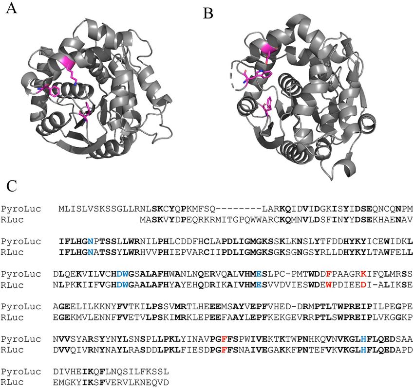
www.nature.com/scientificreports/
the intake siphon10, making light production tightly linked with colony size21. The blue-green light emitted by P.
atlanticum has been reported to have a peak emission at 475 nm22, 485 nm23, and 493 n m24.
Given their propensity to respond to light, pyrosomes are the only known colonial organisms where biolumi-
nescence is associated with communication between the zooids in a c olony25. Furthermore, pyrosome colonies
have been shown to respond to the bioluminescence of c onspecifics7. This way of using light for intraspecific
communication is well described in non-colonial marine species, such as polychaetes, ostracods, and fi shes26,27.
The serial photic excitation of pyrosome zooids results in a wave of bioluminescence that travels at 2.1–4.1 mm/s
across the colony22. This phenomenon was first noted in the 1800s6 and can be seen in Supp. Video 1 and 2 in
P. atlanticum and Pyrosomella verticillata. When the light flash is absorbed by the eyes of neighbouring zooids,
they both emit light and arrest ciliary movement, which ceases propulsion10. While it can be presumed that this
response enables zooids to close down and stop filtering when exposed to harmful stimuli, this behavior has not
been confirmed by observation in a natural setting. One possible explanation as to why such behavior might
be beneficial is that pyrosomes could use their light emissions as ‘burglar alarms’; similarly densely populating
organisms sometimes appear to use bioluminescence to prompt second order predators to come after their
attackers8. Given the propensity of pyrosomes to form dense blooms, such a tactic might be aided by nearby
colonies producing their own bioluminescence.
Regardless of the function behind pyrosome bioluminescence and the tissue localization, the exact mecha-
nism has not been determined. Like other bioluminescent organisms, pyrosomes rely on a chemical reaction
between a substrate (luciferin) and an enzyme (luciferase) to produce their light; however, the specific luciferin
and luciferase have yet to be i dentified8. Bacterial-bodies have sometimes been implicated as the causative agent
behind pyrosome light emission, but that explanation has been debated since the early 1900s10,28. The results we
present below advance this debate, suggesting that P. atlanticum has an endogenous luciferase that is related to
the presumed haloalkane dehalogenases of other invertebrates. The type of endogenous enzyme is also found in
both bacteria and eukaryotes, and appears to have evolved into luciferases in two other invertebrate lineages29.
Their more typical function is to break carbon–halogen bonds30.
Results
Transcriptomic sequencing and analysis. Assembled transcriptomes (Illumina HiSeq sequences) for
Brazilian sample 2B had 152,084 contigs with a total of 75,635 ORFs while sample 2C had 134,746 contigs with
a total of 70,340 ORFs; the Canadian sample P2 had 227,360 contigs with a total of 112,334 ORFs while sample
P3 had 206,824 contigs with a total of 104,057 ORFs. The large number of ORFs corresponds to the fact that we
used a 5 amino acid minimum to allow for searches for other proteins of interest that may be short.
Of the transcriptomes, one from Brazil (2B; identity = 48%; e = 3.8−46) and one from Canada (P3; iden-
tity = 48%; e = 6.67−97) had ORFs that matched the “Chain A, Crystal Structures Of The Luciferase And Green
Fluorescent Protein” of the sea pansy, Renilla reniformis (PDB accession = 2PSF), also known as RLuc. However,
when comparing the 2B sequence to nr in GenBank rather than Swissprot/Uniprot, the sequence was less clearly
a luciferase than a haloalkane dehalogenase (48% vs. > 50% identity). The Canadian sequence P3 that matches
RLuc is hereafter referred to as PyroLuc; the Brazilian sequence is named PyroB.
For generating homology models, Swiss-Model utilized Renilla luciferase accession 2PSJ (Fig. 2). The models
were rendered using PyMol 2.3 (pymol.org). This RLuc luciferase is evolutionarily related to the α/β hydrolase
family with close homology31,32. Sequence alignment of PyroLuc and Renilla luciferase (2PSJ33) showed conser-
vation of the catalytic triad and the active site (Fig. 2).
The alignment of PyroLuc with 2PSJ shows the secondary structure around the binding pocket of colentera-
mide to be similar to that of 2PSJ and the colenteramide molecule seems to fit well in the binding pocket (Fig. 2).
For PyroB there is a shift in the secondary structure as compared to the 2PSJ, which may cause a shift in the bind-
ing pocket of colenteramide. Accordingly, PyroLuc was used for downstream expression, while PyroB was not.
Samples P2 (65% identity and e = 1.66−50) and P3 (62% identity and e = 8.03−89) also match a luciferase from
a Pleuromamma sp. (AAG54096), which is known to exhibit bioluminescent properties (Patent: US 6232107-B
15-MAY-2001). While these are rather good quality matches, neither one possessed start codons and were
accordingly not used for downstream analysis or expression testing.
Pyrosome luminescence experiments. Mixing coelenterazine with P. atlanticum homogenate pro-
duced a luminescent reaction (Supp. Figure 3). Purified protein was used for luminescence experiments. Fig-
ure 3 shows a representative trial of the PyroLuc luminescence: 3.2 µM PyroLuc was used with 24.54 µM of
coelenterazine, resulting in a luminescence reading of 1.5 × 10⁶ relative light units (RLU). To confirm enzymatic
activity, we conducted several controls. We boiled the purified PyroLuc sample, which resulted in 5.4 × 10² RLU.
In addition, we purified a protein, matrix metalloproteinase-7 (MMP7), unrelated to bioluminescence under
the same conditions and did not observe significant light emission (Supp. Figure 4). Buffer controls were also
performed, using buffers involved in all purification steps. NanoLuc, an optimized luciferase (Promega), was
expressed in our lab and was used as a positive control in all experiments. We used matrix metalloproteinase-7
as a control for the luminescence experiments given it has no known luminescent properties. The expressed
PyroLuc produces significantly more light than in controls. For the control of matrix metalloproteinase-7, we
saw values of ~ 1.5 × 10⁴. For PyroLuc we saw a peak luminescence reading of 1.4 × 10⁷. The concentrations were
4.32 μM MMP7 for elution 1 and 0.57 μM MMP7 for elution 2 in PBS, pH 7.4.
RACE PCR. RACE PCR on a Canadian P. atlanticum sample recovered the majority of the PyroLuc sequence
with 100% identity, confirming the presence of PyroLuc in a second sample.
Scientific Reports | (2020) 10:17724 | https://doi.org/10.1038/s41598-020-73446-w 3
Vol.:(0123456789)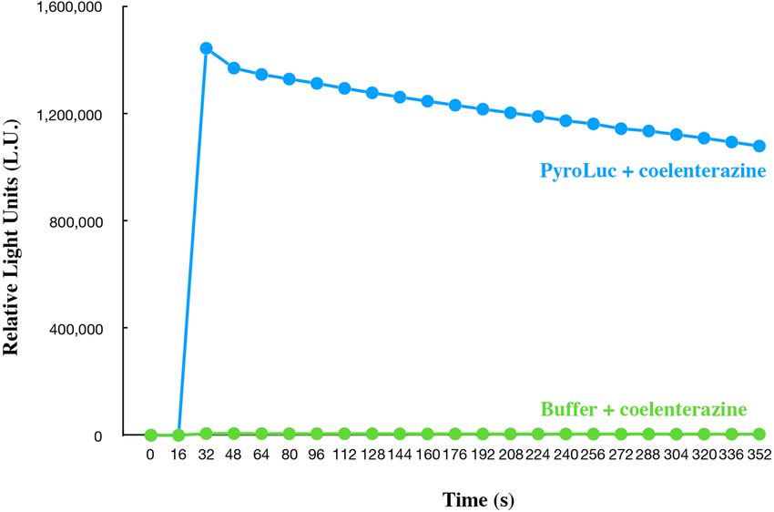
www.nature.com/scientificreports/
Figure 2. (A) Predicted model of PyroLuc created using SWISS-Model based on Renilla luciferase and (B)
model of Renilla luciferase ( 2PSJ33). Both models were rendered in PyMOL 2.3 (https://pymol.org). Magenta
sticks show conserved active site residues in the coelenterazine binding site. (C) Alignment of PyroLuc and
RLuc. The residues highlighted in blue make up the catalytic triad in RLuc and those in red represent those in
the coelenterazine binding site. Bold type represents identical residues.
Luciferase phylogenetics. A maximum likelihood phylogenetic reconstruction (Fig. 4) resulted in high
support (100% bootstrap) for a eukaryote clade being seperated from a bacterial clade. Within the eukary-
otic clade, the luciferases were not phylogenetically sister to one another. The pyrosome luciferase (PyroLuc)
sequence was found to be within the eukaryote clade. More specifically, it was phylogenetically sister to Corella
inflata (a tunicate), albeit with low support in maximum likelihood (51% bootstrap). All tunicates formed a
clade.
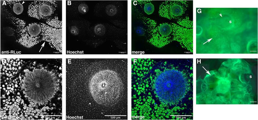
Immunolocalization of a Renilla‑like luciferase protein. To attempt to detect a Renilla-like luciferase
protein in pyrosome tissue, whole mount samples were fixed in 4% paraformaldehyde, treated with 1% Triton
in PBS to permeabilize tissues, and incubated with an antibody that recognizes Renilla luciferase. This antibody
was previously used to detect a Renilla-like luciferase in a brittle star29. Compared to samples incubated with pre-
immune serum as a control, a strong signal was detected in each zooid in a circular area underlying the incurrent
siphon, which is an average of 511 µm in diameter (n = 6) (Fig. 5A,C–D,F). The location and size of this circular
structure is in the region of the luminous organ in samples examined by Mackie and Bone10. Tissue that was
positive for RLuc was nucleated (staining using Hoechst stain), as would be predicted for a eukaryotic luciferase
(Fig. 5B–C,E–F), but not a bacterial luciferase. Non-specific staining was detected as small, circular patches on
the tunic (Fig. 5A,G–H) and low-level autofluorescence was observed (data not shown). While we have not gen-
erated a pyrosome-specific antibody, this data suggests that an RLuc-like protein is present in pyrosome tissues.
Scientific Reports | (2020) 10:17724 | https://doi.org/10.1038/s41598-020-73446-w 4
Vol:.(1234567890)www.nature.com/scientificreports/
Figure 3. In blue, luminescence reading of purified PyroLuc (3.2 μM) following the addition of coelenterazine
(24.5 μM). PyroLuc and coelenterazine were diluted in PBS with 300 mM imidazole, pH 7.4. In green,
PBS buffer control with addition of 24.54 μM coelenterazine. Coelenterazine was injected at 16 s for both
experiments.
Discussion
There are dozens of known bioluminescent systems, consisting of nine known natural luciferins, as well as dozens
of complementary luciferases and photoproteins that have independently e volved8,34,35. Based on transcriptom-
ics, phylogenetics, protein expression, and immunohistochemistry data, we present the first luciferase sequence
putatively used by a chordate (P. atlanticum). This luciferase (PyroLuc) appears to be highly convergent with
luciferases from two other phyla: Cnidaria (RLuc) and Echinodermata. Furthermore, like RLuc, PyroLuc reacts
with coelenterazine. The first luciferase isolated and found to interact with coelenterazine was from a deep-water
shrimp (Oplophorus gracilirostris)36. Renilla luciferase (RLuc) was among the first luciferases to be cloned37, and
is the closest described enzyme to PyroLuc. RLuc is popular for bioimaging and other bioluminescent studies,
as it can be expressed in numerous cell types. The properties and applications of PyroLuc in an experimental
biology context are yet to be determined.
Pyrosome bioluminescence. Bioluminescence is extremely common among marine animals. The most
thorough quantitative study found that 76% of organisms in an area from shallow environments to the deep-sea
emitted light38. This was similarly the case in the phylum Chordata: within tunicates, appendicularians were esti-
mated to have 94% bioluminescent individuals; within vertebrates, fishes may have around 70% bioluminescent
species38. However, putative molecular machinery behind luciferase production had not previously been indi-
cated for any chordate. Given that our experiments mixing coelenterazine with P. atlanticum homogenate and
this newly discovered PyroLuc both produced light, it appears probable that P. atlanticum uses coelenterazine as
a luciferin. Coelenterazine is an imidazolopyrazinone luciferin found in bioluminescent organisms from at least
nine phyla (from protozoans to vertebrates; not bacteria), and is of great importance in the evolutionary history
of bioluminescence across the tree of life8,29,39.
Coelenterazine is found in many non-bioluminescent organisms, possibly obtained via d iet40, and has strong
antioxidative properties41. Most organisms that use coelenterazine as a luciferin do not synthesize it themselves,
but they do typically produce their luciferases endogenously8. Pyrosoma atlanticum’s putative use of coelent-
erazine is congruent with the widespread use of this luciferin. Larvacean tunicates have also been shown to use
coelenterazine8.
The debate on bacterial bioluminescence in pyrosomes. It was initially proposed that pyrosome
bioluminescence is due to bacterial symbionts, as the intracellular sources of light (“luminous cell”) resemble
bacteria42. This hypothesis was furthered due to electron microscopy o bservations10 and by some associated
43
bacterial luciferase a ctivity . However, our data provides some evidence supporting an endogenous coelent-
erazine-based luciferase as the source of bioluminescence in pyrosomes (bacteria do not use coelenterazine).
Furthermore, our immunohistochemistry work highlights nucleated cells, which would not be present in bac-
teria. Still, these data are not sufficient to determine that bacteria are not used for pyrosome bioluminescence.
Some other studies have rejected the hypothesis of symbiotic bacteria as the source of pyrosome luminescence
due to consistently failed efforts to cultivate bacteria from luminous cells of Pyrosoma, as well as it being dif-
ficult to explain the wave-like mechanism of bioluminescence spreading across the colony34. Bioluminescence in
Scientific Reports | (2020) 10:17724 | https://doi.org/10.1038/s41598-020-73446-w 5
Vol.:(0123456789)www.nature.com/scientificreports/
Figure 4. Maximum likelihood phylogeny of RLuc-like luciferases (bolded) and haloalkane dehalogenases.
PyroLuc from Pyrosoma atlanticum is accentuated with a box. Support values are summaries of 1000 bootstrap
replicates. The luciferases from A. filiformis have yet to be functionally confirmed, but are highly probable29.
bacteria is controlled via the lux operon and all other known examples of bacterial bioluminescence (flashlight
fish, bobtail squid, etc.) exhibit non-modulatable bioluminescence, unlike that of P. atlanticum (Supp. Video 1).
Convergent bioluminescence evolution across phyla. One of the most interesting points of discov-
ery in this work is that a third phylum (Chordata) has at least one member (P. atlanticum) that putatively evolved
a Renilla-like luciferase from the more widespread haloalkane dehalogenase gene family. A similar luciferase
was recently predicted (yet to be expressed) from the brittle star Amphiura filiformis29. Based on our findings
and others’29, the luciferases from these three phyla (Cnidaria, Echinodermata, and Chordata) appear to use
coelenterazine as the luciferin.
The authors of the A. filiformis work elegantly connected the dots between the cnidarian and echinoderm
luciferases with bacterial haloalkane dehalogenases, noting, for instance, a conserved amino acid triad in the
luciferases29. Within animal RLuc-like genes, there also appears to be a conserved cysteine site that indeed is
important for RLuc a ctivity29,33. These other RLuc-like genes are from non-bioluminescent animal species, but
they do share a notable level (> 44%) of similarity. While the function is not known for all of these species, at
least one (DspA) to date has been confirmed to act as a haloalkane d ehalogenase44. A few vertebrate sequences in
GenBank are annotated as putative matches to haloalkane dehalogenase, but we are unaware of further research
assessing the presence of these proteins in vertebrates.
Luciferases are oxygenases, and it is interesting that a light producing oxygenase could be derived multiple
times from an enzyme with a rather different function (i.e., dehalogenases). However, this seems to be the case.
Our phylogenetic results build on past w ork29 and indicate that, within animal RLuc-like genes, luminescence
has evolved independently. PyroLuc is phylogenetically sister to a sequence from C. inflata and more generally
nested within a clade of tunicate sequences. This helps show that PyroLuc is most likely of tunicate origin. It
also indicates that this luciferase’s function evolved independently from the other RLuc-like genes, as the other
Scientific Reports | (2020) 10:17724 | https://doi.org/10.1038/s41598-020-73446-w 6
Vol:.(1234567890)www.nature.com/scientificreports/
Figure 5. Expression of Renilla-like luciferase protein in Pyrosoma atlanticum. (A–F) Extended focus confocal
projections of pyrosomes immunostained with an antibody to Renilla luciferase (anti-RLuc, green) and Hoechst
(blue) to label nuclei. External views show incurrent siphons (s) of multiple (A–C) or single (D–F) zooids.
Greyscale shown for A, B, D, and E; fluorescence shown for C and F. RLuc-like protein immunolocalizes to a
large, circular structure underlying the incurrent siphon (A, arrowhead). (G–H) Fluorescent stereomicroscope
images of a sample incubated with RLuc antibody (G) or rabbit pre-immune serum as a control (H). Individual
patches of staining outside the siphon (arrows) appear to be localized to the tunic, and were shown to be non-
specific using pre-immune serum (G, H and data not shown). Internal circular staining is specific to the RLuc
antibody (G, arrowhead). Scale bars, 500 μm.
tunicates examined are non-bioluminescent. (Although it is possible that this gene also has a bioluminescent
capacity even if it is not used for this purpose in one or more of the other examined tunicates.) Furthermore,
not only do these systems rely on coelenterazine, but their light emissions have roughly similar wavelengths:
the blue-green light from P. atlanticum ranges from 475 to 493 n m22–24, while the brittle star light peaks around
472 nm and RLuc peaks at 480 n m37. Sequence variation in these convergent luciferases presumably cause these
wavelength differences. Recent work has found that a single amino acid change next to the catalytic site of RLuc
can make RLuc have both bioluminescence and dehalogenase f unctioning45. It would similarly not be surprising
if one or a few amino acids could shift an ancestral dehalogenase to have luciferase functionality. However, we
do not find any sites that converge between bioluminescent RLuc-like sequences in our study that are not found
elsewhere, so we do not currently propose a specific site for future mutational work.
Other studies have found similar types of convergence from a single common gene source. Even within
luciferases, the firefly luciferases are similar to those found in a sponge and a squid, and likely emerged from the
widespread acyl-CoA l igases29,46.
Areas of further investigation. It is worth noting that even the most comprehensive studies to date have
later determined that transcripts were from potential prey items47. Along these lines, while transcriptomics and
RACE PCR showed PyroLuc in two samples, it was not found in all transcriptomes. Since transcriptome analysis
is dependent on the genes that are being expressed at a given time-point, it is possible that some samples were
not producing the bioluminescent gene at the time of collection; however, it is hard to be sure about this. Fur-
thermore, whereas our results indicate the luciferase system in P. atlanticum is likely coelenterazine-based, there
is always the possibility that some other symbiont, such as bacteria, is at play in pyrosome luciferase production.
In addition, immunohistochemistry in this study was performed using an Anti-RLuc antibody. While this anti-
body appears to be cross-reactive with PyroLuc, a specific anti-PyroLuc antibody does not yet exist, but would
be useful.
Furthermore, it is still possible that many dehalogenases across non-bioluminescent animals may be capable of
light production when exposed to coelenterazine, but are surely not using this luciferin. Additional work should
be conducted into whether dehalogenases from non-bioluminescent animals produce light when exposed to
coelenterazine. If these animals produce light, despite not being exposed to this luciferin in their environment,
it would explain how easily this protein could be co-opted for light production if an animal consumes another
organism that produces coelenterazine. However, it might also suggest that PyroLuc may not be used by the
organism for light production.
It would be useful to identify the exact wavelength of PyroLuc luminescence using microspectrophotometry.
In addition, mass spectrometry could be used to identify coelenterazine in the homogenate of the pyrosome.
Scientific Reports | (2020) 10:17724 | https://doi.org/10.1038/s41598-020-73446-w 7
Vol.:(0123456789)www.nature.com/scientificreports/
Lastly, designing a specific antibody for PyroLuc would be useful for further microscopy studies as well as analysis
of the crude tissue by Western Blot.
Conclusion
We believe this work adds important information regarding chordate bioluminescence for at least one species:
the pyrosome P. atlanticum. Evidence for our conclusion—that this species likely uses a coelenterazine-based
luciferase, similar to RLuc from a cnidarian—comes from transcriptomics, phylogenetics, coelenterazine experi-
ments, expression data, and immunohistochemistry. We describe a novel luciferase that might be of utility in the
growing molecular biology toolkit, given the usefulness of other structurally and functionally similar cnidarian
RLuc luciferases.
Methods
Specimen collection. Seven specimens of P. atlanticum were collected on May 2017 in SE Brazil, near
Alcatrazes Archipelago, using an Isaacs-Kidd Midwater Trawl as well as a Triton 3300/3 submarine with a soft
robotic arm operated via a haptic g love11 (Fig. 1E and Supp. Video 4); soft robotics appear not only to reduce
physical damage, but also to cause less stress-induced transcriptional changes48. These specimens were collected
under Permit # Sisbio 57721 from the Instituto Chico Mendes de Conservação da Biodiversidade (ICMBio),
Brazilian Ministry of the Environment.
Hundreds of P. atlanticum specimens were collected using the CCGS John P Tully between July 21–26, 2017
off Vancouver Island, Canada (Supp. Figure 2), as part of the ongoing Line P Monitoring and La Perouse Zoo-
plankton Monitoring programs run by Institute of Ocean Sciences (IOS)—Ocean Science D ivision12,13. Bongo
nets were deployed off the aft deck, lowered at a rate of 0.5 m/s and retrieved at 1 m/s. The bongo net consists of
two black cylindrical–conical nets mounted on a central towing frame and weight. Each net has a 0.25 m2 mouth
area, a filtering area/mouth area ratio of 11.5, and 0.23 mm aperture black mesh. Volume filtered is measured by
a TSK flowmeter mounted in the mouth of one net. Tow depths (determined from wire out and wire angle) fol-
lowed established time series protocols for the offshore and continental margin regions: near-bottom-to-surface
or 250 m-to-surface. Time from the net to the − 80 °C freezer was kept to less than 10 min for all pyrosomes. The
P. altlanticum specimens used in this study represent a disparate geographic range (Supp. Figure 2).
Transcriptomic sequencing and analysis. An RNeasy Fibrous Tissue Mini Kit (Qiagen #74704) was
used to extract RNA from these P. atlanticum samples. The two highest quality extractions from Brazil and the
two highest quality extractions from Canada were then used for transcriptomic sequencing at the New York
Genome Center using a HiSeq 25000 (125 × 125 bp). Sequences are in the Short Read Archive under BioProject
PRJNA667300.
Sequences were processed following our prior w ork49,50. In short, assemblies were produced using Trinity 2.4
with sequences first being trimmed with T rimmomatic51. Transdecoder 3.052 was then used to call open reading
frames (ORFs); a 5 amino acid minimum was established to allow for searches of possibly short luciferins. We
used ORF sequences as blastp queries against the local databases of luciferases and photoproteins from our prior
work50,53. Queries using blastp against these local databases used an e-value minimum cutoff of e −5. Matches
meeting this cutoff were then reciprocally used as blastp queries against Swissprot/Uniprot to confirm that no
better match was found in a well-curated database. If bitscores were better or equal for our local blastp searches,
the sequence was considered a putative match. Bitscores were used instead of e-values, as they do not rely on
database size which is highly skewed between local searches and large databases. Any identified proteins of inter-
est (e.g., luciferases) were modeled for homology with Swiss-Model54 using the default parameters.
Novel luciferase expression and bioluminescence assays. PyroLuc was successfully synthesized
and expressed in E. coli Origami DE3 (Novagen). The gene for PyroLuc was cloned into a pET-45b( +) vector
with an N-terminal His tag. Cloning was done by Genscript U.S.A. A starter culture of Origami DE3 (Novagen)
was grown at 37° C overnight. Larger cultures were inoculated with the starter culture and 100 mM IPTG was
used for induction once cells reached an O.D. 600 of 0.6. Following induction, the culture was grown at 37° C for
3 h. Induction pellets were washed in 1% PBST buffer (1X PBS and 1% Triton X-100), pH 7.4, and centrifuged at
6000 rpm for 20 min. The supernatant was discarded and the pellet was then resuspended in 15 mL of 1% PBST
buffer with 10 mM DTT, pH 7.4. The resuspension was sonicated at 100% amplitude for 10 min (30 s bursts with
1-min breaks), and the lysate was then centrifuged at 8000 rpm for 20 min. Supernatant was again discarded and
the pellet was resuspended in 25 mL 1% PBST with 8 M Urea and 10 mM DTT, pH 7.4. The solubilized super-
natant was then run through the column. We refolded the protein on the Nickel NTA column using a series of
refolding buffers with decreasing urea concentration (8 M Urea, 6 M Urea, 4 M Urea, 2 M Urea, and 0 M Urea)
in 1X PBS, pH 7.4. PBS with 300 mM imidazole, pH 7.4, was used to elute the protein. A gel of the protein puri-
fication is shown in Supp. Figure 5. The identity of PyroLuc was confirmed by mass spectrometry analysis (MS
BioWorks, Ann Arbor MI) of a gel band following protein purification. Bioluminescent assays were conducted
on a Spectra Max-L Microplate reader (Molecular Devices, San Jose CA) using an emission of wavelength of
480 nm, consistent with coelenterazine based luciferases.
RACE PCR. RACE PCR was used to validate the presence of our PyroLuc in another Canadian sample. Spe-
cifically, we performed 3′ RACE System for Rapid Amplification of cDNA Ends (ThermoFisher# 18373-019) and
5′ RACE System for Rapid Amplification of cDNA Ends (ThermoFisher# 18374-058).
Scientific Reports | (2020) 10:17724 | https://doi.org/10.1038/s41598-020-73446-w 8
Vol:.(1234567890)www.nature.com/scientificreports/
Luciferase phylogenetics. The luciferase found in P. atlanticum was combined with a matrix of luciferases
and haloalkane dehalogenases. We compiled these sequences using those from a study focussing on A. fili-
formis luciferase29. The sequences on ANISEED55 were taken by conducting a tblastn search against each tunicate
genome that had gene models available, with a query of PyroLuc and the similar Ciona robusta sequence from
the prior study29 (C. intestinalis in that study; please see the following paper regarding taxonomic changes in this
lineage56). Putative matching sequences were then searched for reciprocally as blastp queries against Swissprot/
Uniprot. Only hits that did not find better matches in this search or better matched a known dehalogenase or
luciferase sequence were kept. In essence, we kept sequences that appeared to be dehalogenases or luciferases.
The same was done for Branchiostoma belcheri from GenBank. The matrix was processed following our prior
work50: alignments (Supp. Data 1) were produced via MUSCLE v3.8.3157, and then a phylogenetic reconstruc-
tion of the data was produced using 1000 bootstrap replicates for support with the LG + I + G4 model (picked
using automatic model selection) in IQ-Tree multicore version 2.0.558 in the CIPRES Science Gateway59. Bacte-
rial haloalkane dehalogenases were used as outgroup taxa.
Imaging of bioluminescence. Immediately following collection in Brazil, P. atlanticum was brought into a
dark aquarium room and stimulated (either mechanically or photically with a Nikon Speedlight SB-910 strobe)
to initiate bioluminescence. Videos and stills were taken on a Sony A7s II camera.
Immunohistochemistry. Pyrosomes were fixed in 4% paraformaldehyde in PBS for 20 min and then incu-
bated in 0.5% Triton in PBS for 1 h at room temperature on a rocking nutator. Samples were then blocked in
PBT-BSA (0.5% Triton X-100, 0.5% bovine serum albumin) for 30 min at room temperature. An anti-Renilla
luciferase antibody (1:250, GTX125851, GeneTex) or rabbit pre-immune serum (1:250, ab37415, Abcam) was
added and samples were incubated at 4 °C overnight. GTX 125851 is a polyclonal antibody with specificity for
Renilla luciferase. Samples were then washed in PBS and incubated with AlexaFluor-488-conjugated anti-Rabbit
secondary (1:200, Invitrogen) for 1 h at room temperature. Following secondary antibody incubation, samples
were washed in PBS and Hoechst 33342 (Molecular Probes, 1:2000) was added during a 10 min wash in PBS.
Samples were dissected and placed in a glass-bottomed petri dish with PBS for imaging. In addition to the pre-
immune serum control, other control samples were incubated without primary or secondary antibody, and no
specific signal was observed (data not shown).
Confocal imaging. Confocal images were acquired on a Zeiss 880 Airyscan Live Cell laser-scanning confo-
cal microscope equipped with a 10 × 0.30 NA M27 EC Plan-Neofluor objective and ZEN Black software. Maxi-
mum intensity projections were rendered using Fiji (Image J) software. Fluorescent images were obtained on a
Zeiss Pentafluor Discovery V8 stereomicroscope equipped with a 0.63X Achromat FWD 107 mm objective lens
and ZEN Blue software. Images were processed using Adobe Photoshop CC.
Received: 9 April 2020; Accepted: 10 September 2020
References
1. van Soest, R. W. M. A monograph of the order Pyrosomatida (Tunicata, Thaliacea). J. Plankton Res. 3, 603–631 (1981).
2. Archer, S. K. et al. Pyrosome consumption by benthic organisms during blooms in the northeast Pacific and Gulf of Mexico. Ecol-
ogy 99, 981–984 (2018).
3. Kuo, C.-Y. et al. An unusual bloom of the tunicate, Pyrosoma atlanticum, in southern Taiwan. Bull. Mar. Sci. 91, 363–364 (2015).
4. Huxley, T. H. Observations upon the anatomy and physiology of Salpa and Pyrosoma. Philos. Trans. R. Soc. Lond. 141, 567–593
(1851).
5. Péron, F. Mémoire sur le nouveau genre Pyrosoma. Annales du Museum d’histoire naturelle, Paris 4, 437–446 (1803).
6. Panceri, P. The luminous organs and light of Prosoma. Q. J. Microsc. Sci. 13, 45–51 (1873).
7. Burghause, F. Kreislauf und Herzschlag bei Pyrosoma giganteum nebst Bemerkungen zum Leuchtvermögen. Zeitschrift für Wis-
senschaftliche Zoologie 108, 430–497 (1914).
8. Haddock, S. H. D., Moline, M. A. & Case, J. F. Bioluminescence in the sea. Ann. Rev. Mar. Sci. 2, 443–493 (2010).
9. Pierantoni, U. Gli organi luminosi simbiotici ed il loro ciclo ereditario in Pyrosoma giganteum. Pubblicazioni della Stazione zoo-
logica di Napoli3, 191–222 (1921).
10. Mackie, G. O. & Bone, Q. Luminescence and associated effector activity in Pyrosoma (Tunicata: Pyrosomida). Proc. R. Soci. B Biol.
Sci. 202, 483–495 (1978).
11. Phillips, B. T. et al. A dexterous, glove-based teleoperable low-power soft robotic arm for delicate deep-sea biological exploration.
Sci. Rep. 8, 14779 (2018).
12. Brodeur, R. et al. An unusual gelatinous plankton event in the NE Pacific: the great pyrosome bloom of 2017. PICES Press 26,
22–27 (2018).
13. Sutherland, K. R., Sorensen, H. L., Blondheim, O. N., Brodeur, R. D. & Galloway, A. W. E. Range expansion of tropical pyrosomes
in the northeast Pacific Ocean. Ecology 99, 2397–2399 (2018).
14. Kocot, K. M., Tassia, M. G., Halanych, K. M. & Swalla, B. J. Phylogenomics offers resolution of major tunicate relationships. Mol.
Phylogenet. Evol. 121, 166–173 (2018).
15. Lebrato, M. & Jones, D. O. B. Mass deposition event of Pyrosoma atlanticum carcasses off Ivory Coast (West Africa). Limnol.
Oceanogr. 54, 1197–1209 (2009).
16. Drits, A. V., Arashkevich, E. G. & Semenova, T. N. Pyrosoma atlanticum (Tunicata, Thaliacea): grazing impact on phytoplankton
standing stock and role in organic carbon flux. J. Plankton Res. 14, 799–809 (1992).
17. Perissinotto, R., Mayzaud, P., Nichols, P. D. & Labat, J. P. Grazing by Pyrosoma atlanticum (Tunicata, Thaliacea) in the south Indian
Ocean. Mar. Ecol. Prog. Ser. 330, 1–11 (2007).
18. Bone, Q. The Biology of Pelagic Tunicates (Oxford University Press on Demand, Oxford, 1998).
Scientific Reports | (2020) 10:17724 | https://doi.org/10.1038/s41598-020-73446-w 9
Vol.:(0123456789)www.nature.com/scientificreports/
19. Sweeney, B. M., Fork, D. C. & Satoh, K. Stimulation of bioluminescence in dinoflagellates by red light. Photochem. Photobiol. 37,
457–465 (1983).
20. Mauchline, J. IX.—the biology of the euphausiid crustacean, Meganyctiphanes norvegica (M. Sars). Proc. R. Soc. Edinb. Biol. 67,
141–179 (1959).
21. Widder, E. Bioluminescence and the pelagic visual environment. Mar. Freshw. Behav. Physiol. 35, 1–26 (2002).
22. Bowlby, M. R., Widder, E. A. & Case, J. F. Patterns of stimulated bioluminescence in two pyrosomes (Tunicata: Pyrosomatidae).
Biol. Bull. 179, 340–350 (1990).
23. Herring, P. J. The spectral characteristics of luminous marine organisms. Proc. R. Soc. B Biol. Sci. 220, 183–217 (1983).
24. Swift, E., Biggley, W. H. & Napora, T. A. The bioluminescence emission spectra of Pyrosoma atlanticum, P. spinosum (Tunicata),
Euphausia tenera (Crustacea) and Gonostoma sp. (Pisces). J. Mar. Biol. Assoc. UK 57, 817 (1977).
25. Mackie, G. O. Unconventional signalling in tunicates. Mar. Freshw. Behav. Physiol. 26, 197–205 (1995).
26. Verdes, A. & Gruber, D. F. Glowing worms: biological, chemical, and functional diversity of bioluminescent annelids. Integr.
Comput. Biol. 57, 18–32 (2017).
27. Davis, M. P., Holcroft, N. I., Wiley, E. O., Sparks, J. S. & Leo Smith, W. Species-specific bioluminescence facilitates speciation in
the deep sea. Mar. Biol. 161, 1139–1148 (2014).
28. Nealson, K. H. & Hastings, J. W. Bacterial bioluminescence: its control and ecological significance. Microbiol. Rev. 43, 496–518
(1979).
29. Delroisse, J. et al. A puzzling homology: a brittle star using a putative cnidarian-type luciferase for bioluminescence. Open Biol. 7,
160300 (2017).
30. Janssen, D. B. Evolving haloalkane dehalogenases. Curr. Opin. Chem. Biol. 8, 150–159 (2004).
31. Loening, A. M., Fenn, T. D., Wu, A. M. & Gambhir, S. S. Consensus guided mutagenesis of Renilla luciferase yields enhanced
stability and light output. Protein Eng. Des. Sel. 19, 391–400 (2006).
32. Woo, J., Howell, M. H. & von Arnim, A. G. Structure-function studies on the active site of the coelenterazine-dependent luciferase
from Renilla. Protein Sci. 17, 725–735 (2008).
33. Loening, A. M., Fenn, T. D. & Gambhir, S. S. Crystal structures of the luciferase and green fluorescent protein from Renilla reni-
formis. J. Mol. Biol. 374, 1017–1028 (2007).
34. Shimomura, O. Bioluminescence Chemical Principles and Methods (World Scientific, Singapore, 2006).
35. Kaskova, Z. M., Tsarkova, A. S. & Yampolsky, I. V. 1001 lights: luciferins, luciferases, their mechanisms of action and applications
in chemical analysis, biology and medicine. Chem. Soc. Rev. 45, 6048–6077 (2016).
36. Shimomura, O., Masugi, T., Johnson, F. H. & Haneda, Y. Properties and reaction mechanism of the bioluminescence system of the
deep-sea shrimp Oplophorus gracilorostris. Biochemistry 17, 994–998 (1978).
37. Lorenz, W. W., McCann, R. O., Longiaru, M. & Cormier, M. J. Isolation and expression of a cDNA encoding Renilla reniformis
luciferase. Proc. Natl. Acad. Sci. USA 88, 4438–4442 (1991).
38. Martini, S. & Haddock, S. H. D. Quantification of bioluminescence from the surface to the deep sea demonstrates its predominance
as an ecological trait. Sci. Rep. 7, 45750 (2017).
39. Shimomura, O., Inoue, S., Johnson, F. H. & Haneda, Y. Widespread occurrence of coelenterazine in marine bioluminescence.
Comp. Biochem. Physiol. Part B Comp. Biochem. 65, 435–437 (1980).
40. Shimomura, O. Presence of coelenterazine in non-bioluminescent marine organisms. Comp. Biochem. Physiol. Part B Comp.
Biochem. 86, 361–363 (1987).
41. Rees, J. F. et al. The origins of marine bioluminescence: turning oxygen defense mechanisms into deep-sea communication tools.
J. Exp. Biol. 201, 1211–1221 (1998).
42. Julin, C. The specific histological characters of the ‘luminous cells’ of Pyrosoma giganteum and of Cyclosalpa pinnata. Rep. Brit.
Ass. 492–493 (1912).
43. Leisman, G., Cohn, D. H. & Nealson, K. H. Bacterial origin of luminescence in marine animals. Science 208, 1271–1273 (1980).
44. Fortova, A. et al. DspA from Strongylocentrotus purpuratus: the first biochemically characterized haloalkane dehalogenase of
non-microbial origin. Biochimie 95, 2091–2096 (2013).
45. Chaloupkova, R. et al. Light-emitting dehalogenases: reconstruction of multifunctional biocatalysts. ACS Catal. 9, 4810–4823
(2019).
46. Viviani, V. R. The origin, diversity, and structure function relationships of insect luciferases. Cell. Mol. Life Sci. 59, 1833–1850
(2002).
47. Haddock, S. H. D., Mastroianni, N. & Christianson, L. M. A photoactivatable green-fluorescent protein from the phylum Cte-
nophora. Proc. Biol. Sci. 277, 1155–1160 (2010).
48. Tessler, M. et al. Ultra-gentle soft robot fingers induce minimal transcriptomic response in a fragile marine animal during handling.
Curr. Biol. 30, R157–R158 (2020).
49. Tessler, M. et al. Marine leech anticoagulant diversity and evolution. J. Parasitol. 104, 210–220 (2018).
50. Tessler, M. et al. Luciferin production and luciferase transcription in the bioluminescent copepod. PeerJ 6, e5506 (2018).
51. Bolger, A. M., Lohse, M. & Usadel, B. Trimmomatic: a flexible trimmer for Illumina sequence data. Bioinformatics 30, 2114–2120
(2014).
52. Haas, B. J. et al. De novo transcript sequence reconstruction from RNA-seq using the Trinity platform for reference generation
and analysis. Nat. Protoc. 8, 1494–1512 (2013).
53. Brugler, M. R., Aguado, M. T., Tessler, M. & Siddall, M. E. The transcriptome of the Bermuda fireworm Odontosyllis enopla
(Annelida: Syllidae): a unique luciferase gene family and putative epitoky-related genes. PLoS ONE 13, e0200944 (2018).
54. Biasini, M. et al. SWISS-MODEL: modelling protein tertiary and quaternary structure using evolutionary information. Nucleic
Acids Res. 42, W252–W258 (2014).
55. Brozovic, M. et al. ANISEED 2017: extending the integrated ascidian database to the exploration and evolutionary comparison of
genome-scale datasets. Nucleic Acids Res. 46, D718–D725 (2018).
56. Gissi, C. et al. An unprecedented taxonomic revision of a model organism: the paradigmatic case of Ciona robusta and Ciona
intestinalis. Zool. Scr. 46, 521–522 (2017).
57. Edgar, R. C. MUSCLE: a multiple sequence alignment method with reduced time and space complexity. BMC Bioinform. 5, 113
(2004).
58. Nguyen, L.-T. et al. IQ-TREE: a fast and effective stochastic algorithm for estimating maximum-likelihood phylogenies. Mol. Biol.
Evol. 32, 268–274 (2014).
59. Miller, M. A., Pfeiffer, W. & Schwartz, T. Creating the CIPRES Science Gateway for inference of large phylogenetic trees. In 2010
Gateway Computing Environments Workshop (GCE) (2010). https://doi.org/10.1109/gce.2010.5676129.
Acknowledgements
We thank the Dalio Family Foundation, OceanX, the crew of the M/Y Alucia, and the submarine pilots of the
Triton 3300/3 for pyrosome collections in Brazil. Thanks to Peter Kragh and John Cullum for assistance in
filming bioluminescent pyrosomes. We thank Sunita Subramanian for assistance with molecular work. For the
Canadian collections, we thank the captain and crew of the CCGS JP Tully for their generous help in collecting
Scientific Reports | (2020) 10:17724 | https://doi.org/10.1038/s41598-020-73446-w 10
Vol:.(1234567890)www.nature.com/scientificreports/
zooplankton. Special thanks to Marie Robert, chief scientist (Line P Monitoring Program) and Doug Yelland,
chief scientist (La Perouse Zooplankton Monitoring Program). The authors would like to acknowledge the Live
Imaging and Bioenergetics Facility of CUNY Advanced Science Research Center for instrument use, scientific,
and technical assistance. This work was supported by DARPA-NESD Grant #N66001-17-C-4012 to D.F. Gru-
ber, J.P. Gaffney and V. Pieribone; NSF Instrument Development for Biological Research Award #s 1556164 to
R.J. Wood and #1556213 to D.F. Gruber; NSF DEB award #1257555 to J.S. Sparks; FAPESP 2017/26279-2 to A.
Oliveira; FAPESP 2014/18541-0 to J. Mirza; and DOE EvoNet Grant #DE-SC0014377 supported Michael Tessler’s
work on this project. The funders had no role in study design, data collection and analysis, decision to publish,
or preparation of the manuscript.
Author contributions
M.T., J.P.G., and D.F.G. conceptualized the project and wrote the manuscript. All authors reviewed and revised
the manuscript. D.F.G., A.G.O., J.D.M., M.G., J.S.S., V.A.P. and R.J.W. were involved with the collection of speci-
mens. D.F.G., J.P.G., V.A.P. and R.J.W. obtained funding for this work. Experiments and computational analyses
were conducted by M.T., J.P.G., A.G., and N.G. Immunostaining and confocal imaging was performed by A.G.
and K.C.D.
Competing interests
The authors declare no competing interests.
Additional information
Supplementary information is available for this paper at https://doi.org/10.1038/s41598-020-73446-w.
Correspondence and requests for materials should be addressed to M.T., J.P.G. or D.F.G.
Reprints and permissions information is available at www.nature.com/reprints.
Publisher’s note Springer Nature remains neutral with regard to jurisdictional claims in published maps and
institutional affiliations.
Open Access This article is licensed under a Creative Commons Attribution 4.0 International
License, which permits use, sharing, adaptation, distribution and reproduction in any medium or
format, as long as you give appropriate credit to the original author(s) and the source, provide a link to the
Creative Commons licence, and indicate if changes were made. The images or other third party material in this
article are included in the article’s Creative Commons licence, unless indicated otherwise in a credit line to the
material. If material is not included in the article’s Creative Commons licence and your intended use is not
permitted by statutory regulation or exceeds the permitted use, you will need to obtain permission directly from
the copyright holder. To view a copy of this licence, visit http://creativecommons.org/licenses/by/4.0/.
© The Author(s) 2020
Scientific Reports | (2020) 10:17724 | https://doi.org/10.1038/s41598-020-73446-w 11
Vol.:(0123456789)You can also read