Insulin and the insulin receptor in experimental models of learning and memory
←
→
Page content transcription
If your browser does not render page correctly, please read the page content below
European Journal of Pharmacology 490 (2004) 71 – 81
                                                                                                                          www.elsevier.com/locate/ejphar

                 Insulin and the insulin receptor in experimental models of
                                    learning and memory
                        Wei-Qin Zhao a,*, Hui Chen b, Michael J. Quon b, Daniel L. Alkon a
  a
      Blanchette Rockefeller Neurosciences Institute, 3rd floor, Academic and Research Building, 9601 Medical Center Drive, Rockville, MD 20850, USA
                                  b
                                    Diabetes Unit, LCI, NCCAM, National Institutes of Health, Bethesda, MD 20862, USA
                                                               Accepted 27 February 2004

Abstract

   Insulin is best known for its action on peripheral insulin target tissues such as the adipocyte, muscle and liver to regulate glucose
homeostasis. In the central nervous system (CNS), insulin and the insulin receptor are found in specific brain regions where they show
evidence of participation in a variety of region-specific functions through mechanisms that are different from its direct glucose regulation in
the periphery. While the insulin/insulin receptor associated with the hypothalamus plays important roles in regulation of the body energy
homeostasis, the hippocampus- and cerebral cortex-distributed insulin/insulin receptor has been shown to be involved in brain cognitive
functions. Emerging evidence has suggested that insulin signaling plays a role in synaptic plasticity by modulating activities of excitatory and
inhibitory receptors such as glutamate and GABA receptors, and by triggering signal transduction cascades leading to alteration of gene
expression that is required for long-term memory consolidation. Furthermore, deterioration of insulin receptor signaling appears to be
associated with aging-related brain degeneration such as the Alzheimer’s dementia and cognitive impairment in aged subjects suffering type 2
diabetes mellitus.
D 2004 Elsevier B.V. All rights reserved.

Keywords: Insulin receptor; Learning and memory; Signal transduction; Receptor trafficking; Diabetes mellitus; Alzheimer’s disease

1. Introduction                                                                 basis for the CNS insulin/insulin receptor action on learning
                                                                                and memory.
    The presence of insulin and insulin receptors in the brain
suggests that the brain is a target organ for insulin. However,
unlike the classic peripheral insulin target tissues such as                    2. Memory-improving effects of insulin
adipocyte, muscle and liver, where the primary function of
insulin is to regulate glucose homeostasis, insulin in the                         The presence of insulin and insulin receptor in the
central nervous system (CNS) exhibits more diverse actions,                     hippocampus and cerebral cortex suggests a functional
most of which have not been clearly understood. In addition                     involvement in brain cognition phenomena such as learn-
to its central role in food intake and weight control, a direct                 ing and memory. Insulin has been shown to exert a
role of CNS insulin/insulin receptor signaling in improving                     memory-enhancing action on both humans and experi-
cognitive functions, including learning and memory, and the                     mental animals. Administration of insulin into the third
association of insulin receptor deterioration with brain                        cerebral ventricles of rats shortly after a passive avoid-
degenerative dementia (e.g., Alzheimer’s disease) have                          ance training experience resulted in higher memory
attracted increasing interest. Although still at an early stage,                retention levels compared to rats that received saline
efforts in behavioral, electrophysiological and biochemical                     and a heat-inactivated insulin injection (Park et al.,
studies have begun to uncover the cellular and molecular                        2000). Because insulin was given after acquisition of
                                                                                the experience, it most likely contributed to processes
                                                                                underlying memory consolidation. Extracranial delivery of
   * Corresponding author. Tel.: +1-301-294-7179; fax: +1-301-294-              insulin has also shown effects on memory. However, the
7007.                                                                           outcomes appear to be complicated by alteration of blood
   E-mail address: zhaow@brni-jhu.org (W.-Q. Zhao).                             glucose levels. For example, intraperitoneal (i.p.) injection

0014-2999/$ - see front matter D 2004 Elsevier B.V. All rights reserved.
doi:10.1016/j.ejphar.2004.02.045
72 W.-Q. Zhao et al. / European Journal of Pharmacology 490 (2004) 71–81
of insulin without simultaneous administration of glucose neuronal necrosis caused by ischaemia, whereas increased
to maintain normal circulating glucose levels caused glucose supply alone did not have such effects (Voll et
impaired retention in mice that were trained on either al., 1989). Lesions to the hippocampus of rats produced
an inhibitory avoidance or a habituation learning task severe loss of learning and memory ability (Horel, 1978;
(Kopf and Baratti, 1995, 1996, 1999). Because a periph- Huppert and Piercy, 1979; Spiers et al., 2001). However,
eral delivery of insulin would certainly lower circulating pretreatment with insulin of those rats that received
glucose levels in the absence of insulin resistance, the dorsal hippocampal lesions significantly reduced memory
memory impairment induced by insulin i.p. injection was deficits for an active avoidance learning experience in-
more likely to be caused by hypoglycemia rather than a duced by hippocampal lesions (De Castro and Balagura,
direct CNS effect of insulin. Indeed, when these inves- 1976). In pharmacological experiments, insulin was
tigators gave concomitant administration of glucose and shown to overcome scopolamine-induced memory impair-
insulin, the memory impairment was overcome in a ment in rats trained in a radial arm maze task (Blanchard
glucose dose-dependent manner (Kopf and Baratti, and Duncan, 1997), indicating insulin’s positive action on
1995, 1999). On the other hand, systemic infusion of adrenergic transmission in the brain.
insulin under euglycemic conditions (maintained by glu- The significance of understanding roles of insulin receptor
cose clamp) produced significant memory improvement in in memory has been highlighted by the hypothesis that
verbal memory and selective attention (Kern et al., 2001). deterioration of CNS insulin receptor functions is associated
Recently, an intranasal administration of insulin and other with the pathogenesis of aging-related brain degenerative
hormonal peptides has been reported to be an advanta- dementias such as sporadic Alzheimer’s disease (Hoyer,
geous delivery route. Delivery of insulin to human 1998, 2002; Hoyer and Lannert, 1999; Watson and Craft,
subjects via this method induced a sharp and rapid 2003). Clinical and experimental evidence supporting this
increase in cerebrospinal fluid insulin concentration with- notion have been shown by different investigators. For
out affecting blood insulin and glucose levels (Kern et example, while glucose and insulin improve memory in
al., 1999; Born et al., 2002). When tested in a cognitive young animals and humans, only insulin but not glucose
task responding to an auditory stimulus, those subjects exhibits a memory enhancing effect in aged subjects (Long et
showed a negative shift in auditory-evoked potentials in al., 1992). Brain insulin and insulin receptor are reduced in
specific cortical areas (Kern et al., 1999), which are the brains of sporadic Alzheimer’s disease patients (Frolich et
thought to be associated with facilitation of working al., 1998). Consistent results have also been found in rodents
memory processing (Pelosi and Blumhardt, 1999; Wolach whose brains show aging-reduced insulin receptor numbers
and Pratt, 2001; Shucard et al., 2003). An aged popula- (Zaia and Piantanelli, 2000) and insulin receptor mRNA
tion with impaired memory showed increased latencies levels (Fig. 1). Systemic administration of insulin under
and amplitudes in components of auditory-evoked poten- euglycemic or hyperglycemic conditions was shown to
tials (Pelosi and Blumhardt, 1999). A negative shift of improve memory performance of Alzheimer’s disease
auditory-evoked potentials was also correlated with the patients whereas hyperglycemia alone did not have such an
insulin-induced memory enhancement tested in a similar effect (Craft et al., 1999, 2000). More recent results from
task (Kern et al., 2001; Fehm et al., 2000; Born et al., those authors have shown that different doses of insulin are
2002). Consistent with these findings, gene expression of required to produce memory improvement in elderly people
insulin receptor has been upregulated in the CA1 region and Alzheimer’s disease patients that were associated with
of the rat brain associated with short-term memory the presence of the apoE-4 allele in tested subjects (Craft et
formation after a spatial learning experience (Zhao et al., 2000, 2003).
al., 1999). Increased insulin receptor at protein levels Cognitive impairments associated with diabetes melli-
have also been found in the hippocampal synaptic mem- tus caused by inadequate insulin/insulin receptor functions
brane fractions correlated with short-term memory forma- have also been documented. In this category, memory
tion (Zhao et al., 1999). Taken together, these results deficits are not evident in insulin-dependent type 1
suggest that insulin/insulin receptor signaling is activated diabetes in humans (Ryan and Williams, 1993), and if
in the early stage of memory formation and may play a they occur, are often associated with hypoglycemia
role in memory sorting for long-term storage. (Kaufman et al., 1999; Sommerfield et al., 2003). In
Insulin appears to exert a more significantly beneficial diabetic animals most commonly induced by streptozocin,
effect on preventing memory loss from brain damage only subtle memory impairment was detected (Biessels et
induced by ischaemia, lesions and pharmacological inhi- al., 1996, 1998; Popovic et al., 2001), which was
bition. When insulin treatment was given with a con- accompanied by altered synaptic plasticity (Biessels et
comitant glucose supply to rats immediately following al., 1996; Kamal et al., 1999). On the other hand,
ischaemia, it prevented ischaemia-induced learning defi- cognitive deficits have been reported more consistently
cits when those rats received water maze navigation in non-insulin dependent diabetes (type 2 diabetes) often
training 1 –2 months after experiencing ischaemia. The found concurrent with increased insulin resistance. Cog-
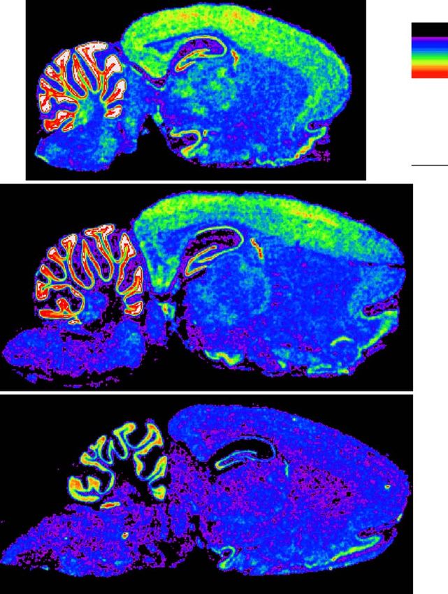
insulin treatment also significantly reduced the CA1 nitive decline in type 2 diabetes depends on duration ofW.-Q. Zhao et al. / European Journal of Pharmacology 490 (2004) 71–81 73 Fig. 1. Reductions of the brain insulin receptor mRNA by aging. Cryosections were prepared from rat brains of different ages. Distribution of insulin receptor mRNA was examined by in situ hybridization using an insulin receptor riboprobe (Zhao et al., 1999). Insulin receptor mRNA signals were revealed by film autoradiography. The brain insulin receptor mRNA level showed a significant reduction correlating with aging. Brain regions were defined according to The Rat Brain Stereotaxic Coordinates by Paxinos and Watson (1998). AO: olfactory bulb, anterior; CrCtx: cerebral cortex; CCtx: cerebellar cortex; chP: choroid plexus; HP: hippocampus; ThNuc: thalamic nuclei. the disease (Cosway et al., 2001), correlates highly with apoE4 allele (Haan et al., 2003). All of these results aged subjects (Perlmuter et al., 1984, 1987; Mooradian et suggest a link between aging-related insulin receptor al., 1988; Ryan and Geckle, 2000) and is worsened by failure and Alzheimer’s dementia. brain vascular complications such as stroke (Wu et al., 2003). In addition, type 2 diabetes has been more commonly reported to affect verbal learning and memory, 3. Mechanisms underlying roles of insulin/insulin tests of which generally require subjects to repeat verba- receptor in learning and memory tim a short story or to recall a list of words (Mooradian et al., 1988; Ryan and Geckle, 2000). These patients also 3.1. The CNS insulin receptor is functionally different from show poor performance in the mini-mental state exami- those in classical insulin target tissues. Therefore, memory nation (MMSE), which is a general test for concentration, improvement by insulin/insulin receptor is not due to a language and other memory components, used to screen direct effect on glucose metabolism for demented subjects (Folstein and McHugh, 1978). In parallel with poor memory performance, these patients Although glucose is the major nutrient and energy source also showed abnormal brain electroencephalograms for brain cells and plays critical role in brain cognitive (EEGs) in the cerebral cortex and subcortical areas that functions (McNay and Gold, 2002), its uptake, transport and were not a result of hyperglycemia (Mooradian et al., utilization in the majority of brain regions do not depend on 1988). Furthermore, the rate of cognitive decline associ- insulin. Firstly, the adult brain appears to express two main ated with diabetes was increased by the presence of the glucose transporters (GLUTs) that are not insulin-sensitive.
74                                W.-Q. Zhao et al. / European Journal of Pharmacology 490 (2004) 71–81

While GLUT-1 is expressed in the endothelium of cerebral               uptake and metabolism in brain cells (Goodner et al.,
microvessels and astrocytes, GLUT-3 is predominantly                   1980; Gorus et al., 1984; Hertz et al., 1981). For example,
distributed in neurons (Vannucci et al., 1998; Simpson et              insulin infusion under euglycemia showed no effect on
al., 1999; Duelli and Kuschinsky, 2001). Different local-              glucose transport across the blood – brain barrier, and net
izations of glucose transporter subtypes suggest different             brain glucose uptake in humans (Hertz et al., 1981; Hassel-
regulatory mechanisms underlying glucose uptake into                   balch et al., 1999). Neither did it affect cerebral metabolism
different cell types. Low abundance of the insulin-sensitive           (Hasselbalch et al., 1999) and oxidation in cultured cortical
GLUT-4 has been found in the brain associated with early               cells (Gorus et al., 1984). Although some investigators
development of the brain (Vannucci et al., 2000; Royer et              report that insulin increased glucose uptake by glial but
al., 2000) and is predominantly expressed in the cerebellum            not neuronal cells (Werner et al., 1989), the extracellular
of both developing and adult brains (Rayner et al., 1994;              glucose level exerted much stronger effects on glucose
Kobayashi et al., 1996; Vannucci et al., 1998) This suggests           uptake into glial cells (Walker et al., 1988). Exposure of
that insulin may directly mediate glucose transport to                 neurons to increased extracellular glucose also elevated
cerebellar neurons. A recent study showed that although                glucose transport to neurons although to a much lesser
GLUT-4 gene expression could be detected in the hippo-                 extent compared to glial cells (Walker et al., 1988). Lastly,
campus, its protein level, however, was very low compared              while the whole body homozygous insulin receptor knock-
with the level in cerebellar neurons (El Messari et al., 2002).        out is lethal, specific deletion of the brain insulin receptor
Because the cerebellum controls balance and coordinates                did not show an evident disruption in brain development
movement of the body, the insulin-sensitive glucose trans-             and neuronal survival (Bruning et al., 2000).
port in the cerebellum may partially explain why type 1                    However, accumulated evidence from a variety of experi-
diabetic subjects often suffer psychomotor (Ryan and Wil-              ments together with the results of brain specific deletion of
liams, 1993; Bohannon et al., 1995), and postural impair-              insulin receptor has clearly indicated that the brain insulin/
ments (Di Nardo et al., 1999; Yamamoto et al., 2001).                  insulin receptor contributes importantly to glucose homeo-
    Secondly, insulin receptor is not homogenously distrib-            stasis. Particularly, the hypothalamic insulin/insulin receptor
uted throughout the brain, but concentrated in rather discrete         is shown to control peripheral insulin secretion and glucose
regions such as the olfactory bulb, hypothalamus, hippo-               levels in the circulation (Amir and Shechter, 1987; Chen et
campus, choroid plexus and cerebellum (Adamo et al.,                   al., 1975; Chowers et al., 1966; Woods et al., 1968; Obici et
1989; Unger et al., 1991; Zhao et al., 1999). Similarly,               al., 2002a, 2002b; Gerozissis, 2003) and plays a role in the
entry of insulin into the brain also shows regional differ-            peripheral insulin receptor resistance (Obici et al., 2002b).
ences (Banks and Kastin, 1998). This brain regional distri-            This action is likely to be mediated via the vagal system, as
bution pattern of and insulin does not correlate well with a           vagotomy and atropine block the CNS action of insulin on
primary role in mediation of brain cell glucose metabolism.            periphery plasma glucose regulation (Woods et al., 1972;
Thirdly, the brain insulin receptor displays structural and            Woods, 1991; Szabo et al., 1983). In addition, centrally
functional differences from that in the peripheral tissues.            administered insulin showed a Pavlovian conditioning effect
The brain insulin receptor has smaller molecular weights in            on peripheral glucose levels in both humans and animals
both alpha- and beta-subunits compared with its peripheral             (Stochhorst et al., 2000; Woods et al., 1968, 1972). Based
counterparts as a result of alternative mRNA splicing of the           on the evidence above, it becomes obvious that the action of
exon-11, and differences in receptor glycosylation (Heiden-            insulin/insulin receptor in the brain is predominantly medi-
reich et al., 1983; Goldstein and Dudley, 1992; Sugimoto et            ated by its roles in neuromodulation.
al., 2000). Unlike in the peripheral tissues, insulin receptor
in the neuron lacks negative cooperativity (receptor down-             3.2. Insulin/insulin receptor modulates synaptic plasticity
regulation in response to prolonged and/or high concentra-             by acting on glutamatergic and GABAergic receptors
tions of ligand reaction), which is otherwise shown in the
peripheral insulin receptor, suggesting different insulin-                 Although hypothalamic insulin signaling may play an
binding specifics of the CNS insulin receptor (Boyd and                indirect role in regulation of peripheral glucose metabolism,
Raizada, 1983; Gammeltoft et al., 1984). Furthermore,                  actions of insulin/insulin receptor on learning and memory,
Heidenreich et al. (1983) reported that an anti-insulin                particularly those located in the hippocampus (see previous
receptor autoantiserum that blocked insulin binding of the             sections), are more likely to be due to direct modulation of
peripheral insulin receptor had no effect on insulin binding           receptor activity in neurons and/or glial cells. Emerging
to the brain insulin receptor. Unlike those from adipocytes,           evidence has shown that insulin receptor signaling plays a
insulin receptor from brain did not bind to wheat germ                 role in synaptic plasticity by acting on both glutamatergic
agglutinin columns. These results again suggest that differ-           and GABAergic transmissions. In Xenopus oocytes
ences in structure and other molecular properties exist                expressed with NMDA receptors, brief insulin exposure
between the peripheral and CNS insulin receptors. Fourthly,            triggered a rapid and significant potentiation of responses
evidence from the majority of experiments has consistently             to NMDA mediated by NMDA receptor subtypes (Liu et al.,
demonstrated insulin’s lack of direct effects on glucose               1995; Chen and Leonard, 1996; Liao and Leonard, 1999).
W.-Q. Zhao et al. / European Journal of Pharmacology 490 (2004) 71–81 75
mediates only NMDA transmission is not functional unless
AMPA receptors are delivered to such synapses (Malinow et
al., 2000, Malinow, 2003). Conversion of a silent synapse to a
functional synapse can be both development dependent (Wu
et al., 1996; Renger et al., 2001) and activity dependent
(Malinow et al., 2000; Malinow, 2003) that have been
hypothesized as a synaptic basis for learning and memory
formation. In cultured differentiating neurons, insulin pro-
moted transfer of silent AMPA synapse to functional synapse
and accelerated reduction of silent synapses (Plitzko et al.,
2001). In the mature brain, insulin facilitated clathrin-depen-
dent internalization of AMPA receptors leading to long-term
Fig. 2. Effect of Ca2 + on in vitro tyrosine phosphorylation of the depression of AMPA receptor-mediated synaptic transmis-
hippocampal insulin receptor and IGF-1 receptor: (A) Tyrosine phosphor- sion in hippocampal CA1 neurons (Man et al., 2000).
ylated synaptic membrane proteins at f 95 kDa detected by an anti- Insulin-mediated receptor trafficking has also been found
phospho-tyrosine antibody. (B) Phosphorylated proteins were immunopre-
cipitated (ip) by the anti-phospho-tyrosine antibody, and then blotted (ib) by in the gamma-aminobutyric acid (GABA) receptor, which
an anti-insulin receptor beta-subunit antibody. (C) Phosphorylated proteins
were precipitated as described in (b), and blotted by an-anti IGF-1 receptor
antibody. The results showed a Ca2 +-inhibited tyrosine phosphorylation of
insulin receptor but not IGF-1 receptor. (1) Tyrosine-phosphorylated IGF-1 IRS-1
receptor overexpressed in NIH3T3 cells; (2) non-phosphorylated synaptic 180kDa
membrane fractions from rat hippocampus; (3) tyrosine phosphorylation in
the absence of Ca2 +; (4) tyrosine phosphorylation in the presence of 1
mMCa2 +; (5) tyrosine phosphorylation of the human liver insulin receptor βActin
that is overexpressed in NIH3T3 cells. 40kDa
N SW T N SW T
This insulin-induced potentiation was blocked by a tyrosine 1 hr after 24 hr after
protein kinase inhibitor, genistein and a broad-spectrum one-day training four-day training
protein kinase inhibitor staurosporine, suggesting involve-
2.0
ment of tyrosine and possibly down-stream serine/threonine
Levels of IRS-1 (IRS/actin)
protein kinases such as protein kinase C (PKC) activities. In
1.5 **
a similar experimental preparation, Skeberdis et al. (2001)
demonstrated that application of insulin increases NMDA
channel activities by recruiting NMDA receptors to the 1.0
membrane surface. This process was blocked by a more
specific insulin receptor tyrosine kinase inhibitor tyrphostin 0.5
*
A47, and may involve function of SNAP-25 (synaptosomal
associated protein 25), but does not seem to require tyrosine 0.0
and serine/threonine phosphorylations at the NMDA recep- N SW T N SW T
tor C-terminus. Other study, however, showed that incuba- 1 hr after 24 hr after
tion of rat hippocampal slices with insulin caused increases one-day training four-day training
in tyrosine phosphorylation of the NR2A and 2B subunits of
NMDA receptors (Christie et al., 1999). Given the important Fig. 3. Changes in amount of IRS-1 in the hippocampal synaptic membrane
fractions after spatial learning: Synaptic membrane fractions were prepared
roles that NMDA receptors may play in synaptic plasticity
from the hippocampus and the amount of IRS-1 was examined with
and learning and memory formation (Huerta et al., 2000; Western blot by an anti-IRS-1 antibody and compared among trained and
Nakazawa et al., 2002), modulation of NMDA transmission control animals. To investigate effects of training intensity (numbers of
may represent one of the synaptic bases for roles of insulin/ training trials that rats experienced) and stages of memory formation
insulin receptor signaling in learning and memory. (short-term vs. long-term) on the amount of IRS-1, animals were subjected
In addition, insulin plays a role in synaptic plasticity by to two experimental conditions: (1) Rats were trained on a four-trial water
maze task for a 1-day and sacrificed 1 h after training. (2) Animals were
acting on alpha-amino-3-hydroxy-5-methylisoxazole-4-pro- trained on four-trial water maze task per day for 4 consecutive days and
pionic acid (AMPA) receptor trafficking. Redistribution of sacrificed 24 h after the 4th day training. For each condition, groups of
AMPA receptors has been proposed to regulate strength of control rats for non-specific behavioral effects were designed. Immuno-
glutamatergic synapses. A mature synaptic connection at reactive signals of IRS-1 were normalized with that of h-actin from the
glutamatergic synapses in the brain requires conversion of same sample for a sample-loading variability control. N: Naı̈ve animals
that were used as the basal control. SW: Animals subjected to swimming
silent glutamatergic synapses into functional synapses during only, but not training activities that were used to control for non-learning
the course of postnatal brain development (Wu et al., 1996; specific effects. T: Trained animals **p < 0.01, *p < 0.05 (one-way
Renger et al., 2001). A silent glutamatergic synapse that ANOVA), n = 4.76 W.-Q. Zhao et al. / European Journal of Pharmacology 490 (2004) 71–81
(Wan et al., 1997). Furthermore, insulin activation of
muscarinic transmission potentiated GABA receptor cur-
rents likely occurs via a phosphoinositide-3 (PI-3) kinase-
dependent mechanism (Ma et al., 2003). Thus, insulin/
insulin receptor plays a role in receptor trafficking during
synaptic maturation and synaptic usage, and it may also
mediate interactions of different neurotransmission systems
during neuronal activation, all of which may underlie
modification of synaptic connections required for higher
Fig. 4. Changes in the amount of Shc in the synaptic membrane during brain functions such as learning and memory.
long-term memory formation: The synaptic membranes were prepared from
hippocampi of different groups of rats after a 4-day water maze training
experience. Shc protein was detected on Western blots with an anti-Shc
3.3. Insulin/insulin receptor-mediated signal transduction
antibody. h-Actin from the same sample was also measured for as a molecular basis underlying learning and memory
normalization of the Shc signal. Control groups were set as described in
Fig. 3, and animals sacrificed 24 h after the 4th day of training. Naı̈ve As in the periphery, insulin’s action in the brain is
animals were used as the basal control, SW: swimming-only animals were mediated by insulin receptor, although at higher concen-
used to control for non-specific effects-induced changes. T: Trained
animals. **p < 0.01, one-way ANOVA, n = 4.
trations insulin also binds to insulin-like growth factor (IGF)
receptors. Under an in vitro phosphorylation system, both
insulin receptor and IGF-1 receptor can be tyrosine phos-
mediates synaptic inhibition important for neuronal func- phorylated. In vitro tyrosine phosphorylation of hippocam-
tions associated with learning (Paulsen and Moser, 1998; pal insulin receptor, but not IGF-1 receptor is inhibited by
Chapouthier and Venault, 2002; McGaugh, 2002). When the presence of Ca2 + (Zhao et al., 1999; Fig. 2), suggesting
applied to HEK 293 cells transfected with the GABAA that insulin receptor and IGF-1 receptor activities are
receptor, insulin caused rapid translocation of the GABAA differential regulated in response to Ca2 + signals such as
receptors to the plasma membrane (Wan et al., 1997). those resulted from glutamatergic and GABAergic trans-
Insulin also recruited functional GABAA receptors onto missions during synaptic activation or inhibition. Binding of
the postsynaptic and dendritic membranes of the CNS insulin activates the protein tyrosine kinase activity of the
neurons, leading to augmented amplitudes of the GABAA insulin receptor h-subunit, which, in turn, triggers cascades
receptor-mediated miniature inhibitory postsynaptic current of signal transduction through its downstream substrate
Fig. 5. Effects of neuronal depolarization and learning on phosphorylation of Erk1/2: (A) Primary cortical neuronal cultures were depolarized with high
concentration (55 mM) KCl for 5 or 20 min. Phosphorylation of Erk1/2 was detected with an anti-phospho-Erk antibody. Phosphorylation extents were
normalized with the total amount of Erk detected with an anti-regular Erk antibody. Erk1/2 was activated by depolarization, but returned to the control level
when depolarization prolonged. (B) Synaptic membranes were obtained from rat hippocampi as described in Fig. 4. A significant learning-specific increase in
Erk1/2 phosphorylation was observed during long-term memory formation. **p < 0.01, one-way ANOVA, n = 4.W.-Q. Zhao et al. / European Journal of Pharmacology 490 (2004) 71–81 77 molecules. Several signaling pathways activated by insulin ing (Fig. 3). However, after exhaustive training experience, receptor have been identified (Olefsky, 1990; White and from which rats normal demonstrated stable spatial memory Kahn, 1996; Bevan, 2001), among which the insulin recep- (Morris, 1984), the amount of IRS-1 in the synaptic mem- tor substrate-1 (IRS-1)/PI-3 kinase/phosphoinositide-depen- branes was reduced (Fig. 3). The increased IRS-1 in the dent kinase (PDK)/protein kinase B (PKB/Akt), and the synaptic membrane may be due to an increase in local SH2 and collagen containing protein (Shc)/growth factor expression, or a translocation of IRS-1 from cytosol to the receptor-bound protein-2 (Grb2)/mitogen-activated protein synaptic membrane. These results suggest that IRS-1 may (MAP) kinase pathways have been most intensively studied. participate in an early stage of memory processing. Tyrosine These two signaling pathways also constitute insulin recep- phosphorylated IRS-1 transduces the insulin signal by tor downstream cascades in the brain (Unger et al., 1991; binding to the p85 subunit of PI-3 kinase, leading to Wozniak et al., 1993; Adamo et al., 1993). When rats were activation of the p110 catalytic subunit (Combettes-Souver- trained in a spatial learning task (Morris water maze ain and Issad, 1998). Both IRS-1 and PI-3 kinase are training), a learning-specific increase in IRS-1 was detected abundantly expressed in the hippocampus, colocalizing with in the hippocampal synaptic membranes shortly after train- insulin receptor (Folli et al., 1994). Involvement of PI-3 Fig. 6. Hypothetic schema for insulin/insulin receptor modulation of memory-associated neuronal activities. During learning, insulin binds to the a-subunit of insulin receptor and cause activation of the tyrosine kinase activity of the b-subunit. Activated insulin receptor may be involved memory formation via several mechanisms: (1) via modulation of glutamatergic and GABAergic transmission. Insulin/insulin receptor potentiates NMDA channel activity, functions of which depend on the presence and activation of AMPA receptor that cause synaptic membrane depolarization and removal of the Mg2 + blockage of the NMDA receptor leading to long-term potentiation. Increased Ca2 + influx via the NMDA receptor and neuronal activities may inhibit tyrosine phosphorylation of insulin receptor via a feedback mechanism. Depending on spatial and temporal specificity of information processing, insulin receptor signaling through PI-3 kinase may be involved in long-term depression via internalization of AMPA receptors. Insulin receptor may also modulate GABA transmission by recruiting functional GABA receptor to the postsynaptic membrane. GABAergic neurons sense the excitatory transmission and regulate synaptic strength by sending feedforward and/or feedback inhibitory inputs to the principal neurons. Regulation of synaptic efficacy by integrated excitatory and inhibitory transmissions within specific neuronal network is thought to underlie memory encoding and retrieval in the hippocampus (Paulsen and Moser, 1998). (2) Activation of nsulin receptor-Shc-MAP kinase pathway after learning may lead to regulation of gene expression that is required for long-term memory storage. (3) Insulin receptor may interact with G-protein coupled receptor and PLC to activate PKC leading to facilitation of short-term memory encoding. (4) The insulin receptor/IRS/PI-3 kinase pathway may trigger synthesis of NO via eNOS activity. NO acts as a retrograde messenger for neurotransmitter release, and may also act intracellularly on memory processing. Furthermore, insulin receptor signaling with the same pathway may promote neuronal survival that is certainly beneficial for long-term memory consolidation.
78                                W.-Q. Zhao et al. / European Journal of Pharmacology 490 (2004) 71–81

kinase has been reported in learning and memory formation              ically sensitized Erk1/2 expression and phosphorylation in
using different experimental paradigms (Lin et al., 2001;              response to insulin, probably due to facilitation of signal
Barros et al., 2001), as well as in regulation of synaptic             transduction involving Shc and other upstream molecules.
plasticity such as long-term potentiation and long-term                   In summary, research in recent years has significantly
depression (Ma et al., 2003; Daw et al., 2002; Sanna et                advanced our knowledge about roles of insulin/insulin
al., 2002; Kelly and Lynch, 2000). Thus, increased IRS-1 at            receptor in functions of the central nervous system, includ-
synaptic locations after learning may activate PI-3 kinase             ing learning and memory at behavioral, synaptic and mo-
leading to regulation of subsequent memory processing.                 lecular levels. Based on evidence accumulated to date, a
Other downstream molecules in the context of insulin                   hypothetical schema for involvement of insulin/insulin re-
receptor-activated IRS-1/PI-3 kinase pathway may include               ceptor in learning and memory consolidation processes is
production of nitric oxide (NO) through nitric oxide syn-              summarized in Fig. 6. However, precise molecular mecha-
thase (NOS) activities. It has been known that insulin                 nisms underlying insulin’s roles in learning and memory,
stimulates NO production (Montagnani et al., 2001; Vincent             and in the pathogenesis of memory degenerative disorders
et al., 2003) via activation of endothelial NOS (eNOS), an             are still far from understood. Many substrates of insulin
IRS-1/PI-3 kinase/Akt pathway mediates this process (Zeng              receptor, and interactions between insulin receptor and other
and Quon, 1996; Zeng et al., 2000; Montagnani et al.,                  receptor and kinase molecules directly associated with
2002). eNOS is expressed in the hippocampus, and has                   learning experiences have not been explored. Future in-
been shown to be involved in learning and synaptic plas-               depth studies at cellular and molecular levels, particularly
ticity (Zhuo et al., 1993; O’Dell et al., 1994; Arancio et al.,        those with gene interference, and specific deletions of
1996; Rickard et al., 1999; Doreulee et al., 2003). However,           insulin receptor and insulin receptor substrates in the brain,
whether activation of eNOS occurs in learning downstream               are expected to provide meaningful answers.
of insulin receptor action has not yet been explored.
    We have also reported learning-induced changes in Shc,
another substrate molecule for insulin receptor that contains          References
a SH2 domain and a collagen-homologous region (Pelicci et
al., 1992) Shc comprises three isoforms, namely Shc66,                  Adamo, M., Raizada, M.K., LeRoith, D., 1989. Insulin and insulin-like
Shc52 and Shc46, due to alternative splicing of the primary                growth factor receptors in the nervous system. Mol. Neurobiol. 3,
                                                                           71 – 100.
Shc transcript (Pelicci et al., 1992; Migliaccio et al., 1997).
                                                                        Adamo, M.L., Shemer, J., Roberts Jr., C.T., LeRoith, D., 1993. Insulin and
Shc can be tyrosine phosphorylated by insulin receptor and                 insulin-like growth factor-I induced phosphorylation in neurally derived
epidermal growth factor (EGF) receptor. While the EGF                      cells. Ann. N.Y. Acad. Sci. 692, 113 – 125.
receptor similarly phosphorylates all three isoforms, insulin           Amir, S., Shechter, Y., 1987. Centrally mediated hypoglycemic effect of
receptor preferentially phosphorylates Shc52, and Shc46 to                 insulin: apparent involvement of specific insulin receptors. Brain Res.
                                                                           418, 152 – 156.
a lesser extent (Sasaoka et al., 1994). Shc52 is a potent
                                                                        Arancio, O., Kiebler, M., Lee, C.J., Lev-Ram, V., Tsien, R.Y., Kandel, E.R.,
activator of Ras21. We have shown that spatial learning                    Hawkins, R.D., 1996. Nitric oxide acts directly in the presynaptic neu-
increases expression of Shc isoforms in the hippocampal                    ron to produce long-term potentiation in cultured hippocampal neurons.
synaptic membrane fraction. While increases in Shc-66 and                  Cell 87, 1025 – 1035.
Shc-52 were seen during short-term memory formation                     Banks, W.A., Kastin, A.J., 1998. Differential permeability of the blood –
                                                                           brain barrier to two pancreatic peptides: insulin and amylin. Peptides
(Zhao et al., 1999), Shc52 was the major isoform that
                                                                           19, 883 – 889.
showed significant increases in long-term memory, although              Barros, D.M., Mello, E., Souza, T., de Souza, M.M., Choi, H., DeDavid, E.,
a small increase was also seen in Shc46 and Shc66,                         Silva, T., Lenz, G., Medina, J.H., Izquierdo, I., 2001. LY294002, an
respectively (Fig. 4). These results suggest that increases                inhibitor of phosphoinositide 3-kinase given into rat hippocampus
of Shc52 and shc46 in the hippocampal synaptic membrane                    impairs acquisition, consolidation and retrieval of memory for one-trial
                                                                           step-down inhibitory avoidance. Behav. Pharmacol. 12, 629 – 634.
may be associated with insulin receptor activity after train-
                                                                        Bevan, P., 2001. Insulin signalling. J. Cell. Sci. 114, 1429 – 1430.
ing. The downstream cascades of Shc52 include activation                Biessels, G.J., Kamal, A., Ramakers, G.M., Urban, I.J., Spruijt, B.M.,
of Ras21 leading to activation of MAP kinases, such as                     Erkelens, D.W., Gispen, W.H., 1996. Place learning and hippocampal
extracellular signal-regulated kinase 1/2 (Erk1/2). In our                 synaptic plasticity in streptozotocin-induced diabetic rats. Diabetes 45,
experimental systems, Erk1/2 has been shown to respond to                  1259 – 1266.
                                                                        Biessels, G.J., Kamal, A., Urban, I.J., Spruijt, B.M., Erkelens, D.W.,
learning and neuronal depolarization (Fig. 5A), with high
                                                                           Gispen, W.H., 1998. Water maze learning and hippocampal synaptic
sensitivity and specificity. Indeed, a lasting activation of               plasticity in streptozotocin-diabetic rats: effects of insulin treatment.
Erk1/2 has been observed after water maze training (Zhao et                Brain Res. 800, 125 – 135.
al., 1999; Fig. 5B). Interestingly, when hippocampal synap-             Blanchard, J.G., Duncan, P.M., 1997. Effect of combinations of insulin,
tic membranes were treated with insulin under an in vitro                  glucose and scopolamine on radial arm maze performance. Pharmacol.
                                                                           Biochem. Behav. 58, 209 – 214.
phosphorylation condition, only those from trained but not
                                                                        Bohannon, R.W., Smith, J., Barnhard, R., 1995. Balance deficits accom-
control animals demonstrated an insulin-induced increase in                panying renal disease are related to diabetic status. Percept. Mot. Skills
total amount of Erk1/2 and its phosphorylation (Zhao et al.,               81, 528 – 530.
1999). These results suggest that learning input has specif-            Born, J., Lange, T., Kern, W., McGregor, G.P., Bickel, U., Fehm, H.L.,
W.-Q. Zhao et al. / European Journal of Pharmacology 490 (2004) 71–81 79 2002. Sniffing neuropeptides: a transnasal approach to the human brain. receptor substrate-1 (IRS-1) distribution in the rat central nervous sys- Nat. Neurosci. 5, 514 – 516. tem. J. Neurosci. 14, 6412 – 6422. Boyd Jr., F.T., Raizada, M.K. 1983. Effects of insulin and tunicamycin on Folstein, M.F., McHugh, P.R., 1978. Defective long-term caloric regulation neuronal insulin receptors in culture. Am. J. Physiol. 245, C283 – C287. in obesity. NIDA Res. Monogr. 20, 182 – 188. Bruning, J.C., Gautam, D., Burks, D.J., Gillette, J., Schubert, M., Orban, Frolich, L., Blum-Degen, D., Bernstein, H.G., Engelsberger, S., Humrich, P.C., Klein, R., Krone, W., Muller-Wieland, D., Kahn, C.R., 2000. Role J., Laufer, S., Muschner, D., Thalheimer, A., Turk, A., Hoyer, S., Zoch- of brain insulin receptor in control of body weight and reproduction. ling, R., Boissl, K.W., Jellinger, K., Riederer, P., 1998. Brain insulin and Science 289, 2122 – 2125. insulin receptors in aging and sporadic Alzheimer’s disease. J. Neural Chapouthier, G., Venault, P., 2002. GABA-A receptor complex and mem- Transm. 105, 423 – 438. ory processes. Curr. Top. Med. Chem. 2, 841 – 851. Gammeltoft, S., Staun-Olsen, P., Ottesen, B., Fahrenkrug, J., 1984. Insulin Chen, C., Leonard, J.P., 1996. Protein tyrosine kinase-mediated poten- receptors in rat brain cortex. Kinetic evidence for a receptor subtype in tiation of currents from cloned NMDA receptors. J. Neurochem. 67, the central nervous system. Peptides 5, 937 – 944. 194 – 200. Gerozissis, K., 2003. Brain insulin: regulation, mechanisms of action and Chen, M., Woods, S.C., Porte Jr., D., 1975. Effect of cerebral intraventric- functions. Cell. Mol. Neurobiol. 23, 1 – 25. ular insulin on pancreatic insulin secretion in the dog. Diabetes 24, Goldstein, B.J., Dudley, A., 1992. Heterogeneity of messenger RNA that 910 – 914. encodes the rat insulin receptor is limited to the domain of exon 11: Chowers, I., Lave, S., Hapern, L., 1966. Effect of insulin administered analysis by RNA heteroduplex mapping, amplification of cDNA, and in intracisternally on the glucose level of the blood and the cerebrospinal vitro translation. Diabetes 41, 1293 – 1300. fluid in vagotomized dogs. Exp. Neurol. 14, 383 – 389. Goodner, C.J., Hom, F.G., Berrie, M.A., 1980. Investigation of the effect of Christie, J.M., Wenthold, R.J., Monaghan, D.T., 1999. Insulin causes a insulin upon regional brain glucose metabolism in the rat in vivo. transient tyrosine phosphorylation of NR2A and NR2B NMDA recep- Endocrinology 107, 1827 – 1832. tor subunits in rat hippocampus. J. Neurochem. 72, 1523 – 1528. Gorus, F.K., Hooghe-Peters, E.L., Pipeleers, D.G., 1984. Glucose metabo- Combettes-Souverain, M., Issad, T., 1998. Molecular basis of insulin ac- lism in murine fetal cortical brain cells: lack of insulin effects. J. Cell. tion. Diabetes Metab. 24, 477 – 489. Physiol. 121, 45 – 50. Cosway, R., Strachan, M.W., Dougall, A., Frier, B.M., Deary, I.J., 2001. Haan, M.N., Mungas, D.M., Gonzalez, H.M., Ortiz, T.A., Acharya, A., Cognitive function and information processing in type 2 diabetes. Dia- Jagust, W.J., 2003. Prevalence of dementia in older latinos: the influ- bet. Med. 18, 803 – 810. ence of type 2 diabetes mellitus, stroke and genetic factors. J. Am. Craft, S., Asthana, S., Newcomer, J.W., Wilkinson, C.W., Matos, I.T., Geriatr. Soc. 51, 169 – 177. Baker, L.D., Cherrier, M., Lofgreen, C., Latendresse, S., Petrova, A., Hasselbalch, S.G., Knudsen, G.M., Videbaek, C., Pinborg, L.H., Schmidt, Plymate, S., Raskind, M., Grimwood, K., Veith, R.C., 1999. Enhance- J.F., Holm, S., Paulson, O.B., 1999. No effect of insulin on glucose ment of memory in Alzheimer disease with insulin and somatostatin, blood – brain barrier transport and cerebral metabolism in humans. Di- but not glucose. Arch. Gen. Psychiatry 56, 1135 – 1140. abetes 48, 1915 – 1921. Craft, S., Asthana, S., Schellenberg, G., Baker, L., Cherrier, M., Boyt, A.A, Heidenreich, K.A., Zahniser, N.R., Berhanu, P., Brandenburg, D., Olefsky, Martins, R.N., Raskind, M., Peskind, E., Plymate, S., 2000. Insulin J.M., 1983. Structural differences between insulin receptors in the brain effects on glucose metabolism, memory, and plasma amyloid precursor and peripheral target tissues. J. Biol. Chem. 258, 8527 – 8530. protein in Alzheimer’s disease differ according to apolipoprotein-E ge- Hertz, M.M., Paulson, O.B., Barry, D.I., Christiansen, J.S., Svendsen, P.A., notype. Ann. N.Y. Acad. Sci. 903, 222 – 228. 1981. Insulin increases glucose transfer across the blood – brain barrier Craft, S., Asthana, S., Cook, D.G., Baker, L.D., Cherrier, M., Purganan, K., in man. J. Clin. Invest. 67, 597 – 604. Wait, C., Petrova, A., Latendresse, S., Watson, G.S., Newcomer, J.W., Horel, J.A., 1978. The neuroanatomy of amnesia. A critique of the hippo- Schellenberg, G.D., Krohn, A.J., 2003. Insulin dose-response effects on campal memory hypothesis. Brain 101, 403 – 445. memory and plasma amyloid precursor protein in Alzheimer’s disease: Hoyer, S., 1998. Is sporadic Alzheimer disease the brain type of non-insulin interactions with apolipoprotein E genotype. Psychoneuroendocrinol- dependent diabetes mellitus? A challenging hypothesis. J. Neural ogy 28, 809 – 822. Transm. 105, 415 – 422. Daw, M.I., Bortolotto, Z.A., Saulle, E., Zaman, S., Collingridge, G.L., Hoyer, S., 2002. The brain insulin signal transduction system and spo- Isaac, J.T., 2002. Phosphatidylinositol 3 kinase regulates synapse radic (type II) Alzheimer disease: an update. J. Neural Transm. 109, specificity of hippocampal long-term depression. Nat. Neurosci. 5, 341 – 360. 835 – 836. Hoyer, S., Lannert, H., 1999. Inhibition of the neuronal insulin receptor De Castro, J.M., Balagura, S., 1976. Insulin pretreatment facilitates recov- causes Alzheimer-like disturbances in oxidative/energy brain metabo- ery after dorsal hippocampal lesions. Physiol. Behav. 16, 517 – 520. lism and in behavior in adult rats. Ann. N.Y. Acad. Sci. 893, 301 – 303. Di Nardo, W., Ghirlanda, G., Cercone, S., Pitocco, D., Soponara, C., Huerta, P.T., Sun, L.D., Wilson, M.A., Tonegawa, S., 2000. Formation of Cosenza, A., Paludetti, G., Di Leo, M.A., Galli, I., 1999. The use of temporal memory requires NMDA receptors within CA1 pyramidal dynamic posturography to detect neurosensorial disorder in IDDM neurons. Neuron 25, 473 – 480. without clinical neuropathy. J. Diabetes its Complicat. 13, 79 – 85. Huppert, F.A., Piercy, M., 1979. Normal and abnormal forgetting in organic Doreulee, N., Sergeeva, O.A., Yanovsky, Y., Chepkova, A.N., Selbach, O., amnesia: effect of locus of lesion. Cortex 15, 385 – 390. Godecke, A., Schrader, J., Haas, H.L., 2003. Cortico-striatal synaptic Kamal, A., Biessels, G.J., Urban, I.J., Gispen, W.H., 1999. Hippocampal plasticity in endothelial nitric oxide synthase deficient mice. Brain Res. synaptic plasticity in streptozotocin-diabetic rats: impairment of long- 964, 159 – 163. term potentiation and facilitation of long-term depression. Neuroscience Duelli, R., Kuschinsky, W., 2001. Brain glucose transporters: relationship 90, 37 – 745. to local energy demand. News Physiol. Sci. 16, 71 – 76. Kaufman, F.R., Epport, K., Engilman, R., Halvorson, M., 1999. Neuro- El Messari, S., Ait-Ikhlef, A., Ambroise, D.H., Penicaud, L., Arluison, M., cognitive functioning in children diagnosed with diabetes before age 10 2002. Expression of insulin-responsive glucose transporter GLUT4 years. J. Diabetes its Complicat. 13, 31 – 38. mRNA in the rat brain and spinal cord: an in situ hybridization study. Kelly, A., Lynch, M.A., 2000. Long-term potentiation in dentate gyrus of J. Chem. Neuroanat. 24, 225 – 242. the rat is inhibited by the phosphoinositide 3-kinase inhibitor, wortman- Fehm, H.L., Perras, B., Smolnik, R., Kern, W., Born, J., 2000. Manipulat- nin. Neuropharmacology 39, 643 – 651. ing neuropeptidergic pathways in humans: a novel approach to neuro- Kern, W., Born, J., Schreiber, H., Fehm, H.L., 1999. Central nervous pharmacology? Eur. J. Pharmacol. 405, 43 – 54. system effects of intranasally administered insulin during euglycemia Folli, F., Bonfanti, L., Renard, E., Kahn, C.R., Merighi, A., 1994. Insulin in men. Diabetes 48, 557 – 563.
80                                       W.-Q. Zhao et al. / European Journal of Pharmacology 490 (2004) 71–81

Kern, W., Peters, A., Fruehwald-Schultes, B., Deininger, E., Born, J.,          Obici, S., Feng, Z., Karkanias, G., Baskin, D.G., Rossetti, L., 2002a. De-
   Fehm, H.L., 2001. Improving influence of insulin on cognitive func-              creasing hypothalamic insulin receptors causes hyperphagia and insulin
   tions in humans. Neuroendocrinology 74, 270 – 280.                               resistance in rats. Nat. Neurosci. 5, 566 – 572.
Kobayashi, M., Nikami, H., Morimatsu, M., Saito, M., 1996. Expression           Obici, S., Zhang, B.B., Karkanias, G., Rossetti, L., 2002b. Hypothalamic
   and localization of insulin-regulatable glucose transporter (GLUT4) in           insulin signaling is required for inhibition of glucose production. Nat.
   rat brain. Neurosci. Lett. 213, 103 – 106.                                       Med. 8, 1376 – 1382.
Kopf, S.R., Baratti, C.M., 1995. The impairment of retention induced by         O’Dell, T.J., Huang, P.L., Dawson, T.M., Dinerman, J.L., Snyder, S.H.,
   insulin in mice may be mediated by a reduction in central cholinergic            Kandel, E.R., Fishman, M.C., 1994. Endothelial NOS and the blockade
   activity. Neurobiol. Learn. Mem. 63, 220 – 228.                                  of LTP by NOS inhibitors in mice lacking neuronal NOS. Science 265,
Kopf, S.R., Baratti, C.M., 1996. Memory modulation by post-training glu-            542 – 546.
   cose or insulin remains evident at long retention intervals. Neurobiol.      Olefsky, J.M., 1990. The insulin receptor. A multifunctional protein. Dia-
   Learn. Mem. 65, 189 – 191.                                                       betes 39, 1009 – 1016.
Kopf, S.R., Baratti, C.M., 1999. Effects of posttraining administration of      Park, C.R., Seeley, R.J., Craft, S., Woods, S.C., 2000. Intracerebroventric-
   insulin on retention of a habituation response in mice: participation of a       ular insulin enhances memory in a passive-avoidance task. Physiol.
   central cholinergic mechanism. Neurobiol. Learn. Mem. 71, 50 – 61.               Behav. 68, 509 – 514.
Liao, G.Y., Leonard, J.P., 1999. Insulin modulation of cloned mouse             Paulsen, O., Moser, E.I., 1998. A model of hippocampal memory encoding
   NMDA receptor currents in Xenopus oocytes. J. Neurochem. 73,                     and retrieval: GABAergic control of synaptic plasticity. Trends Neuro-
   1510 – 1519.                                                                     sci. 21, 273 – 278.
Lin, C.H., Yeh, S.H., Lin, C.H., Lu, K.T., Leu, T.H., Chang, W.C.,              Paxinos, G., Watson, C., 1998. The Rat Brain in Stereotaxic Coordinates,
   Gean, P.W., 2001. A role for the PI-3 kinase signaling pathway in                4th edition Academic Press, San Diego.
   fear conditioning and synaptic plasticity in the amygdala. Neuron 31,        Pelicci, G., Lanfrancone, L., Grignani, F., McGlade, J., Cavallo, F., Forni,
   841 – 851.                                                                       G., Nicoletti, I., Grignani, F., Pawson, T., Pelicci, P.G., 1992. A novel
Liu, L., Brown III, J.C., Webster, W.W., Morrisett, R.A., Monaghan, D.T.,           transforming protein (SHC) with an SH2 domain is implicated in mi-
   1995. Insulin potentiates N-methyl-D-aspartate receptor activity in Xen-         togenic signal transduction. Cell 70, 93 – 104.
   opus oocytes and rat hippocampus. Neurosci. Lett. 192, 5 – 8.                Pelosi, L., Blumhardt, L.D., 1999. Effects of age on working memory: an
Long, J.M., Davis, B.J., Garofalo, P., Spangler, E.L., Ingram, D.K., 1992.          event-related potential study. Brain Res., Cogn. Brain Res. 7, 321 – 334.
   Complex maze performance in young and aged rats: response to glu-            Perlmuter, L.C., Hakami, M.K., Hodgson-Harrington, C., Ginsberg, J.,
   cose treatment and relationship to blood insulin and glucose. Physiol.           Katz, J., Singer, D.E., Nathan, D.M., 1984. Decreased cognitive func-
   Behav. 51, 411 – 418.                                                            tion in aging non-insulin-dependent diabetic patients. Am. J. Med. 77,
Ma, X.H., Zhong, P., Gu, Z., Feng, J., Yan, Z., 2003. Muscarinic potenti-           1043 – 1048.
   ation of GABA(A) receptor currents is gated by insulin signaling in the      Perlmuter, L.C., Tun, P., Sizer, N., McGlinchey, R.E., Nathan, D.M., 1987.
   prefrontal cortex. J. Neurosci. 23, 1159 – 1168.                                 Age and diabetes related changes in verbal fluency. Exp. Aging Res. 13,
Malinow, R., 2003. AMPA receptor trafficking and long-term potentiation.            9 – 14.
   Philos. Trans. R. Soc. Lond., B Biol. Sci. 358, 707 – 714.                   Plitzko, D., Rumpel, S., Gottmann, K., 2001. Insulin promotes functional
Malinow, R., Mainen, Z.F., Hayashi, Y., 2000. LTP mechanisms: from                  induction of silent synapses in differentiating rat neocortical neurons.
   silence to four-lane traffic. Curr. Opin. Neurobiol. 10, 352 – 357.              Eur. J. Neurosci. 14, 1412 – 1415.
Man, H.Y., Lin, J.W., Ju, W.H., Ahmadian, G., Liu, L., Becker, L.E.,            Popovic, M., Biessels, G.J., Isaacson, R.L., Gispen, W.H., 2001. Learning
   Sheng, M., Wang, Y.T., 2000. Regulation of AMPA receptor-mediated                and memory in streptozotocin-induced diabetic rats in a novel spatial/
   synaptic transmission by clathrin-dependent receptor internalization.            object discrimination task. Behav. Brain Res. 122, 201 – 207.
   Neuron 25, 649 – 662.                                                        Rayner, D.V., Thomas, M.E., Trayhurn, P., 1994. Glucose transporters
McGaugh, J.L., 2002. Memory consolidation and the amygdala: a systems               (GLUTs 1 – 4) and their mRNAs in regions of the rat brain: insulin-
   perspective. Trends Neurosci. 25, 456.                                           sensitive transporter expression in the cerebellum. Can. J. Physiol.
McNay, E.C., Gold, P.E., 2002. Food for thought: fluctuations in the brain          Pharm. 72, 476 – 479.
   extracellular glucose provide insight into the mechanisms of memory          Renger, J.J., Egles, C., Liu, G., 2001. A developmental switch in neuro-
   modulation. Behav. Cogn., Neurosci. Rev. 1, 264 – 280.                           transmitter flux enhances synaptic efficacy by affecting AMPA receptor
Migliaccio, E., Mele, S., Salcini, A.E., Pelicci, G., Lai, K.M.V., Sperti-          activation. Neuron 29, 469 – 484.
   Furga, G., Pawson, T., Di Fiore, P.P., Lanfrancone, L., Pelicci, P.G.,       Rickard, N.S., Gibbs, M.E., Ng, K.T., 1999. Inhibition of the endothelial
   1997. Opposite effects of the p52shc and p66shc splicing isoforms on             isoform of nitric oxide synthase impairs long-term memory formation in
   the EGF receptor-MAP kinase-fos signaling pathway. EMBO J. 16,                   the chick. Learn. Mem. 6, 458 – 466.
   706 – 716.                                                                   Royer, C., Lachuer, J., Crouzoulon, G., Roux, J., Peyronnet, J., Mamet, J.,
Montagnani, M., Chen, H., Barr, V.A., Quon, M.J., 2001. Insulin-stimulat-           Pequignot, J., Dalmaz, Y., 2000. Effects of gestational hypoxia on
   ed activation of eNOS is independent of Ca2 + but requires phosphor-             mRNA levels of Glut3 and Glut4 transporters, hypoxia inducible fac-
   ylation by Akt at Ser (1179). J. Biol. Chem. 276, 30392 – 30398.                 tor-1 and thyroid hormone receptors in developing rat brain. Brain Res.
Montagnani, M., Ravichandran, L.V., Chen, H., Esposito, D.L., Quon, M.J.,           856, 119 – 128.
   2002. Insulin receptor substrate-1 and phosphoinositide-dependent ki-        Ryan, C.M., Geckle, M., 2000. Why is learning and memory dysfunction in
   nase-1 are required for insulin-stimulated production of nitric oxide in         type 2 diabetes limited to older adults? Diabetes Metab. Res. Rev. 16,
   endothelial cells. Mol. Endocrinol. 16, 1931 – 1942.                             308 – 315.
Mooradian, A.D., Perryman, K., Fitten, J., Kavonian, G.D., Morley, J.E.,        Ryan, C.M., Williams, T.M., 1993. Effects of insulin-dependent diabetes on
   1988. Cortical function in elderly non-insulin dependent diabetic                learning and memory efficiency in adults. J. Clin. Exp. Neuropsychol.
   patients. Behavioral and electrophysiologic studies. Arch. Intern.               15, 685 – 700.
   Med. 148, 2369 – 2372.                                                       Sanna, P.P., Cammalleri, M., Berton, F., Simpson, C., Lutjens, R.,
Morris, R., 1984. Developments of a water-maze procedure for studying               Bloom F.E., Francesconi, W., 2002. Phosphatidylinositol 3-kinase
   spatial learning in the rat. J. Neurosci. Methods 11, 47 – 60.                   is required for the expression but not for the induction or the mainte-
Nakazawa, K., Quirk, M.C., Chitwood, R.A., Watanabe, M., Yeckel, M.F.,              nance of long-term potentiation in the hippocampal CA1 region. J. Neu-
   Sun, L.D., Kato, A., Carr, C.A., Johnston, D., Wilson, M.A., Tonegawa,           rosci. 22, 3359 – 3365.
   S., 2002. Requirement for hippocampal CA3 NMDA receptors in as-              Sasaoka, T., Rose, D.W., Jhun, B.H., Saltiel, A.R., Draznin, B., Olefsky,
   sociative memory recall. Science 297, 211 – 218.                                 J.M., 1994. Evidence for a functional role of Shc proteins in mitogenic
W.-Q. Zhao et al. / European Journal of Pharmacology 490 (2004) 71–81                                              81

   signaling induced by insulin, insulin-like growth factor-1, and epider-       Watson, G.S., Craft, S., 2003. The role of insulin resistance in the patho-
   mal growth factor. J. Biol. Chem. 269, 13689 – 13694.                            genesis of Alzheimer’s disease: implications for treatment CNS. Drugs
Shucard, D.W., Santa Maria, M.P., Specht, C.M., Podkulski, M., 2003.                17, 27 – 45.
   Single-trial latency variability of auditory evoked potentials may indicate   Werner, H., Raizada, M.K., Mudd, L.M., Foyt, H.L., Simpson, I.A., Rob-
   immediate memory in the albino rat. Int. J. Psychophysiol. 47, 229 – 241.        erts Jr., C.T., LeRoith, D., 1989. Regulation of rat brain/HepG2 glucose
Simpson, I.A., Appel, N.M., Hokari, M., Oki, J., Holman, G.D., Maher, F.,           transporter gene expression by insulin and insulin-like growth factor-I
   Koehler-Stec, E.M., Vannucci, S.J., Smith, Q.R., 1999. Blood – brain             in primary cultures of neuronal and glial cells. Endocrinology 125,
   barrier glucose transporter: effects of hypo- and hyperglycemia revis-           314 – 320.
   ited. J. Neurochem. 72, 238 – 247.                                            White, M.F., Kahn, C.R., 1996. The insulin signaling system. J. Biol.
Skeberdis, V.A., Lan, J., Zheng, X., Zukin, R.S., Bennett, M.V., 2001.              Chem. 269, 1 – 4.
   Insulin promotes rapid delivery of N-methyl-D-aspartate receptors to          Wolach, I., Pratt, H., 2001. The mode of short-term memory encoding as
   the cell surface by exocytosis. Proc. Natl. Acad. Sci. U. S. A. 98,              indicated by event-related potentials in a memory scanning task with
   3561 – 3566.                                                                     distractions. Clin. Neurophysiol. 112, 186 – 197.
Sommerfield, A.J., Deary, I.J., McAulay, V., Frier, B.M., 2003. Short-term,      Woods, S.C., 1991. Conditioned hypoglycemia: effect of vagotomy and
   delayed, and working memory are impaired during hypoglycemia in                  pharmacological blockade. Am. J. Physiol. 223, 1424 – 1427.
   individuals with type 1 diabetes. Diabetes Care 26, 390 – 396.                Woods, S.C., Makous, W., Hutton, R.A., 1968. A new techique for condi-
Spiers, H.J., Maguire, E.A., Burgess, N., 2001. Hippocampal amnesia.                tioned hypoglycemia. Psychon. Sci. 10, 290 – 389.
   Neurocase 7, 357 – 382.                                                       Woods, S.C., Alexander, K.R., Porte Jr., D., 1972. Conditioned insulin
Stochhorst, U., Steingruber, H.J., Scherbaum, W.A., 2000. Classically con-          secretion and hypoglycemia following repeated injections of tolbuta-
   ditioned responses following repeated insulin and glucose administra-            mide in rats. Endocrinology 90, 227 – 231.
   tion in humans. Behav. Brain Res. 110, 143 – 159.                             Wozniak, M., Rydzewski, B., Baker, S.P., Raizada, M.K., 1993. The cel-
Sugimoto, K., Murakawa, Y., Zhang, W., Xu, G., Sima, A.A.F., 2000.                  lular and physiological actions of insulin in the central nervous system.
   Insulin receptor in rat peripheral nerve: its localization and altherna-         Neurochem. Int. 22, 1 – 10.
   tively splicecd isoforms. Diabetes Metab. Res. Rev. 16, 354 – 363.            Wu, G., Malinow, R., Cline, H.T., 1996. Maturation of a central glutama-
Szabo, A.J., Iguchi, A., Burleson, P.D., Szabo, O., 1983. Vagotomy or               tergic synapse. Science 274, 972 – 976.
   atropine blocks hypoglycemic effect of insulin injected into ventrome-        Wu, J.H., Haan, M.N., Liang, J., Ghosh, D., Gonzalez, H.M., Herman,
   dial hypothamoamic nucleus. Am. J. Physiol. 244, E467 – E471.                    W.H., 2003. Impact of diabetes on cognitive function among older
Unger, J.W., Livingston, J.N., Moss, A.N., 1991. Insulin receptors in the           Latinos: a population-based cohort study. J. Clin. Epidemiol. 56,
   central nervous system: localization, signaling mechanisms and func-             686 – 693.
   tional aspects. Prog. Neurobiol. 36, 343 – 362.                               Yamamoto, R., Kinoshita, T., Momoki, T., Arai, T., Okamura, A., Hirao,
Vannucci, S.J., Koehler-Stec, E.M., Li, K., Reynolds, T.H., Clark, R.,              H., Sekihara, H., 2001. Postural sway and diabetic peripheral neurop-
   Simpson, I.A., 1998. GLUT4 glucose transporter expression in rodent              athy. Diabetes Res. Clin. Pract. 52, 213 – 221.
   brain: effect of diabetes. Brain Res. 797, 1 – 11.                            Zaia, A., Piantanelli, L., 2000. Insulin receptors in the brain cortex of aging
Vannucci, S.J., Rutherford, T., Wilkie, M.B., Simpson, I.A., Lauder, J.M.,          mice. Mech. Ageing Dev. 113, 227 – 232.
   2000. Prenatal expression of the GLUT4 glucose transporter in the             Zeng, G., Quon, M.J., 1996. Insulin-stimulated production of nitric oxide is
   mouse. Dev. Neurosci. 22, 274 – 282.                                             inhibited by wortmannin. Direct measurement in vascular endothelial
Vincent, M.A., Montagnani, M., Quon, M.J., 2003. Molecular and physi-               cells. J. Clin. Invest. 98, 894 – 898.
   ologic actions of insulin related to production of nitric oxide in vascular   Zeng, G., Nystrom, F.H., Ravichandran, L.V., Cong, L.N., Kirby, M.,
   endothelium. Curr. Diabet. Rep. 3, 279 – 288.                                    Mostowski, H., Quon, M.J., 2000. Roles for insulin receptor, PI3-
Voll, C.L., Whishaw, I.Q., Auer, R.N., 1989. Postischemic insulin reduces           kinase, and Akt in insulin-signaling pathways related to production
   spatial learning deficit following transient forebrain ischaemia in rats.        of nitric oxide in human vascular endothelial cells. Circulation 101,
   Stroke 20, 646 – 651.                                                            1539 – 1545.
Walker, P.S., Donovan, J.A., Van Ness, B.G., Fellows, R.E., Pessin, J.E.,        Zhao, W., Chen, H., Xu, H., Moore, E., Meiri, N., Quon, M.J., Alkon, D.L,
   1988. Glucose-dependent regulation of glucose transport activity, pro-           1999. Brain insulin receptors and spatial memory. Correlated changes in
   tein, and mRNA in primary cultures of rat brain glial cells. J. Biol.            gene expression, tyrosine phosphorylation, and signaling molecules in
   Chem. 263, 15594 – 15601.                                                        the hippocampus of water maze trained rats. J. Biol. Chem. 274,
Wan, Q., Xiong, Z.G., Man, H.Y., Ackerley, C.A., Braunton, J., Lu, W.Y.,            34893 – 34902.
   Becker, L.E., MacDonald, J.F., Wang, Y.T., 1997. Recruitment of func-         Zhuo, M., Small, S.A., Kandel, E.R., Hawkins, R.D., 1993. Nitric oxide
   tional GABA(A) receptors to postsynaptic domains by insulin. Nature              and carbon monoxide produce activity-dependent long-term synaptic
   388, 686 – 690.                                                                  enhancement in hippocampus. Science 260, 1946 – 1950.
You can also read