Abstract book January 21-22, 2021
←
→
Page content transcription
If your browser does not render page correctly, please read the page content below
Abstract book

January 21-22, 2021

                      1
We are most grateful to our Sponsors and Exhibitors

2
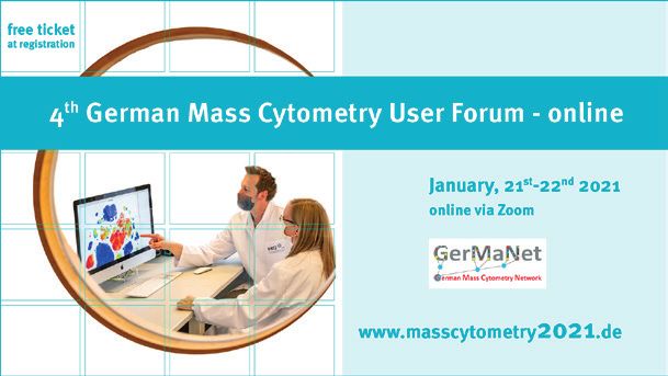
Welcome to the 4th German Mass Cytometry User Forum - online

Dear friends and colleagues,
It is my pleasure to announce the 4th edition of
the German Mass Cytometry User Forum – on-
line.
We have assembled an exciting program with a
focus on the application of mass cytometry in
the study of SARS-Cov2 and COVID-19, and data
analysis techniques.
The meeting will be free of charge for academic
participants.
I am looking forward to an inspiring Mass Cyto-
metry User Forum in 2021!
Best wishes, Henrik Mei
Yours,

Henrik Mei

                                                               3
Thursday, January 21st, 2021
10.30        Welcome, Henrik Mei
                          Getting started... ...with mass cytometry
10:45 Désirée Kunkel, Berlin: Principles of mass cytometry (10)
10:55 Axel Schulz, Berlin: Suspension mass cytometry – basic workflows (20)
11:15 Désirée Kunkel, Berlin: Introduction to Imaging mass cytometry (10)
11:25 Henrike Salié/Bertram Bengsch, Freiburg: Imaging mass cytometry – basic workflows (20)
11:45	Sarah Warth, Ulm, and all presenters: Q&A, tips and tricks (30)
       please send questions to: axel.schulz@drfz.de
12:15   Coffee break

                            Session 1 – Mass Cytometry vs COVID-19

Chairs: Bertram Bengsch & Henrik Mei
13:20	Birgit Sawitzki, Berlin: CyTOF-mediated characterisation of the myeloid cell compartment
       in severe COVID-19 (15+5)
13:40	Henrike Salié, Freiburg: Spatial single-cell mapping reveals an altered local immune res-
       ponse in COVID-19 brains (15+5)
14:00	Short talk: Malte Lehmann, Berlin: Human small intestinal infection by SARS-CoV2 is cha-
       racterized by an activation of CD8+ T cells (12+3)
14:15	Short talk: Dena Panovska, Leuven: Mapping the recovery of critically ill COVID19 patients
       by high-dimensional profiling identifies blood immunotypes following a specific immune
       trajectory(12+3)
14:30   Coffee break

                          Session 2 News from … & selected abstracts

Chairs: Marie Burns & Sarah Warth
15:00	… BIH Berlin: Chotima Böttcher – The uses and limitations of single-cell mass cytometry
       for studying human microglia function (15+5)
15:20	… Ulm: Habib Rahimi: Characterization of acute erythroid leukemia using mass cytometry
15.40 … MPI MG Berlin: Marie-Laure Yaspo, Interpreting bulk RNAseq with single cell technologies
16.00 Short talk: Florian Ingelfinger, Zurich: Single-cell profiling of Myasthenia Gravis identifies a
       pathogenic T cell signature (12+3)

                                         Webinar by OMIQ

Chair: Axel Schulz
16.20	Chris Ciccolella, Santa Clara, USA: Mastering High-Dimensional Analysis with the OMIQ
       Platform
16:40   Coffee break

                                              Session 3

Chair: Henrik Mei
17:00   Chris Tape, London, UK
        Single-Cell Signalling Analysis of Tumour Microenvironment Organoids
17:40   Good night for today

4
Friday, January 22nd, 2021

                                           Speed talks

Chair: Axel Schulz, Désirée Kunkel & Sarah Warth
10.00   Speed talks from selected abstracts (3 min each)
11:45   Coffee break

                                      Webinar by Fluidigm

Chair: Henrik Mei
12:00   Speaker: Andrew Quong, Chief Scientific Officer Fluidigm Corporation
        Preliminary Title: Mass Cytometry – a glance into the future
12:40   Coffee break

                                     Session 4 News from…

Chairs: Marie Burns
13:00   …DRFZ: Axel Schulz: Severe COVID‐19 is characterized by an increased induction of peri-
        pheral plasmablasts with an aberrant CD62L+HLA‐DRlow phenotype (15+5)
13:20 	…Munich: Selina Keppler/Marc Rosenbaum: Establishing CyTOF panels for the analysis of
        murine immune cells (15+5)
13:40	…Freiburg: Lena Sophie Mayer: Alterations in tissue-resident memory and exhausted-like
        CD8+ T cells in active UC. (15+5)
14.00 …Granada: Paulina Rybakowska: Experimental and data processing workflow for large-sca-
        le immune monitoring studies by mass cytometry (15+5)
14:20   Coffee break

                                    Session 5 Data Analysis

Chairs: Henrik Mei, Axel Schulz
14:40 Lucie Rodriguez, Scilifelab, Stockholm, SE: Multi OMICs data integration (15+5)
15:00	Denis Schapiro, Harvard, USA: „Google Maps” for tissue biology – Mapping the tumor
       microenvironment with spatial omics technologies (25+5)
15:30 Mark Robinson, Univ. Zurich, CH: Differential discovery for CyTOF experiments (25+5)

                           Webinar by Beckman Coulter/Cytobank

Chair: Henrik Mei
16:15   Speaker: Dr. Giulia Grazia, Beckman Coulter Life Sciences, Italy
        Title: Navigate safely through a sea of data: management and analysis made smart!
16:35   Coffee break

                                      Virtual round table

Chair: Henrik Mei
16:45   Discussion of hot topics, Denis Shapiro, Chris Tape, Chotima Böttcher - Q&A
17:45   Farewell, Poster price ceremony and goodbye

                                                                                               5
Thursday, January 21th, 2021
                            Getting started... ... with mass cytometry

Désirée Kunkel
Désirée Kunkel, Flow & Mass Cytometry Core Facility, Charité - Universitätsmedizin Berlin & Berlin Institute
of Health (BIH)

Axel Schulz
Mass Cytometry Lab, German Rheumatism Research Center Berlin (DRFZ), a Leibniz Institute

Henrike Salié
University Medical Center Freiburg, Clinic for Internal Medicine II – Gastroenterology, Hepatology,
Endocrinology and Infectious Diseases, Freiburg, Germany

Bertram Bengsch
University Medical Center Freiburg, Clinic for Internal Medicine II – Gastroenterology, Hepatology, Endocri-
nology and Infectious Diseases, Freiburg, Germany

Sarah Warth
Core Facility Cytometry, Ulm University Medical Faculty

Abstract
Our introduction to mass cytometry ensures that        batch normalization. You will also learn about
everyone is at the same level when talking ab-         the advantages of Imaging Mass Cytometry (IMC)
out this technology. Five experts from the field       and how to establish a multiplexed antibody pa-
tell you how mass cytometry works and how it           nel for it. This is complemented by an introduc-
can be used to examine cell suspensions and tis-       tion to current concepts of data analysis, both
sue sections. We will guide you through typical        for imaging and suspension mass cytometry. Fol-
experimental workflows and share our experien-         lowing the introductory talks there will also be
ce with important aspects in the application           time to discuss individual questions concerning
of mass cytometry, such as metal conjugation,          mass cytometry and its application.
sample barcoding, spillover compensation and

Biosketches:
Désirée Kunkel is head of Flow & Mass Cytome-          mass cytometry especially in its application in
try Core Facility of the Berlin Institute of Health    immunology. The core in Ulm is using a Helios
(BIH) for 11 years. As one of the first CyTOF ope-     instrument.
rators in Germany, she and her core facility pro-
vide professional access to suspension mass cy-        Henrike Salié is performing her doctorate stu-
tometry since 2014 and IMC since 2018.                 dies in the lab of Prof. Bengsch in the Clinic for
                                                       Internal Medicine II at the University Medical
Bertram Bengsch is Professor for Translational         Center Freiburg. Her research focus is on un-
Hepatogastroenterology and head of the Mass            derstanding immune responses to malign, auto-
Cytometry Facility at the Clinic for Internal Me-      immune and viral challenges in tissues, with a
dicine II, University Medical Center Freiburg. He      focus on the spatial interactions of exhausted T
has worked with mass cytometry starting in 2014        cells, for which she established and applied an
at the University of Pennsylvania.                     imaging mass cytometry approach.
Sarah Warth coordinates the Core Facility Cy-          Axel Schulz joined the DRFZ mass cytometry la-
tometry in Ulm since 2017. She was formerly            boratory in 2014 after gaining first experience
working in the core of Désirée Kunkel and whe-         with the CyTOF technology at the HIMC in Stan-
re she gained a lot of experience in suspension        ford in 2012. As a postdoc, he works on the tech-

6
nical implementation of the CyTOF technology in
various projects and is currently the operator in
charge of the institute‘s Helios instrument.

                            Session 1 – Mass Cytometry vs COVID-19

CyTOF-mediated characterisation of the myeloid cell compartment in
severe COVID-19
Birgit Sawitzki
Charité University Medicine, Berlin, Germany

Coronavirus disease 2019 (COVID-19) is a mild to      ne cell composition and activation in mild versus
moderate respiratory tract infection. However,        severe COVID-19 over time. HLA-DRhiCD11chi
a subset of patients progresses to severe disease     inflammatory monocytes were elevated in mild
and respiratory failure. The mechanism of pro-        COVID-19. Severe COVID-19 was marked by oc-
tective immunity in mild forms and the pathoge-       currence of neutrophil precursors, as evidence
nesis of severe COVID-19 associated with increa-      of emergency myelopoiesis, dysfunctional ma-
sed neutrophil counts and dysregulated immune         ture neutrophils, and HLA-DRlo monocytes. Our
responses remain unclear. In a dual-center, two-      study provides detailed insights into the systemic
cohort study, we applied single-cell proteomics       immune response to SARS-CoV-2 infection and
of whole-blood and peripheral-blood mononuc-          reveals profound alterations in the myeloid cell
lear cells using mass cytometry and flow cytome-      compartment associated with severe COVID-19.
try, respectively, to determine changes in immu-

Biosketch
Birgit is Professor of “Immune Tolerance” at the      cal trials, sponsored by the European commit-
Institute of Medical Immunology. She has deve-        tee, aiming on e.g. personalized treatment and
loped validated immune monitoring tools for           finally tolerance induction in solid organ trans-
application in investigator-driven clinical trials.   plant patients. In the last month Birgit success-
She is coordinating the immune monitoring of          fully applied her expertise in deciphering the
three big multi-center investigator-driven clini-     pathomechanisms of severe COVID-19.

Keywords
Immune monitoring, COVID-19, Immune cell
composition and functionality

Publications
1. Schulte-Schrepping J, Reusch N, Paclik D, Baß-     Aschenbrenner AC*, Li Y*, Nattermann J*, Sawitz-
ler K, Schlickeiser S, Zhang B, Krämer B, Kram-       ki B*, Saliba AE*, Sander LE*; Deutsche COVID-19
mer T, Brumhard S, Bonaguro L, De Domenico E,         OMICS Initiative (DeCOI). Severe COVID-19 Is
Wendisch D, Grasshoff M, Kapellos TS, Beckstette      Marked by a Dysregulated Myeloid Cell Compart-
M, Pecht T, Saglam A, Dietrich O, Mei HE, Schulz      ment. Cell. 2020 Sep 17;182(6):1419-1440.e23.
AR, Conrad C, Kunkel D, Vafadarnejad E, Xu CJ,        doi: 10.1016/j.cell.2020.08.001. Epub 2020 Aug
Horne A, Herbert M, Drews A, Thibeault C, Pfeif-      5. PMID: 32810438; PMCID: PMC7405822. *shared
fer M, Hippenstiel S, Hocke A, Müller-Redetzky        senior author
H, Heim KM, Machleidt F, Uhrig A, Bosquillon de
Jarcy L, Jürgens L, Stegemann M, Glösenkamp           2. Sawitzki B, Harden PN, Reinke P, Moreau A,
CR, Volk HD, Goffinet C, Landthaler M, Wyler E,       Hutchinson JA, Game DS, Tang Q, Guinan EC, Bat-
Georg P, Schneider M, Dang-Heine C, Neuwinger         taglia M, Burlingham WJ, Roberts ISD, Streitz M,
N, Kappert K, Tauber R, Corman V, Raabe J, Kai-       Josien R, Böger CA, Scottà C, Markmann JF, Hes-
ser KM, Vinh MT, Rieke G, Meisel C, Ulas T, Becker    ter JL, Juerchott K, Braudeau C, James B, Con-
M, Geffers R, Witzenrath M, Drosten C, Suttorp        treras-Ruiz L, van der Net JB, Bergler T, Caldara
N, von Kalle C, Kurth F, Händler K, Schultze JL*,     R, Petchey W, Edinger M, Dupas N, Kapinsky M,
                                                      Mutzbauer I, Otto NM, Öllinger R, Hernandez-Fu-

                                                                                                      7
entes MP, Issa F, Ahrens N, Meyenberg C, Karitzky      1639. doi: 10.1016/S0140-6736(20)30167-7.
S, Kunzendorf U, Knechtle SJ, Grinyó J, Morris
PJ, Brent L, Bushell A, Turka LA, Bluestone JA,        3. Truong KL, Schlickeiser S, Vogt K, Boës D, Stan-
Lechler RI, Schlitt HJ, Cuturi MC, Schlickeiser S,     ko K, Appelt C, Streitz M, Grütz G, Stobutzki N,
Friend PJ, Miloud T, Scheffold A, Secchi A, Crisal-    Meisel C, Iwert C, Tomiuk S, Polansky JK, Pascher
li K, Kang SM, Hilton R, Banas B, Blancho G, Volk      A, Babel N, Stervbo U, Sauer I, Gerlach U, Sawitz-
HD, Lombardi G, Wood KJ, Geissler EK. Regula-          ki B. Killer-like receptors and GPR56 progressive
tory cell therapy in kidney transplantation (The       expression defines cytokine production of human
ONE Study): a harmonised design and analysis of        CD4+ memory T cells. Nat Commun. 2019 May
seven non-randomised, single-arm, phase 1/2A           22;10(1):2263. doi: 10.1038/s41467-019-10018-
trials. Lancet. 2020 May 23;395(10237):1627-           1. PMID: 31118448; PMCID: PMC6531457.

Spatial single-cell mapping reveals an altered local immune response in
COVID-19 brains
Henrike Salié
University Medical Center Freiburg, Clinic for Internal Medicine II – Gastroenterology, Hepatology, Endocri-
nology and Infectious Diseases, Freiburg, Germany

Abstract
COVID-19 causes neurological symptoms that can         croglial clusters, their presence in specific ana-
be potentially life-threatening in up to 67 % of       tomical regions and their cellular interactions.
the patients. To understand the local immune           The analysis further highlights microglial nodu-
response during SARS-CoV-2 infection at a spati-       les and perivascular immune cell clusters as key
ally resolved, high-dimensional single-cell level,     sites of the local immune response in the brain
we performed a 38-biomarker imaging mass cy-           stem. It also demonstrates that disease-associa-
tometry analysis of the brain stem and olfactory       ted neuroinflammation is associated with severe
bulb from COVID-19 patients and additional con-        axonal damage as a structural basis for neurolo-
trols. Importantly, utilizing an unbiased image        gic deficits. Finally, IMC staining for SARS-CoV-2
segmentation and cell classification pipeline,         spike glycoprotein revealed direct evidence of
we observed a significant immune activation in         vasculature-associated viral presence in the ol-
the central nervous system (CNS) and identified        factory bulb and brain stem as well as reactive
novel context-specific CD8 T cell and microglial       astrogliosis. Together these analyses identify the
clusters. Spatially resolved single-cell analysis      immune correlates of a surprisingly high level of
identified distinct phenotypes of T cells and mi-      neuroinflammation in fatal cases of COVID19.

Biosketch
Henrike Salié is performing her doctorate studies      which she established an imaging mass cytome-
in the lab of Prof. Bengsch in the Clinic for Inter-   try approach. During the SARS-CoV-2 pandemic
nal Medicine II at the University Medical Center       she explored the neuroinflammation in brains of
Freiburg. Her research focus is on understanding       COVID-19 patients in a collaborative effort with
immune responses to malign, autoimmune and             the Institutes of Neuropathology in Freiburg (AG
viral challenges in tissues, with a focus on the       Prinz), Hamburg (AG Glatzel) and Göttingen (AG
spatial interactions of exhausted T cells, for         Stadelmann-Nessler).

Keywords
COVID-19, Imaging mass cytometry, T cell ex-
haustion

8
Short talk:
Human small intestinal infection by SARS-CoV2 is characterized by an
activation of CD8+ T cells
Malte Lehmann1
Malte Lehmann1, Kristina Allers1, Claudia Heldt1, Franziska Schmidt2, Yasmina Rodriguez-Silke2, Désirée Kun-
kel2, Michael Schumann1, Chotima Böttcher3, Viktor M. Corman4, Thomas Schneider1, Christoph Loddenkem-
per5, Verena Moos1, Carl Weidinger1,3,7, Anja A. Kühl6,7*, Britta Siegmund1,7*
1
 Charité – Universitätsmedizin Berlin, corporate member of Freie Universität Berlin, Humboldt-Universität
zu Berlin and Berlin Institute of Health, Campus Benjamin Franklin. Medical Department, Division of Gas-
troenterology, Infectiology and Rheumatology (including Nutritional Medicine), Hindenburgdamm 30, 12200
Berlin, Germany
2
 Charité – Universitätsmedizin Berlin, corporate member of Freie Universität Berlin, Humboldt-Universität
zu Berlin and Berlin Institute of Health, Berlin-Brandenburg Center for
Regenerative Therapies BCRT, Charité | BIH Cytometry Core, Campus Virchow Klinikum, Germany
3
 Charité – Universitätsmedizin Berlin, corporate member of Freie Universität Berlin, Humboldt-Universität
zu Berlin and Berlin Institute of Health, Klinik für Psychiatrie und Psychotherapie, Campus Mitte, Germany
Clinician Scientist Program, Berlin Institute of Health
4
 Charité - Universitätsmedizin Berlin, corporate member of Freie Universität Berlin and Humboldt-Universi-
tät zu Berlin, Berlin Institute of Health, and German Centre for Infection Research, Department of Virology,
10117 Berlin, Germany.
PathoTres, Gemeinschaftspraxis für Pathologie und Neuropathologie, Teltowkanalstr. 2, 12247 Berlin, Ger-
5

many
6
 Charité – Universitätsmedizin Berlin, corporate member of Freie Universität Berlin, Humboldt-Universität
zu Berlin and Berlin Institute of Health, iPATH.Berlin, Campus Benjamin Franklin, Germany
7
    The Transregio 241 IBDome Consortium
*
    Contributed equally to this work

Symptoms of COVID-19 suggest a multisystemic            T cells as well as epithelial apoptosis and re-
disease including the gastrointestinal system.          generation. We hypothesize CD8+ T cell activa-
Analysing biopsies of the small intestine from          tion and migration into the intestinal epithelium
COVID-19 patients using imaging mass cytometry          upon infection of intestinal cells as a possible
we identified histomorphological changes of the         cause for gastrointestinal symptoms.
epithelium, characterized by infiltrating CD8+

Short talk:
Mapping the recovery of critically ill COVID19 patients by high-
dimensional profiling identifies blood immunotypes following a specific
immune trajectory
Dena Panovska
Panovska D1#, Penttilä PA2#, Van Gassen S3#, Vanderbeke L4, Van Herck Y5, Quintelier K3, Emmaneel A3, Claeys
A1, Derweduwe M1, Verbeke T1, Chinnaraj R2, Filtjens J6, Malengier-Devlies B6, Ahmadzadeh K6, Van Mol P7,
Borras DM8, Antoranz A3, Bosisio FM9, the CONTAGIOUS consortium$, Wauters E10, Matthys P6, Saeys Y2, Garg
AD8, Wauters J11#, De Smet F3#*.
#
    equal contribution
*
    corresponding author: frederik.desmet@kuleuven.be
1
 Laboratory for Precision Cancer Medicine, Translational Cell and Tissue Research, Department of Imaging &
Pathology, KU Leuven, Belgium
2
    KU Leuven Flow & Mass Cytometry Facility, KU Leuven, Belgium

                                                                                                          9
3
Department of Applied Mathematics, Computer Science and Statistics, Ghent University, Gent, Belgium, and
Data Mining and Modeling for Biomedicine, VIB Center for Inflammation Research, Gent, Belgium.
4
Laboratory of Clinical Bacteriology and Mycology, Department of Microbiology, Immunology and Transplan-
tation, KU Leuven, Belgium
5
Laboratory of Experimental Oncology, Department of Oncology, KU Leuven, Belgium
6
Laboratory of Immunobiology, Department of Microbiology, Immunology and Transplantation, Rega Institu-
te, KU Leuven, Belgium
7
Laboratory of Translational Genetics, Department of Human Genetics, VIB-KU Leuven, Belgium
8
Laboratory for Cell Stress & Immunity (CSI), Department of Cellular and Molecular Medicine (CMM), KU
Leuven, Belgium
9
Translational Cell & Tissue Research, Department of Imaging & Pathology, KU Leuven, Belgium
Laboratory of Respiratory Diseases and Thoracic Surgery (BREATHE), Department of Chronic Diseases and
10
Metabolism, KU Leuven, Belgium
Laboratory for Clinical Infectious and Inflammatory Disorders, Department of Microbiology, Immunology
11
and Transplantation, KU Leuven, Belgium
$
Additional Contagious consortium members: Michael Casaer, Dieter Dauwe, Jan Gunst, Greet Hermans, Ste-
phanie Humblet-Baron, Diether Lambrechts, Adrian Liston, Natalie Lorent, Kim Martinod, Philippe Meersse-
man, Johan Neyts, Paul Proost, Jeroen Raes, Stephen Rex, Sabine Tejpar, Karin Thevissen, Thomas Tousseyn,
Birgit Weynand, Alexander Wilmer, Carine Wouters.
Abstract
The COVID-19 pandemic poses a major burden immune cell type to recover by restoring HLA-
on health-care and economic systems across the DR-positivity and by reducing the immunosup-
globe. Even though a majority of the population pressive CD163+ monocyte population, followed
only develops minor symptoms upon SARS-CoV2 by the recovery of CD8+ and CD4+ T cell, and
infection, a significant proportion are hospita- mDC populations. The determined immunotypes
lized at intensive care units (ICU) requiring cri- also correlated to aberrant cytokine and acute-
tical care. While insights into the early stages phase reactant levels. Finally, integrative analy-
of the disease are gradually expanding, the dy- sis of cytokines and immune cell profiles showed
namic immunological processes occurring in cri- a shift from an initially dysregulated immune
tically ill patients throughout their recovery at response to a more coordinated immunogenic
ICU are far less understood. We have analyzed interplay, highlighting the importance of longi-
longitudinally collected, whole blood samples of tudinal sampling to understand the pathophysio-
40 surviving COVID-19 patients during their re- logy underlying recovery from severe COVID-19.
covery at ICU using high-dimensional cytometry
by time-of-flight (CyTOF) and cytokine multiple-
xing. Based on the neutrophil to lymphocyte ra-
tio (NLR), we defined 4 sequential immunotypes
during recovery that correlated to various clini-
cal parameters, including the level of respirato-
ry support at concomitant sampling times. We
also identified classical monocytes as the first
10Session 2 News from … & selected abstracts
…BIH Berlin:
The uses and limitations of single-cell mass cytometry for studying human
microglia function
Chotima Böttcher
Charité – Universitätsmedizin Berlin
Abstract
Microglia, the resident innate immune cells of pression of human microglia (huMG), which can
the central nervous system (CNS), play an im- be combined with scRNA-seq for comprehensive
portant role in brain development and homeos- analysis, as it allows single-cell analysis of post-
tasis. Studies in animal models reveal the origin translational modifications of proteins, which
and development of microglia, and how these provides insights into cell signalling dynamics in
cells alter their transcriptional and phenotypic targeted cells. In addition, imaging mass cyto-
signatures during CNS pathology. However, little metry (IMC) has recently been demonstrated for
is known about their human counterparts. Recent analysing multiple cell types in human brain sec-
studies in human brain samples have harnessed tions. IMC leverages mass spectrometry to acqui-
the power of mass cytometry (CyTOF) to provide re spatial data of cell-cell interactions on brain
a comprehensive molecular view of human mi- sections. Here, the use and limitations of CyTOF
croglia in healthy and diseased brains. CyTOF is in studing huMG are discussed.
a powerful tool to study single-cell protein ex-
Biosketch
Chotima Böttcher is a group leader and principal loid cells including monocytes and brain microg-
investigator at the Charité – Universitätsmedizin lia/macrophages. The main goal is to identify
Berlin, Germany. Dr. Böttcher obtained her PhD cellular complexity and heterogeneity of the
at Institute of Pharmacy, at Martin-Luther-Uni- myeloid compartment of the human central ner-
versity Halle-Wittenberg, Halle/Saale, Germany. vous system and to further investigate how the-
Her research focuses on systems immunology in se signatures alter during neurodegeneration/
neuroscience, with particular emphasis on mye- neuroinflammation.
Publications
1. Böttcher C*, Schlickeiser S*, Sneeboer MAM*, 3.Böttcher C*, van der Poel M*, Fernández-Za-
Kunkel D, Knop A, Paza E, Fidzinski P, Kraus L, pata C*, Schlickeiser S, Leman JKH, Hsiao CC,
Snijders GJL, Kahn RS, Schulz AR, Mei HE, NBB- Mizee MR, Adelia, Vincenten MCJ, Kunkel D, Hui-
Psy, Hol EM, Siegmund B, Glauben R, Spruth EJ, tinga I*, Hamann J*, Priller J*. Single-cell mass
de Witte LD, Priller J: Human microglia regio- cytometry reveals complex myeloid cell com-
nal heterogeneity and phenotypes determined position in active lesions of progressive multip-
by multiplexed single-cell mass cytometry. Nat le sclerosis. Acta Neuropathol Commun 8, 136
Neurosci 22, 78–90 (2019). (*equal contribution) (2020). (*equal contribution)
2.Sankowski R*, Böttcher C*, Masuda T, Geirs-
dottir L, Sagar, Sindram E, Seredenina T, Muhs
A, Scheiwe C, Shah MJ, Heiland DH, Schnell O,
Gru¨n D*, Priller J*, Prinz M*: Mapping microglia
diversity in the human brain through the integ-
ration of high-dimensional techniques. Nat Neu-
rosci 22, 2098–2110 (2019). (*equal contribution)
11… Ulm
Characterization of acute erythroid leukemia using mass cytometry
Habib Rahimi
Institute of experimental cancer research, University Hospital Ulm
Acute erythroleukemia (AEL) is a rare subtype motherapy treatment and 3) the development of
of acute myeloid leukemia (AML) which accounts innovative targeted therapies are missing. Along
for less than 5% of all de novo AML cases. There with a comprehensive multiomics approach,
have been several efforts to characterize AEL at flow cytometry and murine bone marrow trans-
a molecular level, describing recurrent altera- plantation experiments, we developed a mass
tions in TP53 and in the NPM1 and FLT3 gene cytometry marker panel for an in-depth charac-
(Iacobucci et al. Nat Genet 2019). A comprehen- terization of human AEL bone marrow samples
sive genomic analysis of AEL cases confirmed the in comparison to other AML subtypes and bone
complexity of this AML subtype. Despite these marrow (BM) from healthy donors. A total of 8
advances, the underlying biology of AEL is still AELs, 30 AMLs and 5 BM controls were success-
not precisely defined and the prognosis is dismal fully analyzed. Marker combinations, identified
with a median survival of only 2-3 months for and validated by conventional flow cytometric
pure erythroid leukemia. Marker combinations analysis, were able to separate erythroid from
suitable for 1) the identification and characteri- myeloid blast populations and might help in
zation of leukemic stem cell (LSC) candidates, 2) identifying MRD.
monitoring minimal residual disease during che-
Biosketch
Habib Rahimi is currently a PhD Student at the He is a member of the Cellular and Molecular Me-
Institute of Experimental Cancer Research and chanisms in Aging (CEMMA)-research group (GRK
the International Graduate School in Molecular 1789) and is involved in projects within the CRC
Medicine in Ulm, Germany. Experimental Models and Clinical Translation in
Leukemia (SFB 1074) and the Aging-related epi-
His research mainly focuses on acute myeloid genetic remodeling in acute myeloid leukemia
leukemia, hematopoietic stem cells and aging. consortium (FOR 2674).
Keywords
Acute erythroleukemia (AEL), Minimal residual disease (MRD), Leukemic stem cells (LSC)
… MPI Molecular Genetics Berlin
Interpreting bulk RNAseq with single cell technologiest
Marie-Laure Yaspo
MPI Molecular Genetics, Berlin
12Short talk:
Single-cell profiling of Myasthenia Gravis identifies a pathogenic T cell
signature
Florian Ingelfinger
Florian Ingelfinger1,2, Sinduya Krishnarajah1, Michael Kramer3, Sebastian G. Utz1, Edoardo Galli1, Mirjam
Lutz1, Pascale Zwicky1, Ayse U. Akarca4, Nicole Puertas Jurado1, Corinne C. Widmer5, Luca Piccoli3, Federica
Sallusto3,6, Nicolás G. Núñez1, Teresa Marafioti4, Didier Schneiter7, Isabelle Opitz7, Antonio Lanzavecchia3,
Donatella De Feo1, Sarah Mundt1, Hans H. Jung2, Bettina Schreiner1,2,†,*, Burkhard Becher1,†,*.
1
Institute of Experimental Immunology, University of Zurich, Zurich, Switzerland.
2
Department of Neurology, University Hospital Zurich, Zurich, Switzerland.
3
Institute for Research in Biomedicine, Università della Svizzera italiana, Bellinzona, Switzerland.
4
Department of Cellular Pathology, University College London Hospital, United Kingdom.
5
Department of Medical Oncology and Hematology, University Hospital Zurich and University of Zurich, Zu-
rich, Switzerland.
6
Institute of Microbiology, ETH Zurich, Zurich, Switzerland.
7
Department of Thoracic Surgery, University Hospital Zurich, Zurich, Switzerland.
†
These authors jointly supervised this work.
*
Correspondence to: becher@immunology.uzh.ch and bettina.schreiner@uzh.ch.
One Sentence Summary
Single-cell immunophenotyping reveals a pathogenic T cell signature accumulating in the inflamed thymus
of Myasthenia gravis patients.
Abstract
Myasthenia Gravis (MG) is an autoimmune disea- two dysregulated subsets of inflammatory cir-
se characterized by impaired neuromuscular sig- culating memory T helper (Th) cells. These sig-
naling due to autoantibodies targeting the ace- nature ThCD103 and ThGM cells populated the
tylcholine receptor. Although its auto-antigens diseased thymus, were reduced in the blood of
and effector mechanisms are well defined, the MG patients, and were inversely correlated with
cellular and molecular drivers underpinning MG disease severity. Both signature Th subsets re-
remain elusive. Here we employed high-dimen- bounded in the blood of MG patients after sur-
sional single-cell mass and spectral cytometry of gical thymus removal, indicative of their role as
blood and thymus samples from MG patients in cellular markers of disease activity. Together,
combination with supervised and unsupervised this in-depth analysis of the immune landscape
machine-learning tools to gain insight into the of MG provides valuable insight into disease pa-
immune dysregulation underlying MG. By crea- thogenesis, suggests novel biomarkers and iden-
ting a comprehensive immune map we identified tifies new therapeutic targets for treatment.
Biosketch
Florian Ingelfinger is a PhD student in the lab understand the contribution of genetic and en-
of Prof. Dr. Burkhard Becher at the Institute of vironmental drivers in the development of neu-
Experimental Immunology, University of Zurich, roimmunological disorders like Multiple Sclerosis
Switzerland. His research interest focusses on or Myasthenia Gravis.
the integration of single cell technologies in cli-
nical studies to decipher complex cellular inter-
actions leading to inflammation and autoimmu-
nity. Utilizing computational tools he seeks to
13Webinar by OMIQ
Mastering High-Dimensional Analysis with the OMIQ Platform
Chris Ciccolella
Co-Founder & CEO, Omiq, Inc., Santa Clara, USA
Abstract
Before biology can be controlled, it must be un- and is often a challenging obstacle for resear-
derstood. To be understood, it must be measu- chers. In this talk, the principles of effective
red. Mass Cytometry gives us incredible power to Mass Cytometry data analysis will be presented
measure biology. The process of understanding, in context of the OMIQ Data Science Platform
i.e., data analysis, is a rich and evolving field (www.omiq.ai), an advanced cloud software for
with a considerable amount of detail to learn, cytometry data analysis
Biosketch
Chris is originally a cell biologist and cytome- 2018 to focus on analytical innovations to
try expert. In 2013 he moved from the bench accelerate the progress of human health.
to the computer to focus on software engi-
neering and data science. Following a te-
nure at Cytobank, he co-founded Omiq in
Keywords
High-dimensional analysis, algorithms, discovery
Session 3
Single-Cell Signalling Analysis of Tumour Microenvironment Organoids
Chris Tape
University College London Cancer Institute, London, UK
Abstract
Organoids are self-organising stem cell-derived ganoid co-cultures. Integrating single-cell PTM
ex vivo cultures widely adopted as biomime- analysis with thiol-reactive organoid barcoding
tic models of healthy and diseased tissues. As in situ (TOBis) enables 35-plex and 126-plex
complex heterocellular systems, organoids are comparison of signalling networks between orga-
especially well-positioned to take advantage of noid co-cultures. Cell-type-specific PTM analysis
emerging high-dimensional single-cell technolo- of colorectal cancer organoid co-cultures revea-
gies. Here we present a Cytometry by Time-Of- led that oncogenic mutations cell-autonomously
Flight (CyTOF) method for single-cell analysis of mimic signalling states normally induced by stro-
post-translational modification (PTM) signalling mal fibroblasts and macrophages.
in organoids and tumour microenvironment or-
Biosketch
Chris received his Ph.D. from Prof. Gillian Mur- (with Dr. Claus Jorgensen and Prof. Chris Mar-
phy’s lab at the CRUK Cambridge Institute (Uni- shall) and Massachusetts Institute of Technology
versity of Cambridge). He was then awarded (MIT) (with Prof. Doug Lauffenburger) to study
a Sir Henry Wellcome Postdoctoral Fellowship how oncogenes signal across multiple cell types
between The Institute of Cancer Research (ICR) in cancer. Chris now leads the Cell-Communica-
14tion Lab at UCL CI under a CRUK Career Develop- Trust).
ment Fellowship (supported by the CRUK Werth
Keywords
Organoids, Barcoding, PTM Signalling
Friday, January 22nd, 2021
Speed talks
Speed talks from selected abstracts
Beckman Coulter/Cytobank donates a year’s licence of the Cytobank Premium SW for
the best Speed Talk at the 4th German Mass Cytometry User Forum!
1. Longitudinal, multi-center study reveals unique immune signatures of
severe COVID-19
Stefanie Kreutmair
Stefanie Kreutmair1,2, Susanne Unger1,3, Nicolás Gonzalo Núñez1,3, Florian Ingelfinger1,3, Chiara Alberti1, Do-
natella De Feo1, Sinduya Krishnarajah1, Manuel Kauffmann1, Ekaterina Friebel1, Benjamin Gaborit4, Sepideh
Babaei5, Mirjam Lutz1, Nicole Puertas Jurado1, Nisar P. Malek5, Siri Goepel5,6, Peter Rosenberger7, Helene A.
Häberle7, Ikram Ayoub8, Sally Al-Hajj8, Manfred Classen5,10, Roland Liblau8,10, Guillaume Martin-Blondel8,9,10,
Michael Bitzer5,10, Antoine Roquilly4,10, Burkhard Becher1,10,*
1
Institute of Experimental Immunology, University of Zurich, Zurich 8057, Switzerland
2
German Cancer Consortium (DKTK) and German Cancer Research Center (DKFZ), Heidelberg, Germany
3
These authors contributed equally
4
Université de Nantes, CHU Nantes, Pôle anesthésie réanimations, Service d’Anesthésie Réanimation chi-
rurgicale, Hôtel Dieu, Nantes, F-44093 France
5
Department Internal Medicine I, Eberhard-Karls University, Tuebingen, Germany
6
German Centre for Infection Research (DZIF), Partner Site Tuebingen, Germany
7
Department of Anesthesiology and Intensive Care Medicine, Eberhard-Karls University, Tuebingen, Germany
8
Centre de Physiopathologie Toulouse-Purpan, Université de Toulouse, Centre National de la Recherche
Scientifique, Institut National de la Santé et de la Recherche Médicale, UPS, 31024 Toulouse, France
9
Department of infectious and tropical diseases, Toulouse University Hospital, Toulouse, France
10
These authors contributed equally
Abstract
The currently ongoing COVID-19 pandemic is con- to SARS-CoV-2 infection.
tinuing to spread around the world, with rising
numbers of deaths. There is clear evidence that For this purpose, we analyzed 150 PBMC samples
severe cases of COVID-19 which require hospita- of a multi-center, longitudinal COVID-19 cohort
lization due to respiratory distress are mediated together with non-SARS-CoV-2 critical pneumo-
by immunopathology. Thus, it is vital to unders- nia patient samples (n=25) and healthy controls
tand how the immune system reacts specifically (n=21). High-throughput, high-dimensional sing-
15le-cell spectral cytometry and algorithm-based Bench-to-bedside translation of the identified
analysis resulted in a complex immune signature immune signatures in severe COVID-19 offered
network underlying COVID-19 severity. By com- potential biomarkers for outcome prediction. On
paring the immune landscape of severe CO- top, we discovered a low but clearly detectable
VID-19 with non-SARS-CoV-2 critical pneumonia, ACE2 expression on a CD4+ T cell subset, which
we were able to extract immune features speci- opens the door for so far unknown potential un-
fic to SARS-CoV-2 infection. While COVID-19 and derlying mechanisms in the immunopathological
non-SARS-CoV-2 pneumonia share an emergency network of COVID-19.
monopoiesis and numerous features of adapti-
ve immune dysfunction, pathological immune Overall, the comparison of two severe infecti-
signatures exclusive to COVID-19 were concen- ous lung diseases driven by different pathogens
trated in the T and NK cell compartment. Inte- allowed us to uncover unique immune signatures
restingly, predominantly but not only the severe in SARS-CoV-2 mediated disease, revealing the
COVID-19 cohort presented several immune al- outlines of a complex immune landscape which
terations which did not recover at the end of can serve as a basis for translational treatment
our study (14 weeks after hospital admission), strategies effectively blocking the origin of the
pointing to a prolonged immune dysregulation. immunopathologic cascade in severe COVID-19.
2. SARS-CoV-2 infection is associated with a pro-thrombotic platelet
phenotype
Melissa Klug
Dario Bongiovanni M.D.*1,2,3, Melissa Klug M.Sc *1,2,4, Olga Lazareva M.Sc4, Simon Weidlich M.D.5, Marina Biasi
B.Sc 1, Simona Ursu Ph.D 6, Sarah Warth Ph.D6, Christian Buske M.D.6,7, Marina Lukas M.D.5, Christoph D. Spin-
ner M.D.5, Moritz von Scheidt M.D.2,8, Gianluigi Condorelli Ph.D3, Jan Baumbach Ph.D4, Karl-Ludwig Laugwitz
M.D.1,2, Markus List Ph.D.4 and Isabell Bernlochner M.D.1,2
*equal contribution
1
Technical University of Munich, School of Medicine, University hospital rechts der Isar, Department of In-
ternal Medicine I, Munich, Germany
2
German Center for Cardiovascular Research (DZHK), Partner Site Munich Heart Alliance, Germany; 3 De-
partment of Cardiovascular Medicine, Humanitas Clinical and Research Center IRCCS and Humanitas Univer-
sity, Rozzano, Milan, Italy
4
Chair of Experimental Bioinformatics, TUM School of Life Sciences Weihenstephan, Technical University of
Munich, Munich, Germany;
5
Technical University of Munich, School of Medicine, University hospital rechts der Isar, Department of In-
ternal Medicine II, Munich, Germany
6
Core Facility Cytometry, Ulm University Medical Faculty, Germany
7
CCC Ulm, Institute of Experimental Cancer Research, University Hospital Ulm, Germany
8
Deutsches Herzzentrum München, Cardiology, Technische Universität München, Munich, Germany;
Abstract
We compared the activation state and the ex- pe in platelets during SARS-CoV-2 infection con-
pression of transmembrane proteins in platelets sisting of highly expressed platelet activation
of 8 hospitalized COVID-19 patients not requiring markers which might contribute to the hyper-
intensive care support and platelets of healthy coagulopathy observed in COVID-19. Additional-
controls, both with and without in-vitro stimula- ly, several transmembrane proteins were higher
tion with thrombin receptor-activating peptide expressed compared to healthy controls.
(TRAP). We detected a hyper-activated phenoty-
163. Deep phenotyping by mass cytometry to define the T cell signature of
childhood allergic asthma
Hartmann Raifer
Hartmann Raifer 1,2
*, Axel R. Schulz3*, Johanna Krusche 4*, Addi Romero 1, Andreas Böck 4,
Wilhelm Bertrams 5, Bernd Schmeck 5, Rolf Müller 6, Michael Lohoff 1, Hyun-Dong Chang 7,
Bianca Schaub 4#, Henrik E. Mei 3#, Magdalena Huber 1#
*These authors contributed equally to this work
#
These authors jointly directed this work
1
Institute for Medical Microbiology and Hospital Hygiene, University of Marburg, 35043 Marburg, Germany
2
Core Facility Flow Cytometry, University of Marburg, 35043 Marburg, Germany
3
German Rheumatism Research Center (DRFZ), 10117 Berlin, Germany
4
Pediatric Allergology, Department of Pediatrics, Dr von Hauner Children‘s Hospital, University Hospital,
LMU Munich, 80337 Munich, Germany
5
Institute for Lung Research, Universities of Giessen and Marburg Lung Center, Philipps-University Marburg,
Member of the German Center for Lung Research (DZL), 35043 Marburg, Germany
6
Institute of Molecular Biology and Tumor Research, Center for Tumor Biology and Immunology, Philipps Uni-
versity ,35043 Marburg , Germany
7
Deutsches Rheuma-Forschungszentrum (DRFZ), an Institute of the Leibniz Association, 10117 Berlin, Ger-
many
Abstract
Allergic asthma (AA) in childhood is characteri- pulation and a specific Th1-clade as well as the
zed by Th2-driven immunity and defects in con- abundance of naïve/resting Tregs were increa-
tra-regulation by Th1 cells and/or Tregs. It can sed in AA, suggesting a modulation of Th sub-
be influenced by genetic mechanisms including set contra-regulation. The abundance of CD8+ T
polymorphisms in interferon regulatory factor 1 cells was decreased in AA, and they displayed
(IRF1), which associates with atopy in childhood. increase in naïve at the expense of memory phe-
We applied single-cell mass cytometry to define notype. This cellular signature associated in part
the T cell signature and assess a possible interre- with IRF1 status, indicating a possible mechanis-
lationship with IRF1 polymorphisms in childhood tic contribution of IRF regulatory elements. Our
AA. Using manual gating and algorithmic analy- approach demonstrates the utility of high-di-
sis we found increased CD4/CD8 T cell ratios in mensional mass-cytometry in combination with
AA which correlated with eosinophilia linked to genetic analysis to interrogate cellular signature
disease severity. Furthermore, a Th2 cell cluster in context of a specific genetic parameter, the-
with high ICOS and TIGIT expression was overre- reby providing basis for further studies to reveal
presented in AA. The ratio between this Th2 po- predictive biomarkers in childhood AA.
4. Deep phenotypical characterization of human CD3+CD56+ T cells by
mass cytometry
Addi Romero-Olmedo
Addi J. Romero-Olmedo1*, Axel R. Schulz2*, Magdalena Huber1, Corinna U. Brehm, M.D.3,4, Hyun-Dong Chang2,
Cristina Chiarolla5, Tobias Bopp6, Chrysanthi Skevaki7, Friederike Berberich-Siebelt5, Andreas Radbruch2,
Henrik E. Mei2**, and Michael Lohoff1**
1
Institute for Medical Microbiology and Hospital Hygiene, University of Marburg, Marburg, Germany
2
German Rheumatism Research Center Berlin (DRFZ), a Leibniz Institute, Berlin, Germany
Comprehensive Biobank Marburg – CBBMR, Member of the DZL, Philipps-University Marburg, Marburg, Ger-
3
many
174
Institute for Pathology, Philipps-University Marburg, University Hospital Marburg, Marburg, Germany
5
Institute of Pathology, Julius-Maximilian University of Wuerzburg, Wuerzburg, Germany
Institute for Immunology, University Medical Center of the Johannes Gutenberg-University Mainz, Mainz,
6
Germany
7
Institute of Laboratory Medicine, Universities of Giessen and Marburg Lung Center (UGMLC), Philipps Uni-
versity Marburg, German Center for Lung Research (DZL), Marburg, Germany
*
These authors contributed equally to this work.
**
These authors jointly directed this work.
Abstract
CD56+ T cells are a group of pro-inflammatory CD56+ T cell phenotypes demonstrating a hither-
CD3+ T lymphocytes with characteristics of natu- to underestimated heterogeneity among these
ral killer cells, and are involved in antimicrobial cells. The novel CD3+CD56+ subset description
immune defense. Here, we performed a deep comprises phenotypes superimposed with naive,
phenotypic profiling of CD56+ T cells of periphe- memory, type 1, type 2, and type 17 differen-
ral blood of normal human donors and individu- tiation stages, in part represented by a pheno-
als sensitized to birch-pollen or/and house dust typical continuum. Frequencies of 2 out of 19
mite by high-dimensional mass cytometry combi- CD3+CD56+ FlowSOM clusters were significantly
ned with manual and computational data analy- diminished in allergic individuals, demonstrating
sis. A co-regulation between major conventional less frequent presence of cells with cytolytic,
T cell subsets and their respective CD3+CD56+ presumably protective, capacity in these donors
cell counterparts which we herein demonstrate, consistent with defective expansion or their
appeared restricted to CD8+, MAIT, and TCRγδ+ recruitment to the affected tissue. Our results
T cell compartments. Interestingly, we find a co- contribute to defining specific cell populations
regulation of several CD3+CD56+ cell subsets in to be targeted during therapy for allergic con-
allergic but not in healthy individuals. Moreover, ditions.
using FlowSOM, we distinguished a variety of
5. Immunity, the beginning: Characterizing immune development in
preterm infants using mass cytometry.
Tomer Salame
Tomer M. Salame1, Shlomit Reich-Zeliger2, Chava Rosen2,3, Lisa Buchauer4, Jacob Rimer2, Tzipora Strauss3
and Nir Friedman2
1
Weizmann Institute of Science, Flow Cytometry Unit, Life Sciences Core Facilities, Rehovot, Israel
2
Weizmann Institute of Science, Department of Immunology, Rehovot, Israel
3
Edmond and Lily Safra Children‘s Hospital Chaim Sheba Medical Center, Neonatology, Ramat Gan, Israel
4
German Cancer Research Center (DKFZ), Heidelberg, Germany
Mortality of preterm infants due to infections that preterm babies born at extreme early week
is a major challenge. An immature immune sys- of gestation (23-26 weeks) show all the immune
tem is a main determinant of susceptibility to cell populations, but in a different composition
pathogens. To better understand the developing compared to more mature preterm babies (32-
immune system, we mapped it in preterm and 36 week of gestation) or full-term babies. PCA
full-term infants, using mass cytometry, which analysis reveals that trajectories tend to have
provides unprecedented information on immune similar starting and end points for babies born in
cell types. Using data from longitudinal samples the same gestation week, and are more similar
and the clinical course of the infants, we gene- among twins. Additionally, a large cell popula-
rated signatures of immune state and repertoire tion which is negative to CD45 and did not sig-
composition. Approximately 20 immune cell po- nificantly express any other markers was found.
pulations were characterized. We demonstrate It was identified by Single-cell RNA-Seq as nuc-
18leated erythrocytes. This population is remarka- treatment. We show here a comprehensive study
bly detected mainly in extreme preterm babies of the neonate immune system components
(or at multiple births) and diminishes over time. which reveals patterns and similarities throug-
Moreover, preliminary analysis links the immu- hout its development and clinical state.
ne state with clinical data, including antibiotics
6. Cell type-specific dysregulation of CD38 expression in Systemic Lupus
Erythematosus
Marie Burns
Marie Burns1*, Lennard Ostendorf1,2*, Andreas Grützkau1, Falk Hiepe1,2, Henrik Mei1*, Tobias Alexander1,2*
1
Deutsches Rheuma-Forschungszentrum (DRFZ Berlin) – a Leibniz Institute
2
Department of Rheumatology and Clinical Immunology – Charité–Universitätsmedizin Berlin
*
These authors contributed equally
Systemic lupus erythematosus (SLE) is an autoim- CD38 expression in SLE. CD38 expression levels
mune disease characterized by pathogenic auto- were not associated with clinical activity of SLE.
antibodies secreted by plasma cells (PC). Among Spearman correlation analysis revealed coor-
novel plasma cell depleting strategies, CD38 has dinated CD38 expression in immune cells, with
been identified as promising target. The monoc- direct associations between most innate cell ty-
lonal anti-CD38 antibody daratumumab is ap- pes and T cell subsets in healthy controls, while
proved for treatment of multiple myeloma and associations between innate and lymphoid cell
provided a therapeutically relevant depletion of CD38 expression levels were rare and limited to
PCs in patients with SLE (Ostendorf et. al, NEJM, naïve T cells. By contrast, SLE patients display-
2020). However, CD38 is widely expressed across ed a heavily reconfigured correlation landsca-
immune cells, and the cellular targets of daratu- pe, including multiple SLE-specific associations
mumab beyond PC, especially in patients with between lymphocyte subsets and innate immune
SLE, are largely unknown. Therefore, we used cells.
mass cytometry to systematically characteri-
ze the expression of CD38 in peripheral blood In conclusion, we provide a comprehensive map
leukocytes to identify potential target cells of of CD38 expression across the peripheral blood
CD38-directed therapies that may contribute to immune cells in SLE and controls. The dysregula-
or limit therapeutic benefits in SLE. ted, commonly moderately increased expression
of CD38 in SLE may establish increased suscep-
In a cohort of 20 SLE patients, CD38 was highly tibility of immune cells to anti-CD38 in patients
expressed on plasmablasts, basophils, NK cells, with SLE. In detail, our study identifies plasma-
monocytes and plasmacytoid dendritic cells cytoid dendritic cells, NK cells, and subsets of
(pDC), the latter being important sources of T and B cells as additional, potential targets
type I interferons. CD38 expression in B and T of anti-CD38 treatment. Reconfigured cell-type
lymphocyte subsets was heterogeneous, ranging specific CD38 expression in SLE suggests the pre-
from absence to very high levels. NK cells, pDC, sence of a common, likely inflammatory mecha-
class-switched memory B cells, marginal zone- nism involved in tuning CD38 expression levels
like B cells, and CD8 central and effector memo- in leukocytes. While mechanistic studies will be
ry T cells expressed significantly increased levels required to evaluate the effects of anti-CD38 on
of CD38 in SLE compared to healthy controls. the different CD38-expressing cells in SLE, our
Naïve CD4 T cells and CD4 TEMRA cells were the overall data support the rationale of anti-CD38
only subsets exhibiting significant reduction of treatment in SLE.
197. Single-cell immune profiling of aged mouse tissues reveals ubiquitous
contraction of the lymphoid compartment and organ-specific phagocyte
adaptation.
Sinduya Krishnarajan
Sinduya Krishnarajah, Florian Ingelfinger, Ekaterina friebel, Dilay Cansever, Ana Amorim, Mirjam Lutz, Sarah
Mundt, Frederike Ridder, Sebastian Stifter, Susanne Unger, Melanie Greter, Sonia Tugues, Donatella De Feo,
Burkhard Becher.
Institute of Experimental Immunology, University of Zurich, CH-8057 Zurich, Switzerland.
Ageing exerts profound and apparently para- aged animals we identified conserved and tissue-
doxical effects on the immune system, at once type-specific patterns of both immune atrophy
impairing cellular proliferation, cytotoxicity and expansion. We uncovered clear phenotypic
and phagocytosis, and inducing chronic inflam- changes in both lymphoid and myeloid lineages
mation. Previous studies have focused on indi- in aged mice, and in particular a contraction in
vidual tissues or cell types, while a comprehen- natural killer cells and plasmacytoid dendritic
sive multi-system study of tissue-resident and cells. These changes correlated with a skewing
circulating immune populations during ageing towards myelopoiesis at the expense of lympho-
is lacking. Here we reveal an atlas of age-re- cyte production in aged mice. Taken together,
lated changes in the abundance and phenotype this atlas represents a systematic and thorough
of immune cell populations across twelve mouse resource of the age-dependent alterations of the
tissues. Using high-parametric CyTOF-based mammalian immune system in lymphoid, barrier
single-cell mapping of samples from young and and solid tissues.
8. Multidimensional spatial profiling of immune contextures in colorectal
cancer reveals heterogeneity within Consensus Molecular Subsets
Marieke Ijsselsteijn
M.E. Ijsselsteijn1, D. Ruano1, A. Somarakis2, N.F. de Miranda1
1
Department of pathology, Leiden university medical center, Leiden, The Netherlands
2
Department of radiology, Leiden university medical center, Leiden, The Netherlands
Colorectal cancers (CRCs), the 3th most com- are characterized by Wnt signaling activation,
monly diagnosed cancer worldwide, can be ca- metabolic adaptations, and dominance of a TGF-
tegorized according to the presence or absence b-related signature, respectively. However, for
of DNA replication repair defects but also by a a full overview of the biological complexity of
cancer’s transcriptional signature. Defects in cancer microenvironments and, specifically, the
the DNA mismatch repair (MMR) system and, less study of anti-tumor immunity, proteomic data is
frequently, in the proof-reading domain of poly- paramount.
merase (POLE), explain the high tumor mutati-
on burden observed in up to 20% of all colorectal We applied a 40-marker imaging mass cytometry
cancers. These cancers are also frequently as- (IMC) panel to characterize the cancer microen-
signed to the consensus molecular subtype (CMS) vironment in a cohort of colorectal cancers. This
1, which is characterized by a prominent contri- analysis reveals additional heterogeneity within
bution of immune cells towards the transcripto- CMS subsets and confirms previous observations
mic signature of this subset. Cancers with DNA that suggest the presence of inflammatory res-
replication repair defects are also more likely to ponses in the CMS4 subtype that could be exploi-
respond to state-of-the-art immunotherapies as ted from a therapeutic point of view. Of note,
consequence of their immunogenic profiles. Ne- while the CMS4 subtype appears to be equipped
vertheless, evidence for the occurrence of anti- with T helper cells and (pro-inflammatory) mye-
tumor immune reactions has also been found in loid subsets that could support an immune res-
other CMS types: CMS2, CMS3, and CMS4, that ponse, most notably, it lacks the presence of cy-
totoxic T cells, in particular intraepithelial CD8+
20T cells. This profile is similar to some tumors important immune escape mechanism adopted
classified in the CMS2 subtype while the CMS3 by these tumors. Finally, we discovered an elu-
subtype appears to be the most deprived from sive immune cell subset, enriched in CMS4 tu-
anti-tumor immune responses. A strong inflam- mors, displaying overexpression of CD38. Among
matory profile was confirmed in the CMS1 subset other proposed functions, CD38 is involved in
that was accompanied by a profuse infiltration the production of adenosine, a potent immune
by granulocytes. Interestingly, the immune sup- suppressive molecule that may play a role in the
pressive molecule IDO was specifically overex- suppression of immune response in colorectal
pressed in CMS1 cancers and may constitute an cancer, most notably in CMS4 tumors.
9. Characterising spatially resolved cell phenotypes in Uveal Melanoma
using Hyperion Imaging Mass Cytometry
Anika Novikov
Anika Novikov¹, Tobias Winterhoff¹, Alexander Kovacsovics¹, Vyacheslav Amstislavskiy¹, Thomas Risch¹, Ma-
rie-Laure Yaspo¹
¹Max-Planck-Institut für molekulare Genetik Berlin
Uveal melanoma is the most common malignan- pixel classification; 2019; available at https://
cy of the eye. Recent therapeutic efforts on raw.githubusercontent.com/BodenmillerGroup/
immunotherapies for metastatic uveal melano- ImcSegmentationPipeline/development/docu-
ma, for example with IMCgp100, are designed mentation/imcsegmentationpipeline_documen-
to bring T lymphocytes in the vicinity of tumour tation.pdf). Signals from single cells are then
cells. It is known that one of the most important denoised, their intensity values are transformed
variables for both disease progression and the- and are clustered using flowSOM. Finally, the
rapy sensitivity is the tumour microenvironment clusters are classified into phenotypes based on
(TME), made out of blood vessels, non-malignant the presence and absence of specific markers.
cells and immune cells. For the development of The pipeline will be made available in a user-
cancer therapies, especially immunotherapies, friendly fashion.
it is therefore crucial to assess the type of cells
present in the TME and their interactions with IMC provided a good extension to NGS analysis
cancer cells. In the T20p project, we have analy- generated from the same tumors, since it vali-
sed 31 metastatic uveal melanoma samples with dated whether high expression of a gene came
Hyperion Imaging Mass Cytometry (IMC), which is from the tumour or the surrounding cells. With
especially suited for TME analysis due to the pos- regards to IMCgp100 therapy, we could confirm
sibility of investigating the spatial distribution the infiltration of the tumour by lymphocytes
of up to 40 antibodies/markers simultaneously. during treatment, which has been described by
Carvajal et al. (“Safety, efficacy and biology of
We have developed a pipeline for identifying the the gp100 TCR-based bispecific T cell redirector
phenotypes of cells observed in the Hyperion IMCgp100 in advanced uveal melanoma”; 2018;
IMC images. The first step is the segmentation of Investigative Ophthalmology & Visual Science).
the image pixels into single cells using Cellprofi- Furthermore, we found an overall negative cor-
ler and Ilastik as proposed in Bodenmiller et al. relation between the level of immune infiltra-
(“A flexible image segmentation pipeline for he- tion and the number of KI67-positive replicating
terogeneous multiplexed tissue images based on cells inside the tumour.
21You can also read