Abstract Management of Arachnoid Cysts: A Comprehensive Review - Cureus
←
→
Page content transcription
If your browser does not render page correctly, please read the page content below
Open Access Review
Article DOI: 10.7759/cureus.2458
Management of Arachnoid Cysts: A
Comprehensive Review
Fatima Mustansir 1 , Sanaullah Bashir 1 , Aneela Darbar 1
1. Surgery, The Aga Khan University
Corresponding author: Fatima Mustansir, fatimamustansir5@gmail.com
Disclosures can be found in Additional Information at the end of the article
Abstract
Arachnoid cysts are non-neoplastic, intracranial cerebrospinal fluid (CSF)-filled spaces lined
with arachnoid membranes. Large arachnoid cysts are often symptomatic because they
compress surrounding structures; therefore, they must be treated surgically. As several surgical
management options exist, we explore the best approach according to each major type of
arachnoid cyst: middle cranial fossa cyst, suprasellar cyst, intrahemispheric cyst, and
quadrigeminal cyst.
Categories: Neurosurgery
Keywords: arachnoid cyst, neuroendoscopy, microsurgical fenestration, cystoperitoneal shunting,
suprasellar cyst, intrahemispheric cyst, middle fossa cyst, arachnoid cyst, quadrigeminal cyst,
neuroendoscopy, microsurgical fenestration
Introduction And Background
Arachnoid cysts can be classified as primary developmental cysts or secondary cysts. Primary
cysts arise from the splitting of the arachnoid membranes in utero, resulting in the
development of anomalous collections of cerebrospinal fluid (CSF). Secondary cysts are less
common, often appearing after trauma, surgery, infection, or intracranial
hemorrhage. Arachnoid cysts comprise 1% of all intracranial space-occupying lesions [1]. The
prevalence in adults is approximately 1.4% with a female preponderance, while the prevalence
in children is 2.6% [2-4].
The signs and symptoms of arachnoid cysts vary according to their size and location. Small
cysts are usually symptomatic, requiring observation and follow up. However, larger cysts can
have a mass effect on neurovascular structures, leading to neurological symptoms [5].
Headaches are the most common symptom, accounting for a share of 66% [6]. Other symptoms
include dizziness, nausea, vomiting, worsening of mood, mental status changes, ataxia,
Received 03/26/2018
Review began 04/03/2018 seizures, and hearing loss [7].
Review ended 04/07/2018
Published 04/10/2018
While arachnoid cysts vary in their location, most are supratentorial and found in the middle
© Copyright 2018 fossa. The remainder may occur in the cerebellopontine angle, suprasellar and quadrigeminal
Mustansir et al. This is an open cisterns, cerebral convexities, and cisterna magna [2, 8].
access article distributed under the
terms of the Creative Commons
Attribution License CC-BY 3.0., Due to the possibility of compression of neurovascular structures by large arachnoid cysts
which permits unrestricted use, (Figure 1), a surgical approach is preferable to passive observation, as is done with smaller
distribution, and reproduction in any cysts.
medium, provided the original
author and source are credited.
How to cite this article
Mustansir F, Bashir S, Darbar A (April 10, 2018) Management of Arachnoid Cysts: A Comprehensive
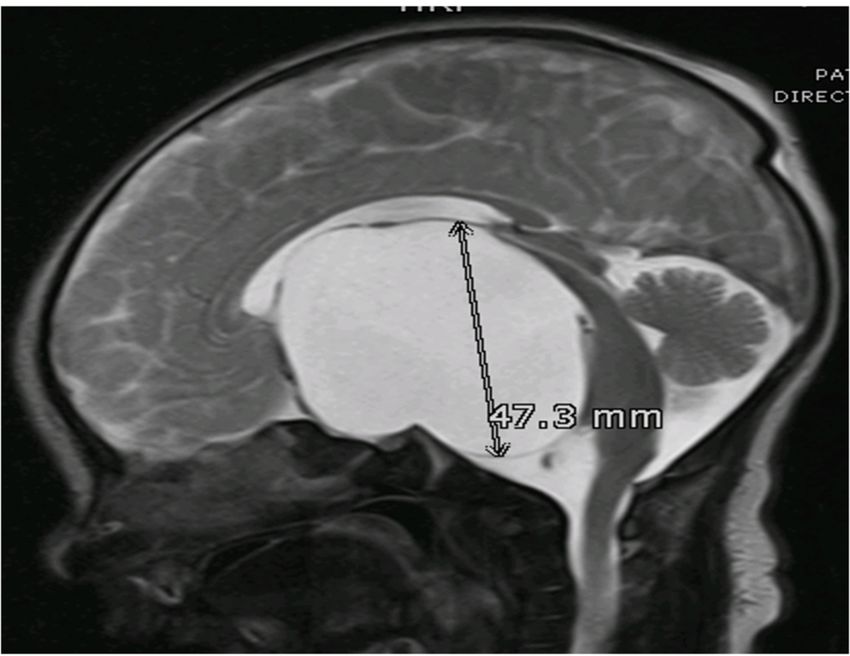
Review . Cureus 10(4): e2458. DOI 10.7759/cureus.2458FIGURE 1: MRI T2WI, Sagittal Section
A large extra-axial, well-defined CSF intensity cystic lesion is identified in the midline in the
sellar and suprasellar region. It measures 48 x 62 x 47 mm in AP transverse and cranio-caudal
dimension. This lesion is causing a compressive effect on the third ventricle and bilateral lateral
ventricles. These findings are consistent with arachnoid cyst.
MRI T2WI: magnetic resonance imaging T2 weighted image; CSF: cerebrospinal fluid.
With regards to large arachnoid cysts, there has been no consensus on the single best
management strategy. The most frequently used methods for treating arachnoid cysts are
microsurgical fenestration via craniotomy, neuroendoscopic fenestration (Videos 1, 2) and
cystoperitoneal shunting [5, 9-10].
VIDEO 1: Endoscopic fenestration of arachnoid cyst: Part 1
View video here: https://youtu.be/nKTUjpyJIEE
2018 Mustansir et al. Cureus 10(4): e2458. DOI 10.7759/cureus.2458 2 of 7VIDEO 2: Endoscopic fenestration of arachnoid cyst: Part 2
View video here: https://youtu.be/dMJWXXZYDPo
Choi et al. propose three categories of cysts on the basis of their clinical presentation and
reasons for surgery [11]. In the first group, the cysts presented with symptoms of hydrocephalus
and intracranial hypertension. In the second group, the cysts presented with vague symptoms
of dizziness, headaches, large head, skull abnormalities, strabismus, seizures, and
developmental delays. There were minimal or no clinical symptoms in the third group, but the
patients were surgically treated anyway, in expectation of improvement of abnormal
radiological and neuropsychological findings. They found that only the first group had a
reasonable improvement rate, while there was a partial improvement in the third group, and
minimal improvement in the second group. Therefore, surgical evaluation is advocated only in
those patients whose symptoms are directly related to the cyst.
Neuroradiological imaging is used to make a choice between neuroendoscopy and shunting.
With the neuroendoscopic approach, imaging is used to identify an area of contiguity between
the ventricular ependyma and the wall of the cyst; this is opened via an endoscope to facilitate
continuous drainage of the cyst. A minimum opening of 10–15 mm has to made, along with
the removal of the cyst wall, in order to prevent the closure of the stoma again [9].
The efficacy of the endoscopic approach with arachnoid cyst fenestration is still a topic of
debate. Several studies have been done to evaluate its importance. A study done by Jung Won
Choi et al. reviewed the surgical outcomes of intracranial arachnoid cysts and concluded that
fenestration of cysts is associated with more surgical complications [11]. However, some studies
suggest that neuroendoscopy remains a superior approach, with a lower risk of surgical
complications associated with craniotomy and shunting [12-13]. In a case series on the
endoscopic approach, a success rate of up to 71–81% has been documented [12]. Furthermore,
with associated hydrocephalus, the possibility of larger spaces makes this treatment option
easier to execute.
The location of the arachnoid cyst also determines the surgical approach and outcome. In terms
of endoscopy, the best results have been reported with suprasellar cysts. In patients with
middle fossa cysts, endoscopy remains controversial; some authors favor microsurgery over
endoscopic surgery for cysts in this location [14]. Furthermore, treating large hemispheric
arachnoid cysts in infants using endoscopic fenestrations have had less favorable outcomes
than the same treatment in older children. However, a neuroendoscopic approach is still
preferred over shunting in this population too, owing to the high risk of shunt failure [15].
Review
2018 Mustansir et al. Cureus 10(4): e2458. DOI 10.7759/cureus.2458 3 of 7Middle cranial fossa arachnoid cyst
Approximately 50-65% of arachnoid cysts occur in the middle cranial fossa [8].
The Galassi classification is used to classify middle fossa cysts into three types [16]. Type I cysts
are typically asymptomatic and are present in the anterior middle cranial fossa. Type II cysts
extend superiorly along the Sylvian fissure, occasionally displacing the temporal lobe. Finally,
Type III cysts are very large and occupy the entire middle cranial fossa, displacing not only the
temporal lobe but also disrupting the parietal and frontal lobes.
Type I cysts are best treated by microsurgical fenestration. The latter two are accessed via
endoscope. The appearance of the chiasmatic and interpeduncular cisterns on magnetic
resonance imaging (MRI) helps to decide between endoscopic and microsurgical fenestration.
Endoscopic cystocisternotomy is advocated when there is ample space between the third
cranial nerve and the tentorial notch and between the optic nerve and the carotid artery within
large cisterns with thin membranes, while microsurgical techniques remain a suitable option in
cases of fenestration of deep thicker membranes in the vicinity of vital structures [17].
Various factors have to be considered before performing endoscopic fenestrations into the basal
cisterns. The medial wall of the cyst is coursed by the arterial vessels of the Sylvian fissure that
can be damaged when stoma is enlarged with sharp instruments. Moreover, the membranes of
these cysts are difficult to penetrate due to their rich collagen content, so a sharp instrument or
scissors are often used [18].
The literature related to middle fossa cyst treatment is not as diverse or as reliable as it is for
other types of cysts. A recent meta-analysis concluded that while all three surgical methods
(endoscopic, microsurgical, and shunting) are effective for the management of middle fossa
cysts, endoscopic fenestration is the preferred primary surgical modality. The latter two
options should only be considered when symptoms are unchanged after endoscopic treatment
[19].
Suprasellar arachnoid cyst
Suprasellar arachnoid cysts are usually found in close proximation to the third ventricle. They
present with hydrocephalus.
Endoscopic fenestration of such cysts is the standard modality of treatment [20]. Open
procedures are usually avoided due to a significantly higher morbidity, and the fact that the
success rate does not go above 70%. When performing endoscopic fenestration, surgeons adopt
different techniques. These range from fenestrating only the apical membrane, usually at the
level of the foramen of Monroe, between the ventricle and the cyst (ventriculocystostomy), to
basilar fenestration toward the prepontine cistern (cyst-cisternostomy), called
ventriculocystocisternostomy (VCC).
A study conducted using MR-imaged CSF flow dynamics shows that fenestration of suprasellar
cysts should be done in both the ventricles and in the basal cisterns, to prevent relapse of
symptoms [21]. In a paper on the management of suprasellar arachnoid cysts, Gui et al.
concluded that endoscopic ventriculocystocisternostomy is far more effective than
ventriculocystostomy [20].
Interhemispheric arachnoid cyst
Mori et al. classified inter-hemispheric cysts into two types: parasagittal and midline [22].
2018 Mustansir et al. Cureus 10(4): e2458. DOI 10.7759/cureus.2458 4 of 7Parasagittal cysts are unilateral; they are found in toddlers and are infrequently associated with
agenesis of the corpus callosum. As these cysts are not near the ventricles, they do not usually
present with hydrocephalus. Hence, the best treatment is excision of the cyst along with its
lining. Midline cysts are complex, multiloculated cysts usually discovered at birth and
associated with agenesis of the corpus callosum. They do present with hydrocephalus. The most
frequent site of contiguity between the cyst and the ventricles is at the level of the roof of the
third ventricle. Therefore, fenestration of the cyst into the third ventricle is required. Moreover,
the septations have to be broken and made to communicate with the nearby cistern [23-24].
Quadrigeminal arachnoid cyst
Quadrigeminal cistern cysts are uncommon; only 79 cases were reported in the literature up to
2008 [25].
Quadrigeminal cysts may be classified into three types. Type I are cysts with supratentorial and
infratentorial extension, Type II are cysts with infratentorial extension (supracerebellar or
supra-retrocerebellar), and Type III includes cysts with lateral extension toward the temporal
lobe. As quadrigeminal arachnoid cysts compress or distort the cerebral aqueduct at an early
stage, they are usually associated with hydrocephalus when symptomatic. Symptoms include
macrocrania, headaches, vomiting, lethargy, papilledema, and impairment of upward gaze and
other ocular disorders [26]. Due to the compressive symptoms produced by these cysts, it is
imperative that they be treated.
Minimally invasive treatment of these cysts is preferred due to their precarious proximity to the
pineal quadrigeminal region. The endoscopic technique used varies according to the extension
of the cyst. It can extend to the trigone superiorly, to the supracerebellar cistern inferiorly, and
to the third ventricle anteriorly. Further endoscopic approaches are cyst fenestration and
removal via the suboccipital supracerebellar approach, lateral ventriclecystostomy, and third
ventriclecystostomy [27].
Two studies that analyzed 14 and 18 cases of quadrigeminal cistern arachnoid cysts treated
with the above mentioned endoscopic approaches, found that patients were shunt independent
in 78% and 93% of the cases respectively [26, 28]. Cinalli et al. concluded that arachnoid cysts
of the quadrigeminal cistern can be effectively treated by endoscopy, with a success rate of 90%
observed in the series if endoscopy was the first line of treatment. Specifically, they stated that
endoscopic third ventriculostomy should be combined with ventriculocystostomy to offer the
highest success rate with a single procedure [26].
Conclusions
As mentioned earlier, the previous techniques used to manage arachnoid cysts were craniotomy
and marsupialization of the cyst or insertion of a cystoperitoneal shunt. While these techniques
still remain useful, neurosurgeons are increasing turning to endoscopic means of management.
The endoscopic management of intracranial arachnoid cysts is a safe and effective therapeutic
modality that results in a high success rate. The approach, trajectory, and site of fenestration
must be planned individually in each case by using preoperative MR imaging and must be
studied carefully intraoperatively. However, the cyst location is important for surgical decision-
making. Arachnoid cysts in the suprasellar and quadrigeminal regions are most amenable to
neuroendoscopy. On the other hand, interhemispheric cysts should be treated by microsurgical
fenestration.
Additional Information
Disclosures
2018 Mustansir et al. Cureus 10(4): e2458. DOI 10.7759/cureus.2458 5 of 7Conflicts of interest: In compliance with the ICMJE uniform disclosure form, all authors
declare the following: Payment/services info: All authors have declared that no financial
support was received from any organization for the submitted work. Financial relationships:
All authors have declared that they have no financial relationships at present or within the
previous three years with any organizations that might have an interest in the submitted work.
Other relationships: All authors have declared that there are no other relationships or
activities that could appear to have influenced the submitted work.
References
1. Albuquerque FC, Giannotta SL: Arachnoid cyst rupture producing subdural hygroma and
intracranial hypertension: case reports. Neurosurgery. 1997, 41:951-955. 10.1097/00006123-
199710000-00036
2. Al-Holou WN, Terman S, Kilburg C, Garton HJ, Muraszko KM, Maher CO: Prevalence and
natural history of arachnoid cysts in adults. J Neurosurg. 2013, 118:222-231.
10.3171/2012.10.jns12548
3. Al-Holou WN, Yew AY, Boomsaad ZE, Garton HJ, Muraszko KM, Maher CO: Prevalence and
natural history of arachnoid cysts in children. J Neurosurg Pediatr. 2010, 5:578-585.
10.3171/2010.2.peds09464
4. Pradilla G, Jallo G: Arachnoid cysts: case series and review of the literature . Neurosurg Focus.
2007, 22:7. 10.3171/foc.2007.22.2.7
5. Wang C, Liu C, Xiong Y, et al.: Surgical treatment of intracranial arachnoid cyst in adult
patients. Neurol India. 2013, 61:60-64. 10.4103/0028-3886.108013
6. Helland CA, Wester K: A population based study of intracranial arachnoid cysts: clinical and
neuroimaging outcomes following surgical cyst decompression in adults. J Neurol Neurosurg
Psychiatry. 2007, 78:1129-1135. 10.1136/jnnp.2006.107995
7. Wojcik G: Intracranial arachnoid cysts in the clinical and radiological aspect . Wiad Lek. 2016,
69:555-559.
8. Goswami P, Medhi N, Sarma PK, Sarmah BJ: Case report: middle cranial fossa arachnoid cyst
in association with subdural hygroma. Indian J Radiol Imaging. 2008, 18:222-223.
10.4103/0971-3026.41831
9. Ahn JY, Chio JU, Yoon SH, Chung SS, Lee KC: Treatment and outcome of intracranial
arachnoid cysts. J Korean Neurosurg Soc. 1994, 23:194-203.
10. Karabatsou K, Hayhurst C, Buxton N, O'Brien DF, Mallucci CL: Endoscopic management of
arachnoid cysts: an advancing technique. J Neurosurg. 2007, 106:455-462.
10.3171/ped.2007.106.6.455
11. Choi JW, Lee JY, Phi JH, Kim SK, Wang KC: Stricter indications are recommended for
fenestration surgery in intracranial arachnoid cysts of children. Childs Nerv Sys. 2015, 31:77-
86. 10.1007/s00381-014-2525-1
12. Lee YH, Kwon YS, Yang KH: Multiloculated hydrocephalus: open craniotomy or endoscopy?. J
Korean Neurosurg Soc. 2017, 60:301-305. 10.3340/jkns.2017.0101.013
13. Di Rocco F, Yoshino M, Oi S: Neuroendoscopic transventricular ventriculocystostomy in
treatment for intracranial cysts. J Neurosurg. 2005, 103:54-60. 10.3171/ped.2005.103.1.0054
14. Kim MH, Jho HD: Endoscopic management of cranial arachnoid cysts using extra-channel
method. J Korean Neurosurg Soc. 2010, 47:433-436. 10.3340/jkns.2010.47.6.433
15. Yadav YR, Parihar V, Pande S, Namdev H, Agarwal M: Endoscopic third ventriculostomy. J
Neurosci Rural Pract. 2012, 3:163-173. 10.4103/0976-3147.98222
16. Galassi E, Tognetti F, Gaist G, Fagioli L, Frank F, Frank G: CT scan and metrizamide CT
cisternography in arachnoid cysts of the middle cranial fossa: classification and
pathophysiological aspects. Surg Neurol. 1982, 17:363-369.
17. Azab WA, Almanabri M, Yosef W: Endoscopic treatment of middle fossa arachnoid cysts . Acta
Neurochir (Wien). 2017, 159:2313-2317. 10.1007/s00701-017-3320-z
18. Cinalli G, Cappabianca P, de Falco R, et al.: Current state and future development of
intracranial neuroendoscopic surgery. Expert Rev Med Devices. 2005, 2:351-373.
10.1586/17434440.2.3.351
19. Chen Y, Fang HJ, Li ZF, et al.: Treatment of middle cranial fossa arachnoid cysts: a systematic
review and meta-analysis. World Neurosurg. 2016, 92:480-490. 10.1016/j.wneu.2016.06.046
2018 Mustansir et al. Cureus 10(4): e2458. DOI 10.7759/cureus.2458 6 of 720. Gui SB, Wang XS, Zong XY, Zhang YZ, Li CZ: Suprasellar cysts: clinical presentation, surgical
indications, and optimal surgical treatment. BMC Neurol. 2011, 11:52. 10.1186/1471-2377-11-
52
21. Wang JC, Heier L, Souweidane MM: Advances in the endoscopic management of suprasellar
arachnoid cysts in children. J Neurosurg. 2004, 100:418-426. 10.3171/ped.2004.100.5.0418
22. Mori K: Giant interhemispheric cysts associated with agenesis of the corpus callosum . J
Neurosurg. 1992, 76:224-230. 10.3171/jns.1992.76.2.0224
23. Mankotia DS, Sardana H, Sinha S, et al.: Pediatric interhemispheric arachnoid cyst: an
institutional experience. J Pediatr Neurosci. 2016, 11:29-34. 10.4103/1817-1745.181258
24. Cappabianca P, Cavallo LM, De Divitiis O, Esposito F: Midline Skull Base Surgery . Springer
International Publishing, Switzerland; 2016. 10.1007/978-3-319-21533-4
25. Almeida JP, Quinino S, Faquini IV, et al.: Neuroendoscopic treatment of quadrigeminal
arachnoid cyst in a two-year-old child. Arq Neuropsiquiatr. 2008, 66:758-760. 10.1590/S0004-
282X2008000500032
26. Cinalli G, Spennato P, Columbano L, et al.: Neuroendoscopic treatment of arachnoid cysts of
the quadrigeminal cistern: a series of 14 cases. J Neurosurg Pediatr. 2010, 6:489-497.
10.3171/2010.8.peds08491
27. Sengul G, Tuzun Y, Cakir M, Duman S, Colak A, Kadioglu HH, Aydin IH: Neuroendoscopic
approach to quadrigeminal cistern arachnoid cysts. Eurasian J Med. 2012, 44:18-21.
10.5152/eajm.2012.04
28. El-Ghandour NM: Endoscopic treatment of quadrigeminal arachnoid cysts in children . J
Neurosurg Pediatr. 2013, 12:521-528. 10.3171/2013.7.peds13155
2018 Mustansir et al. Cureus 10(4): e2458. DOI 10.7759/cureus.2458 7 of 7You can also read