Achondroplasia: a case report and the review of the basics - International Surgery Journal
←
→
Page content transcription
If your browser does not render page correctly, please read the page content below

International Surgery Journal
Agrawal SN. Int Surg J. 2020 Jul;7(7):2420-2424
http://www.ijsurgery.com                                                                     pISSN 2349-3305 | eISSN 2349-2902

                                                                 DOI: http://dx.doi.org/10.18203/2349-2902.isj20202862
Case Report

             Achondroplasia: a case report and the review of the basics
                                                  Sujan Narayan Agrawal*

  Department of Surgery, SBRKM Government Medical College, Jagdalpur (Bastar), Chhattisgarh, India

  Received: 22 April 2020
  Revised: 02 June 2020
  Accepted: 03 June 2020

  *Correspondence:
  Dr. Sujan Narayan Agrawal,
  E-mail: drsujanagrawal@gmail.com

  Copyright: © the author(s), publisher and licensee Medip Academy. This is an open-access article distributed under
  the terms of the Creative Commons Attribution Non-Commercial License, which permits unrestricted non-commercial
  use, distribution, and reproduction in any medium, provided the original work is properly cited.

   ABSTRACT

   The achondroplasia is a variant of short-limbed dwarfism. The word achondroplasia literally means without cartilage
   formation. However, in achondroplasia the problem is not in formation of cartilage but, in its conversion to bone (i.e.
   ossification). This deficient ossification is particularly seen in the long bones of arm and leg. The characteristic
   external appearance of people born with achondroplasia is short stature. The average height of an adult male with
   achondroplasia is 131 centimetres (4 feet, 4 inches), and the average height for adult females is 124 centimetres (4
   feet, 1 inch). The trunk is of average size but the leg and upper arm is of short length. It is because the femur and
   humerus are relatively shorter in length. The range of movement at elbow is limited. The head is enlarged called
   macrocephaly and is with a prominent forehead. People with Achondroplasia are generally of normal intelligence.
   They have bowed legs and abnormal curvature of spine giving rise to lordosis or kyphosis. They may develop spinal
   stenosis, which is associated with pain, tingling and weakness in leg. This may cause difficulty in walking. The other
   health problems associated with Achondroplasia are episodes of apnoea, obesity and recurrent ear infection. The
   purpose of this study is to evaluate the cardinal phenotypic features in patient of Achondroplasia. It is also to assess
   the body physique, anthropometric measurements and to study the typical radiological signs in such patients as the
   main tool of diagnosis.

   Keywords: Achondroplasia, Fibroblast growth factor receptor 3, Rhizomelia, Skeletal dysplasia

INTRODUCTION                                                         literally means ‘without cartilage formation’. However, it
                                                                     is a misnomer, since in achondroplasia, the problem is
The achondroplasia is a type of bony dysplasia, which                not in formation of cartilage but it is, in its ossification. In
results in a short stature. A short stature is defined as the        achondroplasia the cartilage is not converted to bone
height that is less than the third percentile, for the               (ossification), particularly in the long bones of arm and
chronological age of the patient. A clinician can easily             legs. This is the most common type of short limb
diagnose the skeletal dysplasia by history and physical              dwarfism and it occurs in one in 26,000 to 28,000 live
examination of the child. The presenting complaint of the            births.1
parents is short stature of their child, which is established
easily by physical examination. In achondroplasia the                The short stature is further divided into two main patterns
short stature is most prominent in the proximal segment              i.e. proportionate and disproportionate. Achondroplasia is
of the limb i.e. in femur or humerus. This condition is              the most common cause of disproportionate short stature.
called Rhizomelia. The achondroplasia is an example of               Affected individuals have shortening of femur and
the short limb dwarfism. The radiological examination                humerus (rhizomelia). They have macrocephaly, frontal
further confirms the diagnosis. The word achondroplasia              bossing and midface retrusion. The developmental motor

                                                                International Surgery Journal | July 2020 | Vol 7 | Issue 7   Page 2420
Agrawal SN. Int Surg J. 2020 Jul;7(7):2420-2424

milestones are delayed and are accompanied by
hypotonia. The cranio-cervical junction compression
increases the risk of death in infancy. The additional
complications include obstructive sleep apnoea, middle
ear infection, kyphosis or lordosis and spinal stenosis.

CASE REPORT

A patient named X was admitted in our hospital for pain
in joints and vague illness. She is the third child of her
parents. The mother was of 34 years of age, when she
was born. There are three siblings in the family, she is the
youngest. At the time of presentation, her age is 16 years.
Parents and other children are of normal height.

On physical examination her height is 124 CMs. There
was frontal bossing with depressed bridge of nose. The                            Figure 3: X-ray hand in achondroplasia.
upper limb shortening is noted as compare to lower limb.
The extension at elbow is restricted. Fingers are typically         DISCUSSION
short and all are of same length. The middle finger was
also of the same length as others. A provisional diagnosis          The genetics
of achondroplasia was made (Figure 1).
                                                                    Fibroblast growth factor receptor 3 (FGFR-3), is a
                                                                    membrane-spanning tyrosine kinase receptor with an
                                                                    extracellular ligand-binding domain consisting of three
                                                                    immunoglobulin (Ig) subdomains, a transmembrane
                                                                    domain, and a split intracellular tyrosine kinase domain.2
                                                                    FGFR-3 is activated by various fibroblast growth factors
                                                                    (FGFs).3 Binding appear to result in receptor
                                                                    dimerization, transactivation of tyrosine kinase, and
                                                                    transphosphorylation of tyrosine residues.4 These
                                                                    modifications result in activation of a number of
                                                                    downstream signalling pathways, including signal
                                                                    transducer and activator of transcription (STAT),
                                                                    mitogen-activated protein kinase (MAPK) and a number
                                                                    of others.4-6 Overall, these secondary pathways cause
                                                                    slowing of proliferation and differentiation of
                                                                    chondrocyte.7 The G380R mutation in FGFR3
                                                                    transmembrane domain is known as the genetic cause for
     Figure 1: Typical features of achondroplasia.                  achondroplasia.8 Achondroplasia is inherited in an
                                                                    autosomal dominant pattern, which means one copy of
                                                                    the altered gene in each cell is sufficient to cause the
                                                                    disorder. This genetic disorder interferes with the
                                                                    maturation of the cartilage growth plate of long bones.9,10
                                                                    The phenotype is characterized by short stature,
                                                                    narrowing of the lumbar spinal canal, accentuated bowing
                                                                    of the middle and lower part of the back, and trident-
                                                                    shaped hands.11 The affected individuals often exhibit
                                                                    other skeletal as well as neurological complications.

                                                                    Inheritance pattern

                                                                    Achondroplasia is inherited in an autosomal dominant
                                                                    pattern, which means one copy of the altered gene in each
                                                                    cell is sufficient to cause the disorder. About 80 percent
                                                                    of people with Achondroplasia have average-size parents;
                                                                    these cases result from new mutations in the FGFR3
                                                                    gene. Individuals who inherit two altered copies of this
                                                                    gene typically have a severe form of Achondroplasia that
             Figure 2: The spatulated, trident hand.                causes extreme shortening of the bones and an

                                                               International Surgery Journal | July 2020 | Vol 7 | Issue 7   Page 2421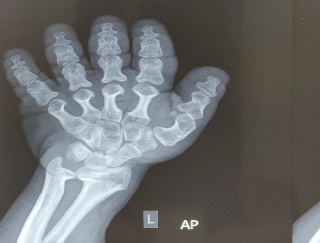
Agrawal SN. Int Surg J. 2020 Jul;7(7):2420-2424

underdeveloped rib cage. These individuals are usually                self-feeding.13 Most children with achondroplasia are
stillborn or die shortly after birth from respiratory failure.        macro cephalic.14 There are midface retrusion and
                                                                      depressed bridge of nose. The head is large
                                                                      (hydrocephalic) may be due to foramen magnum
                                                                      stenosis.15 Middle ear infections is a frequent problem.16
                                                                      It contributes significantly to the hearing loss. In fact,
                                                                      about 40% of individuals with achondroplasia have
                                                                      functional hearing loss. With the hearing loss the ability
                                                                      to learn is also hampered.

                                                                      The skeletal issues

                                                                      There is symmetrical shortening of all long bones, with
                                                                      proximal portions being more affected and lower limb
                                                                      involvement being more than the upper limb
                                                                      (rhizomelia). There’s relative flaring and splaying of
                                                                      metaphyses with normal epiphyses. There is increased
                                                                      gap between 2nd and 3rd digit of hand and inability to
                                                                      approximate them in extension leading to appearance of
                                                                      trident hand (Figure 1-4).17
      Figure 4: Pattern of autosomal dominance.

The physical characters

The children born with achondroplasia have rhizomelic
shortening of arms, hydrocephalic head, narrow chest and
short fingers. The typical physical features are as follows.

Short stature

The average adult height for men with Achondroplasia is
131±5.6 cms. For women it is around 124±5.9 cms.
Obesity is a major problem with this condition.12 Mild to
moderate hypotonia is typical.
                                                                      Figure 4: Pelvis radiograph in achondroplasia, shows,
                Table 1: The skeletal survey.                          square shape of the iliac bone, horizontal acetabular
                                                                        roof (squiggly arrow), and rhizomelic shortening of
 The skeletal
                     Findings in Achondroplasia                                              the femur.
 survey                                                               Also note the trident sign in achondroplasia. The three-pronged
                     Frontal bossing mid face                         pear shape of the sciatic notch is well visualized in this
 Facial
                     hypoplasia, depressed nasal bridge               radiograph (straight arrow).18
 phenotype
                     short neck
                     Lumbar kyphosis exaggerated                      Pelvis
 Trunk               lumbar lordosis protuberant
                     abdomen                                          There is trident pelvis/tombstone shape of iliac bone.
                     Rhizomelic shortening, limited                   Small square shaped ileal wings akin to tombstone, with
                     elbow extension generalized joint                horizontal acetabular roof and telephone handle shaped
 Upper limb
                     laxity brachydactyly and trident                 femur. The spur at the medial and lateral acetabular
                     hand configuration (Figure 2)                     margin and in the centre of the acetabulum gives rise to
                     Bowing of legs or genu varum                     trident sign, due to its resemblance of a three-pronged
 Lower limb          multiple skin creases over the limbs             spear. The pelvic inlet is described as Champagne glass
                     (Michelin tire creases)                          shaped pelvic inlet, because of the flattening of iliac
                     Upper respiratory tract infection,               blades with increased acetabular angles and small Sacro
                     snoring, sleep apnoea otitis media               sciatic notch (Figure 4).
 Miscellaneous
                     muscular hypotonia delayed motor
                     developmental milestones                         Lower limb

The motor development is also slow. There is small joint              Angular deformity of the lower limb usually develops in
hyper motility and with short fingers it may interfere with           the child with achondroplasia. Genu-varum and tibia vera
                                                                      are more common than varus deformities.19 Relative

                                                                 International Surgery Journal | July 2020 | Vol 7 | Issue 7   Page 2422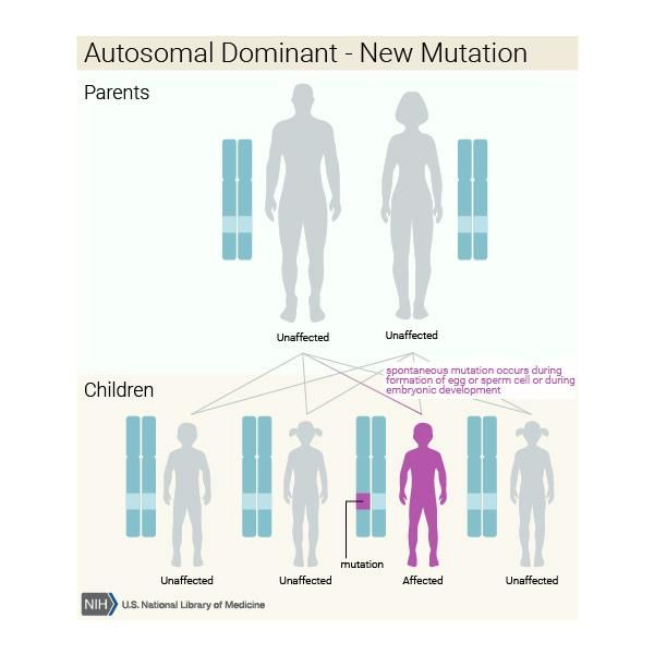
Agrawal SN. Int Surg J. 2020 Jul;7(7):2420-2424

overgrowth of fibula as compare to tibia has been                            selected regions of the US. Am J Med Genet A.
proposed as the cause of varus deformity.                                    2008;146A:2385-9.
                                                                       2.    Laederich MB, Horton WA. Achondroplasia:
Thoracolumbar kyphosis                                                       pathogenesis and implications for future treatment.
                                                                             Curr Opin Pediatr. 2010;22:516-23.
It is seen in slightly older babies when they start sitting. It        3.    Ornitz DM. FGF signalling in the developing
has been proposed that this may be due to the large head,                    endochondral skeleton. Cytokine Growth Factor
reduced muscular tone, lack of trunk control and                             Rev. 2005;16:205-13.
tendency for hip flexion contributes towards kyphotic                  4.    Narayana J, Horton WA. FGFR3 biology and
deformity. There is spinal stenosis due to abnormal                          skeletal disease. Connect Tissue Res. 2015;56:427-
growth of vertebral pedicles. There is interpedicular                        33.
narrowing and thickening of pedicles, hypertrophy of                   5.    Deng C, Boris WA, Zhou F, Kuo A, Leder P.
facets and enlargement of laminae. These abnormalities                       Fibroblast growth factor receptor 3 is a negative
cause spinal stenosis which becomes symptomatic in 3rd                       regulator of bone growth. Cell. 1996;84:911-21.
decade of life or even earlier.20                                      6.    Eswarakumar VP, Lax I, Schlessinger J. Cellular
                                                                             signalling by fibroblast growth factor receptors.
CONCLUSION                                                                   Cytokine Growth Factor Rev. 2005;16:139-49.
                                                                       7.    Klag KA, Horton WA. Advances in treatment of
Traditionally, the term achondroplasia was initially used                    Achondroplasia and osteoarthritis. Hum Mol Genet.
to describe all individuals with short-limbed dwarfing                       2016;25(1):2-8.
disorders. Over the past 50 years diagnostic criteria have             8.    Shiang R, Thompson LM, Zhu YZ, Church DM,
been available to distinguish true achondroplasia from                       Fielder TJ, Bocian M, et al. Mutations in the
other short limb disorders or dwarfism. A condition that                     Transmembrane Domain of FGFR3 Cause the Most
may be confused with achondroplasia includes                                 Common        Genetic     Form     of    Dwarfism,
hypochondroplasia, thanatophoric dysplasia, severe                           Achondroplasia. Cell. 1994;78:335-42.
achondroplasia with developmental delay and acanthosis                 9.    Horton WA, Hall JG, Hecht JT. Achondroplasia.
Nigerians     (SADDAN)        syndrome,       cartilage-hair                 Lancet. 2007;370:162-72.
hypoplasia, pseudoachondroplasia, etc. A good clinical                 10.   Ponseti IV. The Ponseti Technique for Correction of
examination, anthropometric evaluation and radiological                      Congenital Clubfoot. J Bone Joint Surg Am.
findings are usually sufficient to establish the diagnosis                   1970;52:701-16.
of achondroplasia. Achondroplasia is inherited in an                   11.   Vajo Z, Francomano CA, Wilkin DJ. The Molecular
autosomal dominant manner. Approximately 80% of                              and Genetic Basis of Fibroblast Growth Factor
individuals with achondroplasia have parents of average                      Receptor 3 Disorders: The Achondroplasia Family
stature and have achondroplasia as a result of a denovo                      of Skeletal Dysplasias, Muenke Craniosynostosis,
FGFR3 pathogenic variant. Denovo pathogenic variants                         and Crouzon Syndrome with Acanthosis Nigricans.
are associated with advanced paternal age, often defined                     Endocr Rev. 2000;21:23-39.
as older than age 35 years.21 The denovo pathogenic                    12.   Hecht JT, Butler KJ, Cott CL. Long-term
variants causing achondroplasia are exclusively inherited                    neurological sequelae in Achondroplasia. Eur J
from the father.22 The remaining 20% of individuals with                     Pediatr. 1984;143:58-60.
achondroplasia have at least one affected parent. In the               13.   Ireland PJ, Donaghey S, Gill MJ, Zankl A, Ware
families, that have an apparent denovo pathogenic                            RS, Pacey V, et al. Development in children with
variant, they should undergo genetic testing and                             Achondroplasia: a prospective clinical cohort study.
counselling. The optimal time for determination of                           Dev Med Child Neurol. 2012;54:532-7.
genetic risk and discussion of the availability of prenatal            14.   Horton WA, Rotter JI, Rimoin DL, Scott CI, Hall
testing is before pregnancy. It is appropriate to offer                      JG. Standard growth curves for Achondroplasia. J
genetic counselling (including discussion of potential                       Pediatr. 1978;93:435-8.
risks to offspring and reproductive options) to young                  15.   Etus V, Ceylan S. The role of endoscopic third
adults who are affected. Genetic counselling is also                         ventriculostomy in the treatment of tri-ventricular
recommended when both parents have a skeletal                                hydrocephalus seen in children with achondroplasia.
dysplasia.                                                                   J Neurosurg. 2005;103:260-5.
                                                                       16.   Tunkel D, Alade Y, Kerbavez R, Smith B, Hardison
Funding: No funding sources                                                  RD, Fong HJ. Hearing loss in skeletal dysplasia
Conflict of interest: None declared                                          patients. Am J Med Genet A. 2012;158A:1551-5.
Ethical approval: Not required                                         17.   Panda A, Gamanagatti S, Jana M, Gupta AK.
                                                                             Skeletal dysplasias: a radiographic approach and
REFERENCES                                                                   review of common non-lethal skeletal dysplasias.
                                                                             World J Radiol. 2014;6(10):808-25.
1.   Waller DK, Correa A, Vo TM, Wang Y, Hobbs C,                      18.   Jana M, Nair N, Gupta AK, Kabra M, Gupta N.
     Langlois PH, et al. The population-based prevalence                     Pelvic radiograph in skeletal dysplasias: an
     of Achondroplasia and Thanatophoric dysplasia in                        approach. Indian J Radiol Imaging. 2017;27:187-99.

                                                                  International Surgery Journal | July 2020 | Vol 7 | Issue 7   Page 2423
Agrawal SN. Int Surg J. 2020 Jul;7(7):2420-2424
19. Basse TT, GS. Lower extremity abnormalities in growth-factor receptor 3 in sporadic cases of
dwarfing conditions. Instr Course Lect. Achondroplasia occur exclusively on the paternally
1990;30:389. derived chromosome. Am J Hum Genet.
20. Hamamci H, Hawran S, Sorensen BF. 1998;63:711-6.
Achondroplasia and spinal cord lesion: three case
report. Paraplegia. 1993;31:375.
21. Stoll C, Roth MP, Bigel P. A re-examination on
parental age effect on the occurrence of new
mutations for Achondroplasia. Prog Clin Biol Res. Cite this article as: Agrawal SN. Achondroplasia: a
1982;104:419-26. case report and the review of the basics. Int Surg J
22. Wilkin DJ, Szabo JK, Cameron R, Henderson S, 2020;7:2420-4.
Bellus GA, Mack ML, et al. Mutations in fibroblast
International Surgery Journal | July 2020 | Vol 7 | Issue 7 Page 2424You can also read