Alkali metal doping of black phosphorus monolayer for ultrasensitive capture and detection of nitrogen dioxide - Nature
←
→
Page content transcription
If your browser does not render page correctly, please read the page content below
www.nature.com/scientificreports
OPEN Alkali metal doping of black
phosphorus monolayer
for ultrasensitive capture
and detection of nitrogen dioxide
Azam Marjani1,2, Mehdi Ghambarian3 & Mohammad Ghashghaee4*
Black phosphorus nanostructures have recently sparked substantial research interest for the rational
development of novel chemosensors and nanodevices. For the first time, the influence of alkali metal
doping of black phosphorus monolayer (BP) on its capabilities for nitrogen dioxide (NO2) capture and
monitoring is discussed. Four different nanostructures including BP, Li-BP, Na-BP, and K-BP were
evaluated; it was found that the adsorption configuration on Li-BP was different from others such that
the NO2 molecule preferred a vertical stabilization rather than a parallel configuration with respect
to the surface. The efficiency for the detection increased in the sequence of Na-BP < BP < K-BP < Li-BP,
with the most significant improvement of + 95.2% in the case of Li doping. The Na-BP demonstrated
the most compelling capacity (54 times higher than BP) for NO2 capture and catalysis (− 24.36 kcal/
mol at HSE06/TZVP). Furthermore, the K-doped device was appropriate for both nitrogen dioxide
adsorption and sensing while also providing the highest work function sensitivity (55.4%), which was
much higher than that of BP (10.4%).
In recent decades, chemosensors and biosensors have been studied intensely due to the increasing importance
of safety precautions and environmental protections. Sensors have been used in many industries for the detec-
tion of harmful compounds. Nitrogen dioxide (NO2) with a biting odor is considered as one of the most vital
noxious gases in terms of air pollution. Also, it is used for the commercial production of nitric acid. Hence,
modern society has a strong motivation to explore more sensitive NO2 detectors for controlling its impact on
the environment and protection of human health and s afety1. Various types of sensors are actively studied for
this purpose. Some well-established classes of detectors including solid electrolytes, electrochemical sensors,
graphene-based systems, as well as metal oxides have been i ntroduced2.
Novel nanostructured and two-dimensional (2D) materials are actively pursued toward different optical
and sensing a pplications3–11. Investigations of black phosphorus structures have been carried out by many
researchers after its successful fabrication for different optical, biomedical, and environmental applications12–17.
These 2D nanomaterials have outstanding heat as well as electron conductivities due to their great anisotropic
properties18–21. Moreover, black phosphorus possess appropriate chemical and fire r esistance22,23.
With regard to NO2, efficient detectors have been introduced, which have been reviewed elsewhere1.
The research on such sensors is of great interest in different research groups exploring the properties of 2D
materials18,24–27. Black phosphorus itself has provided physisorption capabilities for several gases, such as NO218,28.
However, BP has been recorded to outperform molybdenum disulfide (MoS2) for the nitrogen dioxide analy-
sis in terms of the sensitivity threshold and quick regeneration29. An increased conductance sensitivity down
to 5 ppb of nitrogen dioxide has been recorded for the multilayer BP detector30. In another study of nitrogen
dioxide adsorption capabilities, Si embedding into graphene was quite effectual31. another study has shown
that graphene/NiO heterostructures were helpful for N O2 sensing32. Similarly, the NiO-decorated BP slab has
shown (116 times) stronger adsorption relative to the pristine layer for nitrogen dioxide capture and c atalysis18.
Aluminum decoration of BP has also shown useful for the capture of this toxic molecule with the adsorption
energy of 3.96 eV33. Recently, the SnO monolayer slab has been presented to be auspicious for nitrogen dioxide
1
Department for Management of Science and Technology Development, Ton Duc Thang University, Ho Chi
Minh City, Viet Nam. 2Faculty of Applied Sciences, Ton Duc Thang University, Ho Chi Minh City, Viet Nam. 3Gas
Conversion Department, Faculty of Petrochemicals, Iran Polymer and Petrochemical Institute, P.O. Box 14975‑112,
Tehran, Iran. 4Department of Petrochemical Synthesis, Faculty of Petrochemicals, Iran Polymer and Petrochemical
Institute, P.O. Box 14975‑112, Tehran, Iran. *email: m.ghashghaee@ippi.ac.ir
Scientific Reports | (2021) 11:842 | https://doi.org/10.1038/s41598-020-80343-9 1
Vol.:(0123456789)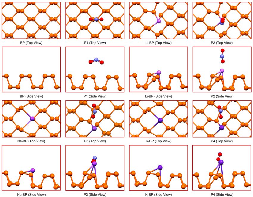
www.nature.com/scientificreports/
Figure 1. Relaxed configurations of the intact and working chemosensors obtained at the PBE/SVP
computational level. The color indicators include orange for the P atoms, red for the O atoms, blue for the N
atoms, and purple for the alkali metal (Li, Na, K) atoms. The images were drawn using Mercury 3.6.
capture and monitoring34. Among 2D nanomaterials, gallium nitride demonstrated significant bandgap altera-
tions of 1.65 eV after the exposure to nitrogen d ioxide35. Recently, the effectiveness of defect and ZnO species
incorporation into the BP monolayer was shown for the NO2 monitoring and removal, r espectively19.
Doping is a useful tool for the modulation of the anisotropic characteristics of the 2D materials for the sensing
applications. For instance, Zhou et al. have found that the embedding of transition metals, particularly Ti and
Au, can significantly enhance the chemical reactivity of graphene, thus leading to activation of the NO2 molecule
for the graphene-based catalysis a pplications36. One may reasonably postulate that the effective doping of black
phosphorus can enhance its capabilities toward the adsorption of NO2 molecule. In spite of the intriguing char-
acteristics of black phosphorus, still little is known about the possible effects of such modifications on either the
adsorption strength or sensitivity. In the same line, we explored the effects of alkali metal (Li, Na, and K) doping
of BP monolayer on the sensitivity to the NO2 molecule. The nitrogen dioxide detection using black phosphorene
has not been studied conclusively. The current research study deals with investigation of different BP structures
in terms of the electronic as well as energetic properties for nitrogen dioxide adsorption.
As will be shown in the following, the suggested modifications can make substantial improvements in both
the adsorption and sensing of N O2 on black phosphorene depending on the alkali metal employed. In a broader
sense, the reported data would help supply a deeper understanding for the rational development of novel phos-
phorene-based nanomaterials for the adsorption and sensing of gaseous pollutants.
Results and discussion
Several BP-based sensors have been inspected here, which include the pristine (BP) and alkali-doped M-BP
nanomaterials (M = Li, Na, K). The methodology for the construction of the sensors is given in the Supple-
mentary Information. Several modifiers, including transition metal (TM) elements, have been examined in the
phosphorene functionalization and doping, as reviewed elsewhere13. Here, we have chosen the lightest members
of the alkali metals, being among the ten most abundant elements in Earth c rust37. As such, the modifications
at hand would be less expensive and environmentally benign.
The sensors with optimized geometries at the rest and operating conditions are shown in Fig. 1. From Fig. 1,
we observe that the nitrogen dioxide molecule interacted through its two O atoms with the P atoms of the
unmodified pristine layer while stabilizing in the armchair orientation with the O–N–O plane (P1). The stabi-
lization of the N
O2 molecule on the M-doped sensors was different. Notably, the related structure with Li-BP
was the most different such that the molecule preferred a vertical arrangement with respect to the surface while
interacting through an O atom (P2). This observation is in correspondence with the absence of the 3d orbitals in
lithium and the lower protrusion of the Li atom compared to the other dopants (vide infra). The N O2 molecule
on both Na-BP and K-BP was optimized more similarly to P1 except that the O–N–O angle was stabilized in
Scientific Reports | (2021) 11:842 | https://doi.org/10.1038/s41598-020-80343-9 2
Vol:.(1234567890)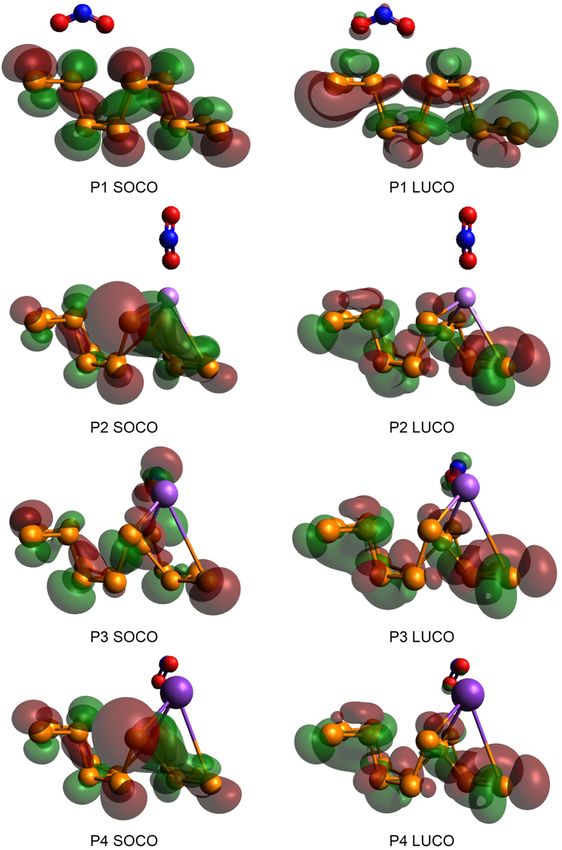
www.nature.com/scientificreports/
Sensor d Eform ∆Eads
BP 2.79 – − 0.45
Li-BP 1.82 − 1.63 − 9.68
Na-BP 0.31 − 1.17 − 24.36
K-BP 0.50 − 1.27 − 20.59
Table 1. Adsorption distances (d, Å), formation energies (Eform, eV), and the corresponding sorption energies
(∆Eads, kcal/mol) of the NO2 molecule on the original and modified sensors at the HSE06/TZVP level of
theory.
Structure r(M–P) r(N–O) ε τ μ η ω
BP – – – – 5.00 0.73 17.18
P1 – 1.21 40.5 19.3 fs 5.07 0.60 21.57
Li-BP 2.81 ± 0.45 – – – 4.53 0.83 12.37
P2 3.01 ± 0.57 1.23 ± 0.03 79.1 14.8 ns 5.47 0.53 28.07
Na-BP 3.08 ± 0.17 – – – 4.43 0.71 13.88
P3 3.58 ± 0.70 1.25 22.4 33.2 s 5.28 0.78 17.89
K-BP 3.37 ± 0.06 – – – 3.97 0.64 12.32
P4 3.82 ± 0.62 1.24 ± 0.01 43.5 132.1 ms 5.29 0.64 21.71
Table 2. Various specifications of the investigated sensors at the HSE06/TZVP level: the mean M–P bond
length [r(M–P), Å], the mean N–O bond length [r(N–O), Å], the efficiency of the analysis (ε, %), the
regeneration time of the sensor (τ, fs–s), the chemical potential factor (μ, eV), the global hardness of the
structure (η, eV), and the electrophilicity indicator (ω, eV). The efficiency was defined using Eq. (7). Also, the
visible light radiation at 343 K was considered for the calculation of the recovery time.
the zigzag direction, with one of the oxygen atoms tending to approach the dopant center (P3 and P4). In both
cases, the adsorption of N O2 molecule also led to the more projection of the metal atom away from the surface
(vide infra); but, the solid matrix integrity was not lost in either case owing to the small distortions.
The formation energy values for the M-BP materials were calculated as follows:21,38
Eform = (EM−BP + µP ) − (EBP + µM ). (1)
Here, EBP and EM-BP denote the total energies of the unmodified and alkali-doped BP slabs.; the symbol µP
denotes the chemical potential of phosphorus. The pure phosphorene chemical potential was considered in this
study. The symbol µM refers to the chemical potential of the M dopant. The energetics of the nitrogen dioxide
molecule on various phosphorene-based structures was determined as follows:21,39–41
(2)
Eads = Eop − Egas + Esensor
in which the subscript op refers to the adsorption configuration (the operating device), and gas denotes the
nitrogen dioxide molecule.
The energetic data have been supplied in Table 1. The formation energy for the incorporation of alkali metal
was predicted to be negative in all cases indicating that the incorporation of these metal elements into the struc-
ture would be highly favorable, and high doping concentrations are plausible thermodynamically. The sequence
of Na-BP < K-BP < Li-BP was found for the energetic favorability of the modification. In spite of the fact that the
adsorption of nitrogen dioxide on the pristine layer was weak, the gaseous molecule was almost tightly chem-
isorbed on the alkali-modified sensor, and the adsorption strength was sequenced as BP < Li-BP < K-BP < Na-BP
(up to − 24.36 kcal/mol at HSE06/TZVP). The equilibrium distance for the N O2 molecule varied in the range of
0.31–2.79 Å, inversely correlating with the adsorption strength (Table 1). It was concluded that the NO2 adsorp-
tion strength had been increased 54 times upon sodium doping. Such enhancement implies that the alkali-doping
is a useful means of increasing the capability of black phosphorene for N O2 capture.
One can see from the geometrical data that the average distance for the M–P bonds increased in the order of
Li-BP < Na-BP < K-BP (Table 2). Furthermore, the adsorption of NO2 led to an elongation of the r(M–P) values by
7.1%, 16.2%, and 13.4%, correlating with the adsorption strength. In the same line, the r(N–O) values increased
slightly from 1.20 Å for the free molecule to 1.21, 1.23, 1.25, and 1.24 Å in P1, P2, P3, and P4, respectively. The
geometrical features demonstrate that the stronger the adsorption, the shorter the adsorption distance, and the
longer the M–P and N–O bonds at the adsorption site.
The induced magnetic moment in the operating mode was found to be 1.0 µB due to the stabilization of
the nitrogen dioxide molecule on the surface. The spin moment (µs) contribution from nitrogen dioxide was
sequenced as P3 (0.03 µB) < P4 (0.27 µB) < P2 (0.63 µB) < P1 (0.93 µB). The charge gaps of the non-magnetic
structures were determined as follows:
Scientific Reports | (2021) 11:842 | https://doi.org/10.1038/s41598-020-80343-9 3
Vol.:(0123456789)
www.nature.com/scientificreports/
Figure 2. Electronic bandgaps of the unmodified and alkali-modified sensors at the off state and working
conditions for nitrogen dioxide detection at the HSE06/TZVP computational level.
Eg = ELUCO −EHOCO , (3)
where ELUCO refers to the lowest unoccupied crystal orbital (LUCO) energy level with the unit of eV. Also, EHOCO
denotes the highest occupied crystal orbital (HOCO) energy level (eV). Similarly, the spin-conserving gaps for
both up and down spin channels could be determined using the following equations:15
Eg↑ = ELUCO↑ −ESOCO↑ (4)
and
Eg↓ = ELUCO↓ −ESOCO↓ (5)
in which ESOCO denotes the single occupied crystal orbital (SOCO) energy level (eV). The nanosensor signal is
determined by the alteration in the electrical conductance defined as follows:15,42,43
−E g
σ = AT 3/2 exp (6)
2kB T
in which T signifies the working temperature (K), kB signifies the Boltzmann constant (eV K–1), and A represents
the constant of proportionality (in electrons m –3 K–3/2). The sensors bandgaps at the off state as well as during
operation are provided in Fig. 2. As evinced by these charts, all sensors kept their semiconducting nature through
modification and operation, and no half-metallic behavior was induced. The bandgap of the pristine layer was
obtained as 1.46 eV, which indicates that there is a great agreement between the obtained value and the experi-
mental range (1.0–1.5 eV) for the single-layer BP nanodevice44. This observation confirms the suitability of the
method applied h ere15,38.
The original bandgap (BP) was slightly expanded after lithium doping, but it was slightly shrunk after sodium
and potassium doping (Fig. 2). However, it should be pointed out that the amount of bandgap in the up-spin
channel was enlarged with the NO2 sensitivity, while it was decreased in the down-spin gap after the interaction
of the analyte for all sensors. A huge variation in the bandgap was observed in the case of Li doping compared
to the other cases, which implied the higher sensitivity of this sensor to the NO2 molecule. The electronic band
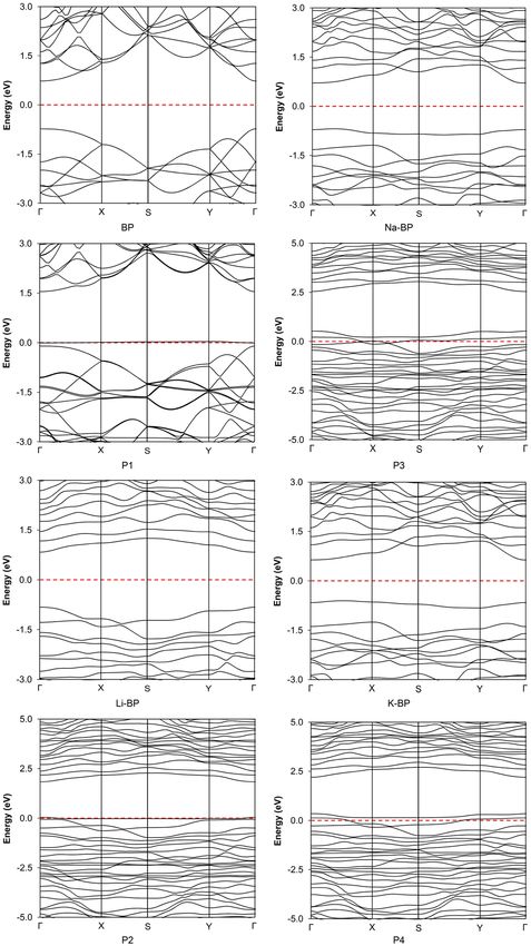
structures of the sensors are presented in Fig. 3. The determined band structure for the pristine phosphorene
was fully consistent with the Brillouin zone (BZ) data reported in the literature45, indicating the existence of a
direct bandgap at the Γ point of the BZ15. A similar type of (direct) bandgap was obtained in the case of Li-BP
and Na-BP. Nonetheless, the electronic band structure for the K-BP nanodevice exhibited an indirect X → Y
nature. In terms of band alignment, the energy levels of the conduction band minimum (CBM) and the valence
band maximum (VBM) both shifted upward upon alkali doping with the magnitude of the changes following
the sequence of Li-BP < Na-BP < K-BP. The relative energy position of the Fermi level is discussed under the
work function (vide infra). When the type of semiconduction was concerned, almost all metal impurities were
neutral, thus retaining the intrinsic behavior of the original BP material intact. However, the analyte behaved as
an acceptor and led to a p-type semiconductor in all operating cases.
The detector quality or efficiency was evaluated from the relative alterations of the bandgap:18
Eg −E0
ε = 100 (7)
Eg
in which ε represents the detector efficiency, while the initial state of the sensor was indicated by 0 and the symbol
Eg is the bandgap at the operating conditions18. Based on this definition, the amount of Eg is negative when there is
a shortening bandgap due to the interactions with the nitrogen dioxide. Table 2 reports the central operating data
for the unmodified and alkali-doped sensors for the NO2 molecule analysis19. The absolute efficiency changed in
the order of Na-BP < BP < K-BP < Li-BP, indicating the efficiency changes relative to the unmodified BP of + 95.2,
− 44.6, and + 7.4% with the Li, Na, and K doping, respectively. Therefore, the Li-doped sensor has registered the
highest efficiency (79.1%) for NO2 detection. So, it could be concluded that the pristine phosphorene became
Scientific Reports | (2021) 11:842 | https://doi.org/10.1038/s41598-020-80343-9 4
Vol:.(1234567890)
www.nature.com/scientificreports/

                                 Figure 3.  Band structures of the unmodified and alkali-modified sensors at the off and operating states at the
                                 HSE06/TZVP computational level. The outputs have been obtained from Burai 1.3.

Scientific Reports |   (2021) 11:842 |               https://doi.org/10.1038/s41598-020-80343-9                                                    5

                                                                                                                                            Vol.:(0123456789)
www.nature.com/scientificreports/
more capable of NO2 monitoring and removal with the incorporation of K and Li in its structure. On the basis
of the intermediate adsorption energy (− 9.68 kcal/mol) released on Li-BP, it would be an ideal choice for NO2
detection (vide infra).
The sensors reusability is one of the most critical parameters in order to appraise its performance. Conven-
tional transition state theory (TST) was applied to the calculation of the recovery time using the amount of
adsorption energy:43,46
−�Eads
τ = ν0−1 exp (8)
kB T
in which ΔEads shows the adsorption energy (kcal/mol) determined from Eq. (2), and ν0 refers to the attempt
frequency (s−1)18. The calculated data provided in Table 2 delineate the speed of regeneration. Although BP
provided the lowest recovery time (19.3 fs), it should be mentioned that the obtained recovery period was
extremely short for stable monitoring. Furthermore, the alkali-doped sensors were capable of being regenerated
under visible radiation at 343 K. Interestingly, the most sensitive sensor (Li-BP) had a recovery time of 14.8 ns at
these conditions. Even the calculated recovery time in the slowest case (33.2 s for Na-BP) was quite reasonable
compared to the performance of novel tellurene and borophene m aterials47,48. Therefore, the findings point to the
conclusion that the alkali doping can adjust the electronic properties of the phosphorene layer to be a sensitive
and reusable sensor for nitrogen dioxide detection and monitoring. In addition, the Na-doped material would
be more appropriate for N O2 removal and catalysis owing to the relatively high retention time and the stronger
adsorption. Moreover, the K-BP material would be considered as an ultrasensitive sensor with reasonable reus-
ability at mild conditions.
Table 2 shows more indicators for the evaluation of the sensors in terms of reactivity of the structures. The
indicators were selected based on Koopman’s t heorem43,49,50. The chemical potential is the first indicator, and
can be expressed as follows:15
µ = −χ, (9)
χ = (I + A)/2 ≈ −(EHOCO + ELUCO )/2 (10)
in which I represents the ionization potential, χ is the electronegativity, and A signifies the electron affinity.
Another expression can be given for the magnetic complexes:18,51,52
χ = −[(ESOCO↑ + ELUCO↑ ) + (ESOCO↓ + ELUCO↓ )]/4. (11)
Also, the following equations were used for the estimation of the global hardness (Eq. 12), the spin potential
at zero net spin transfer (Eq. 13), and the electrophilicity (Eq. 14):18
η = (ELUCO −EHOCO )/2, (12)
η = [(ELUCO↑ + ELUCO↓ ) − (ESOCO↑ + ESOCO↓ )]/4, (13)
ω = µ2 /(2η). (14)
15
Table 2 contains such indicators for the four sensors at the HSE06/TZVP computational level . The data
showed that the chemical potential and electrophilicity values increased with the adsorption of the N
O2 molecule.
As indicated, the sensor efficiency correlated well with the electrophilicity of the operating sensor.
Another consideration is that the adsorption of nitrogen dioxide may alter the work function of the chemire-
sistive nanodevice. In such cases, the material would be a work function detector. Equation (15) was used for the
evaluation of the changes in the work function during the detection process:19
ϕ = Evac − EF , (15)
where EF denotes the Fermi energy level, and Evac is the vacuum energy (the electrostatic potential in the vac-
uum)15. The obtained magnitudes are shown in Fig. 4. The base-case work function was 5.14 eV at the HSE06/
TZVP level of theory, which was in excellent agreement with the theoretical magnitude of 5.03 eV53 and the
experimental measurement of 5.30 e V54 for a single-layer phosphorene. The work function of the sensor itself was
decreased in the order of K-BP < Na-BP < Li-BP < BP. Moreover, the adsorption of N O2 resulted in an enhance-
ment in the work function of all sensors. These changes were sequenced as BP (10.4%) < Li-BP (30.5%) < Na-BP
(35.6%) < K-BP (55.4%), indicating that the K-BP sensor would be an excellent work function sensor (5.3 times
better than the pristine BP at the HSE06/TZVP) for the NO2 detection. Meanwhile, we note that the Na-BP
has shown quite high work function sensitivity to NO2 while requiring higher temperatures for a fast recov-
ery. Higher sensitivity and obvious reusability for nitrogen dioxide detection were obtained with the Li-doped
phosphorene sensor.
The frontier molecular orbital (FMO) dispersions of the adsorption adducts are illustrated in Fig. 5. The alkali
doping changed the orbital distribution, while the density localization did not occur precisely at the dopant
center. Specifically, the HOCO densities of the Li-BP and K-BP sensors were changed substantially. The nitrogen
dioxide molecule was involved mostly in the LUCO of P1; however, a negligible contribution in HOCO could
be found for the analyte. Over the alkali-modified sensors, the NO2 molecule showed the lowest contribution
to the FMO with the Li-BP surface (P2). In terms of Na-BP performance (P3), the amount of HOCO electron
density located on the analyte was more significant, which also exhibited an apparent LUCO involvement by this
Scientific Reports | (2021) 11:842 | https://doi.org/10.1038/s41598-020-80343-9 6
Vol:.(1234567890)www.nature.com/scientificreports/
Figure 4. Work function sensitivity chart of the phosphorene-based materials for nitrogen dioxide capture and
monitoring at the HSE06/TZVP computational level.
Figure 5. HOCO/LUCO dispersions of the working chemosensors at the HSE06/TZVP computational level.
The orbitals have been drawn in Avogadro 1.2.0.
fragment. Finally, the nitrogen dioxide adsorption on K-BP directed to a weak delocalization of LUCO toward
this fragment with almost no density localization in the case of HOCO. These observations qualitatively confirm
the sequence of adsorption energies explained above.
The alkali-doped phosphorene structures can be discussed in terms of the projected (PDOS) and total (TDOS)
density of states, as shown in Fig. 6 for the four sensors after the exposure to nitrogen dioxide. Also shown in
Fig. 6 are the overlap population (OPDOS) curves of the NO2 molecule at the surface. As one can see in these
plots, the partial density plots of the N
O2 molecule were almost the same in the P1 and P2 configurations, due
mainly to the relatively small involvement of the (NO2) analyte in the FMO distributions (vide supra). In the
P3 and P4 structures, however, the corresponding PDOS pattern upshifted by ca. 1.3 eV, thus leading to a more
Scientific Reports | (2021) 11:842 | https://doi.org/10.1038/s41598-020-80343-9 7
Vol.:(0123456789)www.nature.com/scientificreports/
Figure 6. Projected (PDOS) and total (TDOS) profiles of the density of states for nitrogen dioxide monitoring
with the four BP-based chemiresistive nanomaterials at the HSE06/TZVP computational level. The spectra have
been obtained from Multiwfn 3.3.8.
pronounced contribution to the lower edge of the bandgap. This observation further supports our discussion of
the FMO distributions. Further in the same line, the OPDOS curve in P2 indicated the non-bonding nature over
an almost wide energy range around the Fermi level. Moving to P3, however, we observed anti-bonding behavior
near the Fermi level. The orbitals of nitrogen dioxide molecule and phosphorus atoms were slightly hybridized
at − 8.0 eV in the bonding regions of P2. Similar behavior was observed at − 8.2 and − 19.0 eV in the case of P1.
Such observation was not the case for the rest of the sensors. Overall, these explanations describe how the most
sensitive sensors behave differently for N
O2 detection.
Conclusion
In summary, this article studied for the first time the comparative influence of alkali metal doping of the pristine
BP layer on its performance in terms of elimination and detection of nitrogen dioxide with the aid of periodic
quantum-chemical calculations. While nitrogen dioxide was stabilized in a horizontal configuration aligned in
the black phosphorene armchair direction and the zigzag directions of Na-BP and K-BP, the lowest-energy con-
figuration on Li-BP was different such that the molecule preferred a vertical arrangement with an oxygen atom
pointing to the surface. The formation energies indicated the feasibility of alkali doping at high concentrations
following the order of Na-BP < K-BP < Li-BP. However, the Na-BP displayed the strongest analyte chemisorp-
tion (− 24.36 kcal/mol at HSE06/TZVP). Significant correlations were found between the adsorption distance
and the M–P and N–O bond length alterations with the adsorption strength. For all nanosensors investigated
here, the bandgap changed oppositely depending on the spin channel. While the type of bandgap was retained
after Li and Na doping, an indirect X → Y transfer preference was observed in the K-BP band structure. The
Li-doped phosphorene material showed the highest sensitivity (79.1%) toward the N O2 molecule (increased
by 95.2% compared to the pristine BP). Interestingly, the most sensitive material in this series (Li-BP) had an
optimal recovery time of 14.8 ns at 343 K. Meanwhile, the Na-BP material was found to be more appropriate for
nitrogen dioxide capture and catalysis. On the grounds of the global indicators, a connection could be established
between the responsivity and the electrophilicity at the working conditions. While BP was the least effective
for NO2 removal, the K-doped material was applicable for nitrogen dioxide removal and detection, while also
turning out to provide the highest work function sensitivity (55.4%) for the N O2 detection. In summary, we may
conclude that the Li-doped monolayer can be considered as an ultrasensitive and recoverable N O2 chemosensor.
Furthermore, the potassium embedding in the BP pristine layer can transform it into a dual-purpose device for
both nitrogen dioxide removal/catalysis and sensing.
Scientific Reports | (2021) 11:842 | https://doi.org/10.1038/s41598-020-80343-9 8
Vol:.(1234567890)www.nature.com/scientificreports/
Methods
Periodic DFT computations were conducted19 in the environments of CP2K55, NWChem 6.556, Multiwfn 3.3.857,
and Burai 1.3 software58. The pictorial outputs were obtained with Mercury 3.659 and Avogadro 1.2.0.60. Further
details of the computational approach are given in the Supplementary Information.
Received: 10 September 2020; Accepted: 18 December 2020
References
1. Wang, T. et al. A review on graphene-based gas/vapor sensors with unique properties and potential applications. Nano-Micro Lett.
8, 95–119. https://doi.org/10.1007/s40820-015-0073-1 (2016).
2. Hunter, G. W. et al. Editors’ choice—critical review—a critical review of solid state gas sensors. J. Electrochem. Soc. 167, 037570.
https://doi.org/10.1149/1945-7111/ab729c (2020).
3. Qiu, M. et al. Theoretical study on the rational design of cyano-substituted P3HT materials for OSCs: Substitution effect on the
improvement of photovoltaic performance. J. Phys. Chem. C 119, 8501–8511. https://doi.org/10.1021/acs.jpcc.5b01071 (2015).
4. Huang, H. et al. Donor–acceptor conjugated polymers based on thieno[3,2-b]indole (TI) and 2,1,3-benzothiadiazole (BT) for high
efficiency polymer solar cells. J. Mater. Chem. C 4, 5448–5460. https://doi.org/10.1039/c6tc00929h (2016).
5. Qiu, M. et al. Strategy to manipulate molecular orientation and charge mobility in D-A type conjugated polymer through rational
fluorination for improvements of photovoltaic performances. J. Phys. Chem. C 120, 22757–22765. https://doi.org/10.1021/acs.
jpcc.6b03756 (2016).
6. Qiu, M. et al. Toward an understanding of how the optical property of water-soluble cationic polythiophene derivative is altered
by the addition of salts: The Hofmeister effect. J. Phys. Chem. C 117, 21870–21878. https://doi.org/10.1021/jp407430y (2013).
7. Qiu, M. et al. WO3 with surface oxygen vacancies as an anode buffer layer for high performance polymer solar cells. J. Mater. Chem.
A 4, 894–900. https://doi.org/10.1039/c5ta08898d (2016).
8. Marjani, A., Ghashghaee, M., Ghambarian, M. & Ghadiri, M. Scandium doping of black phosphorene for enhanced sensitivity to
hydrogen sulfide: Periodic DFT calculations. J. Phys. Chem. Solids 148, 109765. https://doi.org/10.1016/j.jpcs.2020.109765 (2020).
9. Ghashghaee, M. & Shirvani, S. Catalytic transformation of ethylene to propylene and butene over an acidic Ca-incorporated
composite nanocatalyst. Appl. Catal. A Gen. 569, 20–27. https://doi.org/10.1016/j.apcata.2018.10.017 (2019).
10. Ghashghaee, M., Azizi, Z. & Ghambarian, M. Conductivity tuning of charged triazine and heptazine graphitic carbon nitride
(g-C3N4) quantum dots via nonmetal (B, O, S, P) doping: DFT calculations. J. Phys. Chem. Solids 141, 109422. https://doi.
org/10.1016/j.jpcs.2020.109422 (2020).
11. Ghashghaee, M., Azizi, Z. & Ghambarian, M. Quantum-chemical calculations on graphitic carbon nitride (g-C3N4) single-layer
nanostructures: Polymeric slab vs. quantum dot. Struct. Chem. 31, 1137–1148. https: //doi.org/10.1007/s11224 -020-01496- x (2020).
12. Castellanos-Gomez, A. et al. Isolation and characterization of few-layer black phosphorus. 2D Mater. 1, 025001. https://doi.
org/10.1088/2053-1583/1/2/025001 (2014).
13. Ghambarian, M., Azizi, Z. & Ghashghaee, M. Functionalization and doping of black phosphorus. In Black Phosphorus: Synthesis,
Properties and Applications, 1–30 (Springer, New York, 2020) https://doi.org/10.1007/978-3-030-29555-4_1.
14. Ghashghaee, M., Azizi, Z. & Ghambarian, M. Substitutional doping of black phosphorene with boron, nitrogen, and arsenic for
sulfur trioxide detection: A theoretical perspective. J. Sulfur Chem. 41, 399–420. https://doi.org/10.1080/17415993.2020.1752692
(2020).
15. Ghambarian, M., Azizi, Z. & Ghashghaee, M. Remarkable improvement in phosgene detection with a defect-engineered phos-
phorene sensor: First-principles calculations. Phys. Chem. Chem. Phys. 22, 9677–9684. https: //doi.org/10.1039/d0cp00 427h (2020).
16. An, D. et al. Progress in the therapeutic applications of polymer-decorated black phosphorus and black phosphorus analog nano-
materials in biomedicine. J. Mater. Chem. B 8, 7076–7120. https://doi.org/10.1039/d0tb00824a (2020).
17. Xie, Z. et al. Emerging combination strategies with phototherapy in cancer nanomedicine. Chem. Soc. Rev. https: //doi.org/10.1039/
d0cs00215a (2020).
18. Ghadiri, M., Ghashghaee, M. & Ghambarian, M. Influence of NiO decoration on adsorption capabilities of black phospho-
rus monolayer toward nitrogen dioxide: Periodic DFT calculations. Mol. Simul. 46, 1062–1072. https://doi.org/10.1080/08927
022.2020.1802023 (2020).
19. Ghashghaee, M. & Ghambarian, M. Defect engineering and zinc oxide doping of black phosphorene for nitrogen dioxide capture
and detection: Quantum-chemical calculations. Appl. Surf. Sci. 523, 146527. https://doi.org/10.1016/j.apsusc.2020.146527 (2020).
20. Ghadiri, M., Ghashghaee, M. & Ghambarian, M. Defective phosphorene for highly efficient formaldehyde detection: Periodic
density functional calculations. Phys. Lett. A 384, 126792. https://doi.org/10.1016/j.physleta.2020.126792 (2020).
21. Ghadiri, M., Ghashghaee, M. & Ghambarian, M. Mn-Doped black phosphorene for ultrasensitive hydrogen sulfide detection:
Periodic DFT calculations. Phys. Chem. Chem. Phys. 22, 15549–15558. https://doi.org/10.1039/d0cp02013c (2020).
22. Eswaraiah, V., Zeng, Q., Long, Y. & Liu, Z. Black phosphorus nanosheets: Synthesis, characterization and applications. Small 12,
3480–3502. https://doi.org/10.1002/smll.201600032 (2016).
23. Ghashghaee, M., Ghambarian, M. & Azizi, Z. Chemistry of black phosphorus. In Black Phosphorus: Synthesis, Properties and
Applications, 59–72 (Springer, New York, 2020) https://doi.org/10.1007/978-3-030-29555-4_3.
24. Ghashghaee, M., Ghambarian, M. & Azizi, Z. Theoretical insights into sensing of hexavalent chromium on buckled and planar
polymeric carbon nitride nanosheets of heptazine and triazine structures. Mol. Simul. 46, 54–61. https://doi.org/10.1080/08927
022.2019.1674447 (2019).
25. Ghambarian, M., Ghashghaee, M., Azizi, Z. & Balar, M. Molecular interactions of MeOH and EtOH with black phosphorus mon-
olayer: A periodic density functional study. Phys. Chem. Res. 7, 435–447. https://doi.org/10.22036/pcr.2019.172026.1594 (2019).
26. Khandelwal, A., Mani, K., Karigerasi, M. H. & Lahiri, I. Phosphorene – The two-dimensional black phosphorous: Properties,
synthesis and applications. Mater. Sci. Eng. B 221, 17–34. https://doi.org/10.1016/j.mseb.2017.03.011 (2017).
27. Ghashghaee, M., Azizi, Z. & Ghambarian, M. Adsorption of iron(II, III) cations on pristine heptazine and triazine polymeric carbon
nitride quantum dots of buckled and planar structures: Theoretical insights. Adsorption 26, 429–442. https: //doi.org/10.1007/s1045
0-019-00197-0 (2020).
28. Kou, L., Frauenheim, T. & Chen, C. Phosphorene as a Superior Gas Sensor: Selective Adsorption and Distinct I-V Response. J.
Phys. Chem. Lett. 5, 2675–2681. https://doi.org/10.1021/jz501188k (2014).
29. Cho, S.-Y. et al. Superior chemical sensing performance of black phosphorus: Comparison with MoS2 and graphene. Adv. Mater.
28, 7020–7028. https://doi.org/10.1002/adma.201601167 (2016).
30. Abbas, A. N. et al. Black phosphorus gas sensors. ACS Nano 9, 5618–5624. https://doi.org/10.1021/acsnano.5b01961 (2015).
31. Chen, Y., Gao, B., Zhao, J.-X., Cai, Q.-H. & Fu, H.-G. Si-doped graphene: An ideal sensor for NO- or N O2-detection and metal-free
catalyst for N 2O-reduction. J. Mol. Model. 18, 2043–2054. https://doi.org/10.1007/s00894-011-1226-x (2012).
Scientific Reports | (2021) 11:842 | https://doi.org/10.1038/s41598-020-80343-9 9
Vol.:(0123456789)www.nature.com/scientificreports/
32. Hoa, L. T., Tien, H. N., Luan, V. H., Chung, J. S. & Hur, S. H. Fabrication of a novel 2D-graphene/2D-NiO nanosheet-based
hybrid nanostructure and its use in highly sensitive NO2 sensors. Sens. Actuat. B Chem. 185, 701–705. https://doi.org/10.1016/j.
snb.2013.05.050 (2013).
33. Kuang, A. et al. Acidic gases ( CO2, NO2 and S O2) capture and dissociation on metal decorated phosphorene. Appl. Surf. Sci. 410,
505–512. https://doi.org/10.1016/j.apsusc.2017.03.135 (2017).
34. Yao, Y. et al. Density functional theory insight towards high sensitivity for NO, NO2 and O2 over monolayer SnO. Mater. Res.
Express 6, 095078. https://doi.org/10.1088/2053-1591/ab3016 (2019).
35. Yong, Y. et al. Two-dimensional tetragonal GaN as potential molecule sensors for NO and NO2 detection: A first-principle study.
ACS Omega 2, 8888–8895. https://doi.org/10.1021/acsomega.7b01586 (2017).
36. Zhou, M., Lu, Y.-H., Cai, Y.-Q., Zhang, C. & Feng, Y.-P. Adsorption of gas molecules on transition metal embedded graphene: A
search for high-performance graphene-based catalysts and gas sensors. Nanotechnology 22, 385502. https://doi.org/10.1088/0957-
4484/22/38/385502 (2011).
37. Lide, D. R. CRC Handbook of Chemistry and Physics. 84th edn, (CRC Press LLC, Boca Raton, 2004).
38. Ghambarian, M., Azizi, Z. & Ghashghaee, M. Hydrogen detection on black phosphorene doped with Ni, Pd, and Pt: Periodic
density functional calculations. Int. J. Hydrog. Energy. 45, 16298–16309. https://doi.org/10.1016/j.ijhydene.2020.04.102 (2020).
39. Ghashghaee, M. & Ghambarian, M. Highly improved carbon dioxide sensitivity and selectivity of black phosphorene sensor by
vacancy doping: A quantum chemical perspective. Int. J. Quantum Chem. 120, e26265. https://doi.org/10.1002/qua.26265 (2020).
40. Ghashghaee, M., Ghambarian, M. & Azizi, Z. Molecular-level insights into furfural hydrogenation intermediates over single-atomic
Cu catalysts on magnesia and silica nanoclusters. Mol. Simul. 45, 154–163. https: //doi.org/10.1080/089270 22.2018.154782 0 (2019).
41. Ghashghaee, M. & Ghambarian, M. Protonation of propene on silica-grafted hydroxylated molybdenum and tungsten oxide
metathesis catalysts: A DFT study. Iran. J. Chem. Chem. Eng. 38, 175–187 (2019).
42. Ghashghaee, M., Azizi, Z. & Ghambarian, M. Theoretical insights into hydrogen sensing capabilities of black phosphorene modi-
fied through ZnO doping and decoration. Int. J. Hydrog. Energy. 45, 16918–16928. https://doi.org/10.1016/j.ijhydene.2020.04.138
(2020).
43. Ghadiri, M., Ghambarian, M. & Ghashghaee, M. Detection of CNX cyanogen halides (X = F, Cl) on metal-free defective phos-
phorene sensor: periodic DFT calculations. Mol. Phys. https://doi.org/10.1080/00268976.2020.1819577 (2020).
44. Kulish, V. V., Malyi, O. I., Persson, C. & Wu, P. Adsorption of metal adatoms on single-layer phosphorene. Phys. Chem. Chem.
Phys. 17, 992–1000. https://doi.org/10.1039/c4cp03890h (2015).
45. Qiao, J., Kong, X., Hu, Z.-X., Yang, F. & Ji, W. High-mobility transport anisotropy and linear dichroism in few-layer black phos-
phorus. Nat. Commun. 5, 4475. https://doi.org/10.1038/ncomms5475 (2014).
46. Alwarappan, S. & Kumar, A. Graphene-Based Materials: Science and Technology (Taylor & Francis Group, Abingdon, 2014).
47. Cui, H. et al. Tellurene nanoflake-based NO2 sensors with superior sensitivity and a sub-parts-per-billion detection limit. ACS
Appl. Mater. Inter. 12, 47704–47713. https://doi.org/10.1021/acsami.0c15964 (2020).
48. Xie, Z. et al. Two-dimensional borophene: Properties, fabrication, and promising applications. Research 2020, 2624617. https://
doi.org/10.34133/2020/2624617 (2020).
49. Morell, C., Gazquez, J. L., Vela, A., Guegan, F. & Chermette, H. Revisiting electroaccepting and electrodonating powers: Proposals
for local electrophilicity and local nucleophilicity descriptors. Phys. Chem. Chem. Phys. 16, 26832–26842. https://doi.org/10.1039/
c4cp03167a (2014).
50. Ghashghaee, M. & Ghambarian, M. Ethene protonation over silica-grafted metal (Cr, Mo, and W) oxide catalysts: A comparative
nanocluster modeling study. Russ. J. Inorg. Chem. 63, 1570–1577. https://doi.org/10.1134/S0036023618160015 (2018).
51. Miranda-Quintana, R. A. & Ayers, P. W. Systematic treatment of spin-reactivity indicators in conceptual density functional theory.
Theor. Chem. Acc. 135, 239. https://doi.org/10.1007/s00214-016-1995-5 (2016).
52. Ghambarian, M., Azizi, Z. & Ghashghaee, M. Phosphorene defects for high-quality detection of nitric oxide and carbon monoxide:
A periodic density functional study. Chem. Eng. J. 396, 125247. https://doi.org/10.1016/j.cej.2020.125247 (2020).
53. Hu, T. & Hong, J. First-principles study of metal adatom adsorption on black phosphorene. J. Phys. Chem. C 119, 8199–8207. https
://doi.org/10.1021/acs.jpcc.5b01300 (2015).
54. Takahashi, T., Tokailin, H., Suzuki, S., Sagawa, T. & Shirotani, I. Electronic band structure of black phosphorus studied by
angle-resolved ultraviolet photoelectron spectroscopy. J. Phys. C Solid State Phys. 18, 825–836. https://doi.org/10.1088/0022-
3719/18/4/013 (1985).
55. Hutter, J., Iannuzzi, M., Schiffmann, F. & VandeVondele, J. cp2k: Atomistic simulations of condensed matter systems. WIRES
Comput. Mol. Sci. 4, 15–25. https://doi.org/10.1002/wcms.1159 (2014).
56. Valiev, M. et al. NWChem: A comprehensive and scalable open-source solution for large scale molecular simulations. Comput.
Phys. Commun. 181, 1477–1489. https://doi.org/10.1016/j.cpc.2010.04.018 (2010).
57. Lu, T. & Chen, F. Multiwfn: A multifunctional wavefunction analyzer. J. Comput. Chem. 33, 580–592. https://doi.org/10.1002/
jcc.22885(2012).
58. Giannozzi, P. et al. QUANTUM ESPRESSO: A modular and open-source software project for quantum simulations of materials.
J. Phys. Condens. Mater. 21, 395502. https://doi.org/10.1088/0953-8984/21/39/395502 (2009).
59. Macrae, C. F. et al. Mercury CSD 2.0—new features for the visualization and investigation of crystal structures. J. Appl. Crystallogr.
41, 466–470. https://doi.org/10.1107/S0021889807067908 (2008).
60. Hanwell, M. D. et al. Avogadro: An advanced semantic chemical editor, visualization, and analysis platform. J. Cheminform. https
://doi.org/10.1186/1758-2946-1184-1117 (2012).
Acknowledgements
Technical assistance from Ms Mahboobeh Balar is gratefully acknowledged.
Author contributions
A.M. contributed to formal analysis, writing—original draft, writing—review & editing, and resources. M.G.
(the second author) contributed to conceptualization, data curation, methodology, software, formal analysis,
and resources. M.G. (the third author) contributed to conceptualization, formal analysis, project administration,
validation, visualization, writing—original draft, and writing—review & editing.
Competing interests
The authors declare no competing interests.
Additional information
Supplementary Information The online version contains supplementary material available at https://doi.
org/10.1038/s41598-020-80343-9.
Scientific Reports | (2021) 11:842 | https://doi.org/10.1038/s41598-020-80343-9 10
Vol:.(1234567890)www.nature.com/scientificreports/
Correspondence and requests for materials should be addressed to M.G.
Reprints and permissions information is available at www.nature.com/reprints.
Publisher’s note Springer Nature remains neutral with regard to jurisdictional claims in published maps and
institutional affiliations.
Open Access This article is licensed under a Creative Commons Attribution 4.0 International
License, which permits use, sharing, adaptation, distribution and reproduction in any medium or
format, as long as you give appropriate credit to the original author(s) and the source, provide a link to the
Creative Commons licence, and indicate if changes were made. The images or other third party material in this
article are included in the article’s Creative Commons licence, unless indicated otherwise in a credit line to the
material. If material is not included in the article’s Creative Commons licence and your intended use is not
permitted by statutory regulation or exceeds the permitted use, you will need to obtain permission directly from
the copyright holder. To view a copy of this licence, visit http://creativecommons.org/licenses/by/4.0/.
© The Author(s) 2021
Scientific Reports | (2021) 11:842 | https://doi.org/10.1038/s41598-020-80343-9 11
Vol.:(0123456789)You can also read