Ankyloblepharon Filiforme Adnatum: Case Report and Literature Review Anquilobléfaro Filifome Congénito: Relato de Caso e Revisão da Literatura
←
→
Page content transcription
If your browser does not render page correctly, please read the page content below

CASE REPORT

Ankyloblepharon Filiforme Adnatum: Case
      Report and Literature Review
Anquilobléfaro Filifome Congénito: Relato
     de Caso e Revisão da Literatura
Maria João Vieira1, Sónia Campos1, Fausto Carvalheira1, Henrique Arruda1, Joana Martins1, João Paulo Sousa1,2
                                       1
                                         Ophthalmology Department, Centro Hospitalar de Leiria, Leiria, Portugal
             2
                 Health Sciences Research Centre in Biomedicine, Faculty of Health Sciences, University of Beira Interior, Covilhã, Portugal

                                                              Received/Recebido: 31/3/2021
                                                               Accepted/Aceite: 13/3/2021
                                                             Published/Publicado: 11/3/2021
                   ©
                     Author(s) or their employer(s) and Oftalmologia 2021. Re-use permitted under CC BY-NC. No commercial re-use.
     ©
       Autor(es) ou seu(s) empregador(es) e Oftalmologia 2021. Reutilização permitida de acordo com CC BY-NC. Nenhuma reutilização comercial.

        ABSTRACT
        Ankyloblepharon filiforme adnatum (AFA) is a rare condition defined by a partial or complete
    fusion of eyelids, which can lead to privation amblyopia. A female newborn was referred for oph-
    thalmological examination at her fourth day of life due to the presence of bilateral tissue adhesions
    between upper and lower eyelid. The obstetric history was unremarkable, except for an advanced
    maternal age. The baby underwent surgical excision of the bands at the level of each eyelid mar-
    gin, without complications. Neonatal examination of the new born was normal, without systemic
    alterations or another eye abnormality.
        Ankyloblepharon filiform adnatum can be associated with systemic syndromes and the treat-
    ment should be performed as soon as the diagnosis is done.

       KEYWORDS: Eye Abnormalities/diagnosis; Eye Abnormalities/surgery; Eyelids/surgery; In-
    fant, Newborn

        RESUMO
        O anquilobléfaro filiforme congénito (AFC) é uma condição rara caracterizada por uma fusão
    palpebral que pode ser parcial ou completa, com risco de ambliopia de privação. Um recém-nas-
    cido do sexo feminino com quatro dias de vida foi referenciado ao serviço de Oftalmologia por
    adesão palpebral bilateral. Para além da idade materna avançada, não existiam outros anteceden-
    tes obstétricos relevantes. Foi realizada uma excisão cirúrgica das adesões palpebrais ao nível da
    margem palpebral, sem intercorrências. O exame neonatal do recém-nascido era normal, sem al-
    terações sistémicas ou outras anomalias oftalmológicas. O AFC pode estar associado a síndromes
    sistémicos e o tratamento deve ser realizado assim que o diagnóstico for estabelecido.

       PALAVRAS-CHAVE: Anomalias Congénitas do Olho/diagnóstico; Anomalias Congénitas do
    Olho/cirurgia; Pálpebras/cirurgia; Recém-Nascido

                                                                                                                              Volume 45 - N1 - Janeiro-Março | 49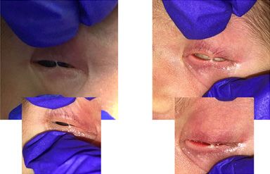
Ankyloblepharon Filiforme Adnatum: Case Report and Literature Review

INTRODUCTION
    Ankyloblepharon filiforme adnatum (AFA) was first
described by Josef von Hasner in 18811 as a congenital ab-
normality with partial or complete adhesion of the ciliary
edges of the upper and lower eyelids. The incidence of AFA
is 4.4 per 100 000 newborns.2 Fusion of eyelids is a normal
stage in fetal development, but it is abnormal at birth. The
developing eyelids fuse during the ninth week of gestation
and remain fused until the fifth month. Between the fifth and
the seventh month of gestation, a spontaneous separation of
upper and lower eyelid happens.3 A disruption of this pro-
cess can occur, due to a temporary arrest of the epithelial
growth,4 or an abnormally rapid proliferation of mesoderm,4
or a combination of the two which allows union of epithelia-
lized mesenchyme at certain points,4 or even due to failure of             Figure 1: Photograph of the new born showing extensile bands of tissue con-
                                                                           necting the eyelid margins of the right and left eye, with Bell’s phenomenon
apoptosis at a critical stage of eyelid development.5 Usually,
                                                                           in the right eye.
AFA constitutes a solitary malformation of sporadic occur-
rence.6 However, AFA may be associated with multisystem
developmental disorders or ophthalmological alterations.

CASE REPORT
    A Caucasian female baby was born at 39 weeks’ and 3
days’ gestation by elective vaginal delivery after an une-
ventful pregnancy, weighing 2870 g, measuring 47.50 cm of
height and 33.40 cm of cephalic perimeter, with an APGAR
index of 10/10/10. The mother was a healthy 40-year-old fe-
male with one successful gestation 5 years before and out of
non-consanguineous relation. There was no family history of
congenital abnormalities, the sibling’s new born was healthy.
On detailed ocular examination of the new born at four days
of age, there was a single band of extensile tissue between
upper and lower eyelid arising from the grey line, with the                Figure 2: Photograph of new born after the treatment, showing a normal
limited interpalpebral aperture, in both eyes (Fig. 1). The de-            eyelid aperture and a normal external ocular aspect.
tailed neonatal examination was normal and did not reveal
any other congenital abnormality. Therefore, the adhesions
were removed with a surgical blade near each eyelid margin,                DISCUSSION
under local anesthesia. Pupillary light reflex, ocular motility,
biomicroscopy and fundoscopy were normal. At the follow-                      AFA can present as an isolated ocular anomaly or as a
-up visit, two weeks postoperatively, no ophthalmological                  manifestation of a multisystem syndrome. Case reports of
abnormality was noted (Fig. 2). The eyelid opening was                     AFA, published from 1979 to 2021 in indexed journals, are
normal, as well as the upper and lower eyelid anatomy. The                 summarized in Table 1.
new born was healthy, without detection of any systemic
anomaly during the Neonatology appointments.

 Table 1: Case reports of ankyloblepharon filiforme adnatum in the literature
                                                                                                                       Ophthal-         Systemic
                  Year of publi-                   Number of     Unilateral /      Number of         Obstretric
 Author                             Journal                                                                           mological        alterations /
                     cation                        AFA cases      Bilateral         bands            alterations
                                                                                                                      alterations      Syndromes
                                                                                                      Maternal
                                                                                                   smoking, me-
                                                                                                   thadone use,                         Edwards’
                                                                  Unilateral         Single                                0
                                    Arch Dis                                                        Intrauterine                        Syndrome
 Williams et al
              9
                      2007                             2                                           growth retar-
                                     Child
                                                                                                    dation (IGR)
                                                                                                    AFA in fami-
                                                                   Bilateral         Single                                0                 0
                                                                                                    liar relatives

50 | Revista da Sociedade Portuguesa de Oftalmologia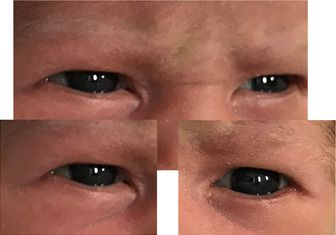
Ankyloblepharon Filiforme Adnatum: Case Report and Literature Review

                                                                                                     AFA, syndac-
                                                                                                                                      Cleft lip and
                                                                     Bilateral             2        tyly in familiar       0
                                                                                                                                         palate
 Akkermans                            Br J                                                             relatives
                      1979                              2
 et al10                           Ophthalmol                                                        AFA, syndac-
                                                                                                                                        Bilateral
                                                                     Bilateral             ?        tyly in familiar       0
                                                                                                                                       syndactily
                                                                                                       relatives
                                                                                                                                     Abnormal tuft
                                   Middle East                       Bilateral          Several            0               0         of hair in the
 Chakraborti
                      2014        Afr J Ophthal-        2                                                                                back
 et al11
                                        mol
                                                                     Bilateral          Several            0               0               0
                                                                                                                                        Cleft lip
                                                                     Bilateral          Several            0               0          without cleft
 Malek et al12        2019          Tunis Med           2                                                                                palate
                                                                                                    Parent consan-                      CHAND
                                                                     Bilateral           Single                            0
                                                                                                       guinity                         Syndrome
 Alami et al13        2013        Pan Afr Med J         1           Unilateral           Single            0               0               0
                                                                                                                                       Edwards’
 Clark et al 7
                      1985       Br J Ophtalmol         1            Bilateral         Single / 2          0               0
                                                                                                                                       Syndrome
                                                                    Unilateral           Single          IGR
                                                                                                                                       Edwards’
 Evans et al   4
                      1990         J Med Genet          3           Unilateral             2               ?               ?
                                                                                                                                       Syndrome
                                                                     Bilateral           Single            0
                                                                                                    Parent consan-                      AEC /
 Kuruvilla                        Ind J Opthal-
                      2016                              1           Unilateral           Single        guinity             0          Hay–Wells
 et al14                               mol
                                                                                                    (Third degree)                    syndrome
 AFA= ankyloblepharon filiforme adnatum, IGR = intrauterine growth retardation, CHAND = curly hair-ankyloblepharon-nail dysplasia, AEC = ankylo-
 blepharon–ectodermal dysplasia–clefting

    AFA was initially subdivided in group I (without as-                     contribution, grant or scholarship.
sociated abnormalities), group II (alterations in cardiac or                     Confidentiality of Data: The authors declare that they
central nervous system), group III (ectodermal syndrome)                     have followed the protocols of their work center on the pu-
and group IV (clef lip and/or cleft palate).7 Other associa-                 blication of data from patients.
tions may include hydrocephalus, meningocele, imper-                             Patient Consent: Consent for publication was obtained.
forate anus, bilateral syndactyly, infantile glaucoma with                       Provenance and Peer Review: Not commissioned; ex-
iridodogoniodysgenesis,8 cardiac problems such as patent                     ternally peer reviewed.
ductus arteriosus and ventricular septal defects.9
    An early separation of the fibrous adhesions is the stan-
dard treatment for AFA, performed with or without local                      RESPONSABILIDADES ÉTICAS
anesthesia and since it is a fibrovascular connective tissue8
there is no major bleeding.                                                      Conflitos de Interesse: OOs autores declaram a inexis-
    This report demonstrates the simplicity in treating the                  tência de conflitos de interesse na realização do presente
condition and the importance of early intervention to avoid                  trabalho.
the development of amblyopia.                                                    Fontes de Financiamento: Não existiram fontes exter-
                                                                             nas de financiamento para a realização deste artigo.
                                                                                 Confidencialidade dos Dados: Os autores declaram ter
CONCLUSION                                                                   seguido os protocolos da sua instituição acerca da publica-
                                                                             ção dos dados de doentes.
    AFA is a very rare condition that requires screening of                      Consentimento: Consentimento do doente para publi-
possible systemic associations, some of them potentially li-                 cação obtido.
fe-threatening. It is important to have a multidisciplinary                      Proveniência e Revisão por Pares: Não comissionado;
team for the management of AFA, including pediatricians,                     revisão externa por pares.
ophthalmologists, oral and maxillofacial surgeons, derma-
tologists, geneticists and psychologists to provide an ade-
quate treatment of patients and family support.                              REFERENCES
                                                                                 1.   Hasner V. Ankyloblepharon filiforme adnatum. Z
ETHICAL DISCLOSURES                                                                   Heilkol. 1888;2:429.
                                                                                 2.   Koubek M, Strakošová K, Timkovič J, Grečmalová D,
    Conflicts of Interest: The authors have no conflicts of                           Orlíková A, Burčková H, et al. A rare form of ankylo-
interest to declare.                                                                  blepharon filiforme adnatum associated with the
    Financing Support: This work has not received any                                 Hay-Wells syndrome and a c.1709T>C mutation on

                                                                                                                 Volume 45 - N1 - Janeiro-Março | 51
the TP63 gene. Ophthalmic Genet. 2018;39:251-4. doi:
10.1080/13816810.2017.1401091.
3. Sharkey D, Marlow N, Stokes J. Ankyloblepharon Fili-
forme Adnatum. J Pediatr. 2008;1524:594. doi:10.1016/j.
jpeds.2007.12.051
4. Evans DG, Evans ID, Donnai D, Lindenbaum RH.
Ankyloblepharon filiforme adnatum in trisomy 18
Edwards syndrome. J Med Genet. 1990;27:720-1.
doi:10.1136/jmg.27.11.720
5. Mohamed YH, Gong H, Amemiya T. Role of apoptosis
in eyelid development. Exp Eye Res. 2003;76:115-23.
doi:10.1016/S0014-4835(02)00269-5
6. Gruener AM, Mehat MS. A newborn with ankylo-
blepharon filiforme adnatum: A case report. Cases J.
2009;2:1-2. doi:10.4076/1757-1626-2-8146
7. D I CLARK AND A PATTERSON. Ankyloblepharon
filiforme adnatum in trisomy 18 (Edwards’s syndro-
me). Br J Ophthalmol. 1985;69:471-473.
8. Scott M, Richard J FB. Ankyloblepharon filiforme ad-
natum associated with infantile glaucoma and irido-
goniodysgenesis. J Pediatr Ophthalmol Strabismus.
1994;31:93-5.
9. Williams MA, White ST, McGinnity G. Ankyloblepha-
ron filiforme adnatum. Arch Dis Child. 2007;92:73-4.
doi:10.1136/adc.2006.103069
10. Akkermans CH, Stern LM. Ankyloblepharon filifor-
me adnatum. Br J Ophthalmol. 1979;63:129-31. doi:
10.1136/bjo.63.2.129.
11. Chandana Chakraborti, Krittika Pal Chaudhury,
Jayanta Das and AB. Ankyloblepharon Filiforme Ad-
natum: Report of Two Cases. Middle East Afr J Oph-
thalmol. 2014;21:200–2.
12. Malek I, Mekni M, Sayadi J, Bezzine A, Zghal I, Na-
cef L. Ankyloblepharon filiform adnatum: Beyond the
eye. Tunis Med. 2019;97:822-5.
13. Alami B, Maadane A, Sekhsoukh R. Ankyloblepha-
ron filiforme adnatum: A case report. Pan Afr Med J.
2013;15:1-3. doi:10.11604/pamj.2013.15.15.2209
14. Kuruvilla, Elizabeth. Simha A. A rare variant of
ankyloblepharon filiforme adnatum associated with
skin hypopigmentation: A case report from South In-
dia. Indian J Ophthalmol. 2016;64:241–243.
*Corresponding Author/
Autor Correspondente:
Maria J Vieira
R. de Santo André
2410-197 Leiria
Portugal
vieiramjp@gmail.com
ORCID: 0000-0001-9554-3427
52 | Revista da Sociedade Portuguesa de OftalmologiaYou can also read