ANTICATARACT ACTIVITY OF ETHANOLIC EXTRACT OF NIGELLA SATIVA
←
→
Page content transcription
If your browser does not render page correctly, please read the page content below
Volume: 2: Issue-4: Oct - Dec -2011                                                ISSN 0976-4550

 ANTICATARACT ACTIVITY OF ETHANOLIC EXTRACT OF NIGELLA SATIVA
        ON GLUCOSE INDUCED CATARACT IN GOAT EYE LENS
       1
        Shabeer Ahmed N, Niyas Ahmed I, Ashar Waheed M P and *Abdul Jaffar Ali H

     Department of Biotechnology and *Department of Biochemistry, Islamiah College,
                        Vaniyambadi -635 752, Tamilnadu, India
                                  Email:shabeer.sanbio@gmail.com

ABSTRACT : The present investigation was aimed to evaluate efficacy of ethanolic extract seeds of
Nigella sativa against glucose induced cataract in goat eye lens. In the in vitro study, goat lenses were
subjected to photographic evaluation and biochemical parameters such as protein, GSH, MDA,
NA+/K+ ATPase and sodium and potassium were also analyzed. Photographic examination of the eyes
showed that treatment with ethanolic extracts of seeds of N. sativa retarded the progression of lens
opacification. Cataract lens treated with N. sativa elevated the activity of Na+ K+ ATPase, total and
water soluble proteins and K+ ions to the level of normal level whereas reduced concentrations of Na+
ions. The MDA levels were significantly less in the N. sativa treated groups whereas, the level of
GSH, in high glucose (55mM), compared to the normal control group was significantly low but N.
sativa treated groups showed higher level of GSH. These results support the view that ethanolic
extract of seeds of N. sativa as seen in this in vitro model may, counteracts the effects of glucose in
inducing cataract to some extent.

Key words: Anticataract, Nigella sativa, goat lens, in vitro
INTRODUCTION
Cataract is one of the complications that diabetic patients are at higher risk of developing. Osmotic
stress imposed by sorbitol accumulation in the ocular lens has long been suggested to be the major
cause of this complication (Harding, 1991 and Nirmalan et al, 2004) since sorbitol was found to be
accumulated to a substantially high level incataractous lenses in diabetic animals like rats, rabbits, and
dogs (Ughade et al, 1998). Under hyperglycemic conditions, sorbitol is formed from the reduction of
glucose by the enzyme aldose reductase (AR) of the polyol pathway (Congdon et al, 2003). There is
accumulating evidence, however, showing the contribution of oxidative stress to the development of
diabetic cataract (Taylor et al, 1995; Kyselova et al, 2004; Mares, 2004). These findings led tothe
conclusion that the major culprit of diabetic cataract is sorbitol accumulation.
Recently, the conversion of sorbitol to fructosevia sorbitol dehydrogenase (SD) has also been
suggested to contribute to redox imbalance in diabetic tissues (Elena et al 2000 & Chandorkar et al,
1981).Na+K+ATPase plays an important role in maintaining the lens transparency and its alteration is
one of the major event leading to cataract formation. Impairment of Na + - K+ ATPase activity causes
accumulation of Na+ and loss of K+ with hydration and swelling of the lens fibers leading to
catactogenesis (Chylack and Kinashita, 1969). Reduced glutathione (GSH) was found to be depleted
in diabetic lenses (Gillis et al, 1992; Gupta et al, 1997) which was accompanied by an increase in the
level of lipid peroxidation products (LPO)
Dietary intervention, particularly the use of traditional food and medicines derived from natural
sources, is the mainstay in the management of diabetes. In this context, there has been a growing
interest in recent times in identifying as many dietary/spice sources as possible for their ability to
control diabetes (Swanstonet al, 1991; Chang, 2000; Suryanarayana et al, 2003). Nevertheless,
exhaust review of literature showed that studies of natural sources focusing on their ability to
maintain blood glucose levels are copious but investigations for their beneficial effects on secondary
complications of diabetes such as cataract; retinopathy, nephropathy and neuropathy are scanty.
Therefore, we have been interested in investigating various dietary sources for their potential to
prevent the secondary complications of diabetes.

   International Journal of Applied Biology and Pharmaceutical Technology                   Page:274
                               Available online at www.ijabpt.com
Shabeer et al ISSN 0976-4550
The surgery has its own limitations; pronounced post-operative inflammation, loss of vitreous humor,
posterior capsule opacification and expensive (Kyselova et al, 2004). So there is a need to look at the
impact of treating cataract and relate it not just to surgery but also to scholastic achievements and
development. In recent times, herbal drugs and natural products are targeted to develop more safe,
effective and economical treatment for prevention or delay the cataract (Gupta and Halder, 2002 &
Gupta and Srinivasa, 2005).
The seed of Nigella sativa is known by different names like black seeds or black cumin. In Latin, it is
called as ‘Panacea’ meaning ‘cure all’, in Arabic it is termed as ‘Habbah Sawda’ or ‘Habbat el
Baraka’ translated as ‘seeds of blessing’. In China it is referred as Hak Jung Chou while in India it is
called as Kalonji and in Persian, it is called as Shoneez. This plant belongs to the Ranunculaceae
family of flowering plants and genus Nigella consists of about 14 species including therapeutically
used seven species such as, Nigella arvensis, Nigella ciliaris, Nigella damascene, Nigella hispanica,
Nigella integrifolia, Nigella nigellastrum Nigella orientalis and Nigella sativa. Among these, Nigella
sativa is the species most exhaustively investigated for therapeutic purposes. The historical references
to use of seeds of Nigella sativa also found in some of the oldest religious and medical texts. The
seeds of Nigella sativa Linn. (Ranunculaceae) are used in herbal medicine all over the world for the
treatment and prevention of a number of diseases.
Several studies have been carried out on the effect of herbal extracts against cataract formation in-
vitro and in-vivo in various animals by various authors (Ho et al, 2000; Rosen et al, 2001; Wolff and
Dean, 1987; Mullarkey et al, 1990; Schmidt et al, 1994). However, there is a paucity of information
about the effects of extracts of seeds of Nigella sativa against cataract lens in goat. With this in
context, the present investigation was aimed to evaluate efficacy of seeds of Nigella sativa against
glucose induced cataract in goat eye lens. Since biochemical parameters such as protein, GSH, MDA,
NA+/K+ ATPase and sodium and potassium are important for reflecting the healthy state of lens, these
parameters were analyzed in the present study.
MATERIALS AND METHODS
Plant material
Plant material consists of dried powdered seeds of Nigella sativa belonging to the family
Ranunculaceae. The seeds were purchased from local market, Vaniyambadi, India, during the month
of November 2009.
Preparation of Ethanolic extract of Nigella sativa
The standard method (Bhargava et al 1998)was followed for the extraction of material. Seeds of
Nigella sativa were dried in shade under room temperature and pulverized to a coarse powder. They
were extracted by percolation at room temperature with 70% ethyl alcohol. The extract was
concentrated under pressure (bath temperature 50°C) and finally dried in vacuum desiccator.
Collection of Eye Balls
Fresh goat eye balls of young and healthy goats were collected from the slaughter house,
Vaniyambadi immediately after the slaughter. These eye balls were immediately transferred to the
laboratory at 0-4°C. Sliced the Cornea from the front of the eye to gain access to the lens.
Lens culture
The lenses were incubated in artificial aqueous humor (NaCl 140mM, KCl 5mM, MgCl2 2mM,
NaHCO3 0.5mM, Na2HPO4 0.5mM, CaCl2 0.4mM, Glucose 5.5mM) for 72 hours at room
temperature at a pH of about 7.8 is maintained. In addition to this 32mg of penicillin and 250mg of
streptomycin were added to prevent bacterial contamination. Glucose 55mM served as cataract
inducer(Chandrokar et al, 1981).
Drug study
Group I : Normal lens glucose 5.5mM (control)
Group II : Glucose 55mM (induced)
Group III : A. Glucose 55mM+Nigella sativa (100µg/ml) (Treated)
B. Glucose 55mM+Nigella sativa (300µg/ml) (Treated)
C. Glucose 55mM+Nigella sativa (500µg/ml) (Treated)
International Journal of Applied Biology and Pharmaceutical Technology Page:275
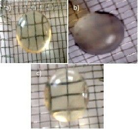
Available online at www.ijabpt.comShabeer et al ISSN 0976-4550 Photographic Evaluation After 72 hours of incubation, lenses were observed for opacity and photographs were taken by placing the lenses on the wire meshes with posterior surface touching the mesh, and the pattern of mesh was observed through the lens as a lens as a measure of lens opacity. Homogenate preparation Lenses were homogenized in Tris buffer (0.23M pH 7.8) and 0.25X10⁻³ M EDTA. The homogenate was adjusted to 10% W/V. The homogenate was centrifuged at 10000 g at 4°C for 1 hour. The supernatant was used for estimation of protein (Lowry et al., 1951), GSH(Moron et al., 1979), MDA(Satohwith modification, 1978), NA+/K+ ATPase(Bonting, 1970) and sodium and potassium (flame photometry). Statistical Analysis All data were expressed as mean ± SD. All data were analyzed with SPSS/10 student software. Hypothesis testing methods included one way analysis of variance (ANOVA) followed by LSD. The values are expressed as mean ± S.D. and results were considered significantly different if P
Shabeer et al                                                                    ISSN 0976-4550

    Table 1. Na+, K+ and Na+ K+ ATPase activity in lens homogenate after 72 hours of
                                     incubation.
                                                                                    Na+K+ATPase activity
      Description
                                            Na+(meq/g m)        K+(meq/gm)          (µg / g lens)
      Normal Goat lens                  153±57.1                11.6±1.2            46.2±4.5
      Goat lens + Glucose55mM           218.4±22.6              6.4±0.3             19.3±2.1
      Goat lens + Glucose55mM +
      extract of seeds of N. sativa
          a) 100µ/ml                    171.8±17.3              8.6±0.94            28.3±3.1
          b) 300µ/ml                    167.5±19.8              9.8±0.9             33.8±4.1
          c) 500µ/ml                    160.5±29.4              10.7±1.8            42.6±3.8
       Values are mean ±S.D. n=5 for each group.

Lower concentration of protein and GSH was observed in the homogenate of glucose induced lenses
whereas, very high MDA (P
Shabeer et al ISSN 0976-4550
In this study, MDA levels were significantly higher in high glucose (55mM) groups, compared with
normal control group. The MDA levels were significantly less in the N. sativa treated groups. The
level of GSH, in high glucose (55mM), compared to the normal control group was significantly low
but N. sativa treated groups showed higher level of GSH.
Incubation in the presence of high glucose (55mM) concentration stimulates a state of clinical
diabetes. A prevention role of Nigella sativa as seen in this in vitro model may, to some extent
suggest in preventing and/or retarding the progression of diabetic cataracts.This in vitro study may not
directly correlate with the in vivo conditions. Therefore in vivo studies in different animal models are
required for further elucidation of the role of Nigella sativa in preventing cataract formation.
Acknowledgement
Authors are grateful to Mr. Ghani Md. Jaweed Sahib, Secretary cum Correspondent and Dr. K. Prem
Nazeer, Principal, Islamiah College, Vaniyambadi for their wise advice and encouragements.
REFERENCES
1. Harding J (1991).Cataract. Biochemistry, Epidemiology and Pharmacology. Chapman &
Hall London.
2. Nirmalan PK, Robin AL, Katz J (2004).Risk factors for age related cataract in a rural
population of southern India. The Aravind Comprehensive Eye Study. Brazilian Journal of
Ophthalmology 88: 989–994.
3. Ughade SN, Zodpey SP, Khanolkar VA (1998). Risk factors for cataract: a case control study.
Indian Journal of Ophthalmology 46:221–227.
4. Congdon NG, Friedman DS, Lietman T (2003). Important causes of visual impairment in the
world today. JAMA290: 2057–2060.
5. Taylor A, Jacques PF, Epstein EM (1995). Relations among aging, antioxidant status, and
cataract. Am J Clin Nutr 62:1439S–1447S.
6. Kyselova Z, Stefek M, Bauer V (2004). Pharmacological prevention of diabetic cataract. J
Diabetes Complications 18: 129–140.
7. Mares JA (2004). High-dose antioxidant supplementation and cataract risk. Nutr Rev.62: 28–
32.
8. Elena MV, de Cavanagh EM, Inserra F, Ferder L, Fraga CG (2000). Enalapril and captopril
enhance glutathione-dependent antioxidant defenses in mouse tissues. AJP-Regulatory,
Integrative and Comparative Physiology 278:R572-77.
9. Chandorkar AG, Albal MV, Bulakh PM,Muley MP (1981). Lens Organ Culture. Indian J
Opthalmol 29:151-2.
10. Chylack LT, Kinashita JH (1969). A biochemical evaluation of a cataract induced in a high
glucose medium. Invest Opthalmol 8: 401-12.
11. Gupta SK, Joshi S, Velpandian T, Awor L, Prakash J (1997). An update on pharmacological
prospectives for prevention and development of cataract. Indian J Pharmacol 29:3-10.
12. Gillis CN, Chen X, Merker MM (1992). Lisinopril and ramiprilat protection of the vascular
endothelium against free radical-induced functional injury. J Pharmacol Exp Ther 262:212-
216.
13. Swanston-Flatt SK, Flatt PR, Day C, Bailey CJ (1991). Traditional dietary adjuncts for the
treatment of diabetes mellitus. Proc Nutr Soc. 50:641–651.
14. Chang J (2000). Medical herbs—drugs or dietary supplements? Biochem Pharmacol. 59:
211–219.
15. Suryanarayana P, Krishnaswamy K, Reddy GB (2003). Effect of curcumin on galactose-
induced cataractogenesis in rats. Mol Vis 9:223–230.
16. Kyselova Z, Stefek M Bauer V (2004). Pharmacological prevention of diabetic cataract. J
Diabetes Complications 18:129–140.
17. Gupta SK, Halder N (2002). Green Tea (Camellia sinensis) protects against selenite-induced
oxidative stress in experimental cataractogenesis. Ophthalmic Res 34:258-63.
International Journal of Applied Biology and Pharmaceutical Technology Page: 278
Available online at www.ijabpt.comShabeer et al ISSN 0976-4550
18. Gupta SK, Srinivasa S (2005).Ocimum sanctum modulates selenite-induced cataractogenic
changes and prevents rat lens opacification. Curr Eye Res. 30:583-91.
19. Ho FM, Liu SH, Liau CS, Huang PJ, Lin-Shiau SY (2000). High glucose-induced apoptosis in
human endothelial cells is mediated by sequential activations of c-Jun NH2-terminal kinase
and caspase-3.Circulation101: 2618 –2624,
20. Rosen P, Nawroth PP, King G, Moller W, Tritschler HJ, Packer L (2001). The role of
oxidative stress in the onset and progression of diabetes and its complications: A summary of a
Congress Series sponsored by UNESCO-MCBN, the American Diabetes Association and the
German Diabetes Society. Diabetes Metab Res Rev 17: 189–212.
21. Wolff SP, Dean RT (1987). Glucose autoxidation and protein modification. The potential role
of autoxidative glycosylation’ in diabetes. Biochem J 245:243–250.
22. Mullarkey CJ, Edelstein D, Brownlee M (1990). Free radical generation by early glycation
products: A mechanism for accelerated atherogenesis in diabetes. Biochem Biophys Res
Commun 173: 932–939,
23. Schmidt AM, Hori O, Brett J, Yan SD, Wautier JL,Stern D (1994). Cellular receptors for
advanced glycation end products. Implications for induction of oxidant stress and cellular
dysfunction in the pathogenesis of vascular lesions Arterioscler Thromb 1521–1528,
24. Bhargava A, Srivastava A, Kumbhare VC (1998). Antifungal activity of polyphenolic
complex of Acacia nilotica bark. Ind Forest.124: 292- 298.
25. Chandorkar AG, Albal MV, Bulakh PM, Muley MP (1981). Lens Organ Culture. Indian J
Opthalmol 29:151-2.
26. Lowry O, Rosebrough A, Farr A, Randall R (1951). Protein measurement with the Folin
phenol reagent. J Biol Chem 193: 265.
27. Moron MS, Depierre JW, Mannervik B (1979). Level of glutathione, glutathione reductase
and glutathione –S- transferase activitie in lungs and liver. Biochem Biophys Acta 82: 67-68.
28. Satoh K (1978). Serum lipid peroxidation in cerebro vascular diseases determined by new
colorimetric method. Clin Chem Acta 90: 37-43.
29. Bonting, S (1970). Membrane and Ion transport, In: Bittar C, Eeeditors W (Eds), London, 1970;
25-28.
30. Lokesh JS, Harikiran H, Sharma (2010). An In vitro prophylactic cataract prevention study on
glucose induced cataract by quercetin and alpha – tocopherol, International Journal of
Pharmaceutical sciences and research 1 (7): 41- 45.
31. Wilbur KM (1949). Estimation of lipid peroxide. Arch Biochem Biophysics 24: 305-15.
32. Chylack LT, Kinashita JH (1969). A biochemical evaluation of a cataract induced in a high
glucose medium. Invest Opthalmol8:401-12.
International Journal of Applied Biology and Pharmaceutical Technology Page: 279
Available online at www.ijabpt.comYou can also read