Antioxidative, Antibacterial and Antibiofilm Activity of Melanin nanoparticles (MN)
←
→
Page content transcription
If your browser does not render page correctly, please read the page content below
International Journal of

                Life Sciences
                                                                                                          Int. J. of Life Sciences, 2020; 8 (2):308-316
                                                                                                                   ISSN:2320-7817(p) | 2320-964X(o)
                International Peer Reviewed Open Access Refereed Journal

 Original Article                                                                                                                   Open Access

 Antioxidative, Antibacterial and Antibiofilm Activity of
 Melanin nanoparticles (MN)
 Tiwari Amit and Pradhan Swapnil*

 Department of Microbiology, Ramnarain Ruia Autonomous College, University of Mumbai, Mumbai, India
 *Corresponding author Email : tiwari.amit8894@gmail.com

Manuscript details:                                                  ABSTRACT

Received: 06.04.2020                                                 Melanin is a complex, diverse, negatively charged, hydrophobic, amorphous
Accepted: 09.06.2020                                                 and high molecular weight hetero-polymeric dark brown photosynthetic
Published: 29.06.2020
                                                                     pigment synthesized by living organisms including animals, humans, and
                                                                     microorganisms in the course of oxidative polymerization of phenolic
Cite this article as:                                                compounds. In this study, melanin production was carried out by
Tiwari Amit and Pradhan Swapnil                                      Pseudomonas sp. which was isolated from soil. After production, Melanin
(2020) Antioxidative, Antibacterial                                  was extracted by acid hydrolysis and characterized based on its solubility i n
and Antibiofilm Activity of Melanin                                  different solvents where it was found to be water-soluble (a unique
nanoparticles (MN), Int. J. of. Life
                                                                     characteristic of melanin); by TLC and by NMR. After the characterizati on of
Sciences, Volume 8(2): 308-316.
                                                                     melanin, melanin nanoparticles were formed by sonication and then
                                                                     characterized and evaluated for antimicrobial and antioxidative properties.
                                                                     The nanoparticle was found to be of discrete sizes. Melanin nanoparticles
                                                                     showed strong antimicrobial activity against Gram-positive organi sms and
Available online on http://www.ijlsci.in                             also higher antioxidative properties than melanin. Melanin Nanoparticles
ISSN: 2320-964X (Online)                                             were found to disrupt formed biofilm of Staphylococcus aureus by 67. 39%.
ISSN: 2320-7817 (Print)                                              Thus, Melanin nanoparticles may find their utility as a coating material in
                                                                     medical implants and as packaging material in food products against
                     Open Access This article is                     oxidation and bacterial contamination.
                     licensed under a Creative
                     Commons Attribution 4.0                         Keywords: Pseudomonas balearica DSM 6083(T), Melanin Nanoparticles,
International License, which permits use,
sharing, adaptation, distribution and
                                                                     KMnO 4, Titanium, Biofilm.
reproduction in any medium or format, as
long as you give appropriate credit to the
original author(s) and the source, provide a                         INTRODUCTION
link to the Creative Commons license, and
indicate if changes were made. The images or
other third party material in this article are                       Melanin is complex, diverse, negatively charged, hydrophobic, amorphous
included in the article’s Creative Commons                           and high molecular weight hetero-polymeric dark black or brown pigments ,
license, unless indicated otherwise in a credit                      a ubiquitous pigment produced in melanosomes, are found in bacteria (s uch
line to the material. If material is not included                    as Brevundimonas sp. SGJ, Shewanella colwelliana D, Aeromonas media,
in the article’s Creative Commons license and
your intended use is not permitted by
                                                                     Pseudomonas putida F6 , Klebsiella sp.GSK, Pseudomonas stutzeri , recombi-
statutory regulation or exceeds the permitted                        nant Escherichia coli W3110, etc), fungi ( such as Aspergillus fumigates,
use, you will need to obtain permission                              Aspergillus Bridgeri, Pneumocystis carinii, Cryptococcus neoformans, Pleurotus
directly from the copyright holder. To view a                        cystidiosus, etc.), plants and in many human organs and tissues, including
copy of this license, visit                                          retina, mucous membrane, ovary, cerebral pia mater and skin (Liu et al.,
http://creativecommons.org/ licenses/by/4.
                                                                     2018; Pal et al., 2015; Shripad et al., 2013; Coon et al., 1994; Wan et al., 2007;

 © 2020 |IJLSCI                                                                www.ijlsci.in                                                 | 308
Antioxidative, Antibacterial and Antibiofilm Activity of Melanin nanoparticles (MN)
Nikodinovic-Runic et al., 2009; Sajjan., 2013; Kumar et study melanin nanoparticles have been used for
al., 2013; Lagunas-Muñoz et al., 2013; Kumar et al., antibiofilm activity. Melanin nanoparticles can be
2011; Youngchim et al., 2004; Plonka et al., 2006; synthesized either by physical and chemical method.
Frases et al., 2006; Selvakumar et al., 2008). M el anin Chemical synthesis of melanin nanoparticles involv es
pigment is produced by the oxidation of tyrosine by reaction between either KMnO4 or NaOH with melanin
tyrosinase in a multistep synthesis via 5,6- under specific set of conditions (Rageh & El-Gebaly,
dihydroxyindole-2-carboxylic acid and 5,6- 2018; Sáez & Mason, 2009).The physical method of
dihydroxyindole (Pezzella et al., 2013). This oxidati ve melanin nanoparticles involves usage of
polymerization of these indoles gives rise to the ultrasonication resulting in breaking of melanin.
melanin biopolymers: black-brown eumelanin and Biofilm formation takes place by quorum sensing
yellow/reddish pheomelanin (Xiao et al., 2015). signals and it has been known that melanin can inhibit
Melanin is one of the most stable, insoluble, and these signals. Melanin inhibit acyl homoserine lactone
resistant pigment (Francisco Solano, 2017). The (AHL)-regulated behaviors by binding competitively to
polymer melanin is known to absorb UV radi ati on by the AHL receptor protein, thus, inhibit or disrupt
redox reactions and electron transfer processes. In biofilm formation (Bin et al., 2012).
addition to photoreactive nature, melanin can also
absorb metal ions, free radicals, etc. thereby act as a In this study, Pseudomonas balearica DSM 6083
cytoprotective agent. They are an antioxidant, isolated from soil have used for synthesis of M el anin.
antitumor, and anti-inflammatory agent, as well as an Synthesized Melanin was extracted by refluxing in
immune-stimulating agent. They also find applicati on strong acid and characterized using its biochemical
in semiconductor and bio-electronics as well as in properties. Melanin nanoparticles have been
polarized sunglass lenses, paints, and varnishes synthesized using ultrasonication method, a non-
(Francisco Solano, 2017). chemical procedure. The anti-oxidant activity of
melanin and melanin nanoparticles have been
Biofilm can be defined as communities of performed using potassium permangnate. Well
microorganisms irreversibly attached to a surface, diffusion assay was performed for determining
encased within a polysaccharide-rich extracellular antibacterial activity of both melanin and melanin
matrix, and exhibiting an altered phenotype which has nanoparticles. Biofilm formation was allowed on
enhanced resistance to antimicrobial drugs (Naik et al., titanium pieces and later it was treated with melanin
2015; Mohammadi et al., 2013). The high density of and melanin nanoparticles to disrupt formed biofilm.
extracellular polysaccharide-rich matrix will not onl y
slow the diffusion of antibiotics but also hinders access MATERIALS AND METHODS:
of immune system defenses such as antibodies and
macrophages (Naik et al., 2015). These biofilms are 1. Sampling Site and Sample Collection
responsible for 60% of Healthcare associated 10 g of soil sample was collected from 19ᴼ01’21” N
infections (HAI) by corroding medical implants. 72ᴼ50’55” E in a sterile glass bottle. The sample was
Various approches have been used to disrupt or inhibit kept in a petri dish at Room Temperature to remove
biofilm such as use of anti-virulence compounds , us e moisture content i.e. clumping of soil..
of mucolytic substances, use of chelating agents, use of
matrix targeting enzymes, use of phages, and recentl y 2. Enrichment of the culture
use of nanoparticles (Percival et al., 2015b; Starkey et 1 g of the processed soil sample was inoculated i n 20
al., 2014; Pérez-Giraldo et al., 1997; Abraham et al., ml of St. Nutrient Broth containing 0.5% Tyrosine and
2012; Chen et al., 2013; Fu at al., 2010; Algburi et al., incubated at Room Temperature for 7 days on a
2017). shaker (adjusted to 150 rpm). The growth of the
Nanomaterials have appeared as one of the most organism was indicated by turbidity in the medium
promising strategy for controlling or treating followed by the brown coloration of the media.
pathogenic biofilms on indwelling medical devices and
implants due to their large surface to volume ratio and 3. Isolation and Culturing of the melanin
unique physico-chemical properties(Naik et al., producing organism
2015)(Ramasamy & Lee, 2016). As melanin i s known After the development of brown coloration of the
to have antimicrobial and antioxidant activity, in this enrichment media, a loopful of growth obtained was
www.ijlsci.in Int. J. of Life Sciences, Volume 8 (2) 2020 | 309Tiwari and Pradhan, 2020
streaked on St. Nutrient Agar Plates containing 0.5% and incubated at 37°C for 3 weeks to produce melani n
Tyrosine (pH 10) and incubated at 37°C for 3 days. pigment (V, V., et al ., 2011).
Colony characteristics of colonies producing brown to After 3 weeks of incubation, chloroform was added
black pigment were selected and sub-cultured on St. into samples at 10 % concentration and kept for 2
Nutrient Agar Slants containing 0.5% Tyrosine (pH 10) days to kill viable cells. Then samples were centrifuged
(Pathan & Pethe, 2016). at 12000 rpm for 15 min. The cell pellet was discarded
and melanin-containing supernatant was then
4. Identification and Characterisation of the acidified to pH 3 with 5 N HCl to ensure precipitation
melanin producing organism of the melanin. The precipitated melanin was
Isolates obtained were identified based on growth centrifuged at 12000 rpm for 15 min, washed three
obtained on the St. Nutrient Agar Plate containing times with deionized water, and allowed it to dry at
0.5% Tyrosine (pH 10) by the production of brown to Room Temperature and then weight was determined
black pigment. Complete characterization of the (Pal et al., 2015).
organism was done based on cultural & morphological
characteristics, and by 16 S rRNA sequencing. Melanin produced was purified using the dialysis
• Cultural characterization was done based on colony method. In the dialysis method, melanin powder was
characteristics of the isolate obtained on the St. kept in the dialysis tube which itself was kept in 2000
Nutrient Agar Plate containing 0.5% Tyrosine (pH ml distilled water which was changed every 4 hours
10). for 2 days.
• The morphological arrangement, Gram nature, and
structure of the cells were identified by performing 6. Characterization of melanin pigment
Gram staining. A smear of culture was taken in a 6. A. Solubility Test
clean glass slide and heated gently over a flame. Purified melanin was identified based on its solubili ty
The smear was covered with a thin film of crystal in different solvents. Purified melanin was dissolved in
violet for 1 min and washed gently in slow runni ng 3 ml of various organic and inorganic solvents (Hou et
tap water. Gram’s iodine solution was flooded over al., 2019).
the smear for 1 min and washed with tap water. Organic solvents: Ethanol, Diethyl ether, Acetone
Alcohol was used to decolorize the smear unti l the (warm and cold), Chloroform (warm and cold),
violet colour ceased to flow away. The slide was Benzene, Dimethyl sulfoxide, Hexane, n-butanol, and
washed with water and counterstain safranin was Phenol.
flooded over the smear for 2 min, then the slide Inorganic solvents: Distilled water, 0.1 N NaOH
was washed, drained, air dried, and viewed under a solution (pH 9) and Conc. HCl.
microscope.
• The genetic makeup of the isolate was determi ned 6. B. Thin Layer Chromatography
by performing 16S rRNA sequencing. 10 mg of Standard melanin (Purchased from Sigma
16S rRNA gene sequence analysis has been wi del y Aldrich) and purified melanin was dissolved in
used for the identification of the species. The Phosphate buffer (pH 8) and applied to silica gel TLC
sequence of 16S rRNA provides a measure of (Thin layer chromatography) plates and a
genetic similarity above the level of species chromatogram was developed using the solvent
allowing comparisons of relatedness across the system of n- butanol: acetic acid: water (70:20:10).
genus, family, etc. In the present study, 700 bp of Plates were then sprayed with the Iodine solution an d
entire 16S rRNA from the selected isolate was kept for drying in a hot air oven. Spots were obtained
sequenced. and the retention factor was calculated (Prajapati et
al., 2017).
5. Pigment Production, Extraction, and Purification
of melanin pigment 6. C. 1H NMR
Isolate was inoculated in 10 ml of St. Nutrient Agar NMR is used in research for determining the content
Broth containing 0.5% Tyrosine (pH 10) and and purity of a sample as well as its molecular
incubated for 24 h at 37°C for activation. 10 ml of structure. The 1H NMR spectrum of melanin using
activated culture was inoculated in 200 ml of St. DMSO as solvent was obtained by Nuclear Magnetic
Nutrient Agar Broth containing 0.5% Tyrosine (pH 10) Resonance spectrometer in 5-mm NMR tubes at 25°C.
310 | Int. J. of Life Sciences, Volume 8 (2) 2020Antioxidative, Antibacterial and Antibiofilm Activity of Melanin nanoparticles (MN)
7. Synthesis of Melanin Nanoparticles of Phosphate buffer solution added in 3.9 ml of KMnO4
The purified melanin was dissolved phosphate buffer and incubated at 25°C for 30 min was used as a
(pH 8) to attain a concentration of 300 µg/ml. 1% control. Vitamin C (300 µg/ml) was used as a posi ti v e
Trisodium citrate was added in melanin solution in a control. The Antioxidant activity percentage (AA %)
ratio of 20:1. Trisodium citrate acts as a capping agent was determined according to Priya et al.:
and prevents the aggregation of Melanin
Nanoparticles. The mixture of trisodium citrate and Antioxidant Activity % = Ac – At/Ac * 100
melanin was sonicated, with the help of probe
sonicator, pulsing for 10 seconds with a gap of 15 11. Antibiofilm Activity of Melanin
seconds between each pulse for a total of 60 min. Nanoparticles
Formed melanin nanoparticles were stirred vigorously After cleaning Ti6Al4V pieces with ethanol, they were
on a magnetic stirrer for 40 minutes at 300 rpm. The transferred in 30 ml of St. Tryptone soy broth
stirring helps in the dispersion of nanoparticles in containing 1% glucose. 3 ml of 18 h old Staphylococcus
solution (Kiran et al.,2017). (Note: Synthesis of aureus culture was inoculated in broth bottles and
nanoparticles should be carried out in 16°C) incubated at 37°C for 5 days to allow the formati on of
biofilm. After incubation, established biofilm on
8. Characterization of Melanin Nanoparticles Ti6Al4V was treated with Melanin and Melanin
The size of the melanin nanoparticles (300 µg/ml) nanoparticle at 37ºC for 1 h. After treatment, each
formed by sonication was determined by the particle piece was washed thrice with St. D/W and then
size analysis. The particle size distribution (volume incubated with 5 mL of crystal violet (O.D. adjusted to
percent) was produced by the computer-controlled 1.0) for 30 min at 37ºC. After incubation, Crystal violet
instrument according to the principle of light was removed and the pieces of the Ti6Al4V were
scattering. washed thrice by St. D/W to remove unbound cells.
After washing, 5mL of ethanol was added and kept at
9. Antibacterial Activity of Melanin room temperature for 15 mins, the reaction mixture
and Melanin Nanoparticles was then read colorimetrically at 590nm and the
Each of Gram-positive (Staphylococcus aureus) and percentage of biofilm disruption was determined using
Gram-negative (Escherichia coli) bacterial culture were (Namasivayam et al., 2013).
inoculated in 10 ml of St. Nutrient broth and incubated
at 37°C for 18 hours. The antibacterial activity of % of disruption=
OD in control -OD in treatment
melanin, as well as melanin nanoparticles, was OD in control
determined by using a well diffusion assay. 0.1 ml of
each 18 h old enriched cultures were spread on St. RESULTS AND DISCUSSION
Nutrient Agar Plate. Wells were made with the hel p of
sterile steel cork-borer and 100 µl of both melanin 1. Isolation and Characterization of Melanin
(300 µg/ml) and melanin nanoparticles (300 µg/ml) Producing Organism
suspension was added to the wells. The plates were A total of 3 distinct bacteria were isolated from the soil
then incubated at 37°C for 24 h, and zone of inhibiti on on Nutrient agar containing 0.5% tyrosine (pH 10)
was monitored (Zerrad et al., 2014). plates. All the isolates were screened for melanin
production, with isolate PP1 identified as the best
10. Antioxidant Activity of Melanin and melanin producer. The isolate PP1 was identified as a
Melanin Nanoparticles rod-shaped Gram-negative bacterium. The 16 S rRNA
The percentage of antioxidant activity (AA %) of sequence of the isolate showed the highest similarity
melanin and melanin nanoparticles was assessed by of 99.76% with Pseudomonas balearica DSM 6083(T).
using KMnO4 described by Amponsah et al. (2016)
with modifications. 0.1 ml each of melanin (300 2. Production and Characterization of Melanin
µg/ml) and melanin nanoparticles (300 µg/ml) was After a week of incubation, it was found that
placed in a test tube with 3.9 ml of 0.25 mg/ml KMnO4 Pseudomonas balearica DSM 6083(T) was able to
solution. The resulting system was incubated at 25°C produce 0.95 mg of Melanin/ml of production media.
for 30 min and the absorbance of the residual KMnO 4 Melanin was characterized based on its chemical
was determined at 530 nm using a colorimeter. 0.1 ml properties. The pigment was found to be insoluble in
www.ijlsci.in Int. J. of Life Sciences, Volume 8 (2) 2020 | 311Tiwari and Pradhan, 2020
ethanol, acetone, chloroform, benzene, hexane, butanol Extracted Melanin was found to be 0.4 ± 0.062 and
and phenol. The pigment was only soluble in al kali ne that of Standard Melanin (purchased from Sigma) was
solvents like sodium hydroxide, Dimethyl sulfoxide found to be 0.38 ± 0.064.
(DMSO), pyridine and water. This insoluble nature of
melanin is because of its aromatic structure. Melanin 1H Nuclear Magnetic Resonance (NMR) Spectroscopy
produced from Pseudomonas balearica DSM 6083(T) was also used to characterize the melanin pigment. 1 H
was found to be water-soluble which is a unique NMR peaks of melanin had shown similarity wi th the
characteristic of melanin. Water insoluble melanin earlier reports (Arun et al., 2015; Guo et al., 2014a).
requires treatment with strong alkali or strong The 1H NMR spectrum of melanin in DMSO showed
oxidants which often damages the structure of signals in both the aromatic and aliphatic regions.
melanin, thus, interferring with antimicrobial and Peaks in the absorption region from 6.5 to 8.5 ppm
antioxidant activity of melanin (Aghajanyan et al., corresponds to the benzene structure in the molecul e.
2011). Peaks in the region between 0.9 to 1.8 ppm
correspond to the protons attached to –CH3 groups.
Melanin was characterized by Thin Layer Absorption at 4.344 and 5.458 ppm is attributable to
Chromatography using the solvent system of n- an alkene group and at 1-3 ppm range probably
butanol: acetic acid: water (70:20:10). The Rf val ue of corresponds to protons attached to an amine group.
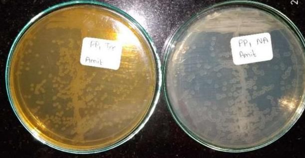
Figure 1: Isolation of isolate PP1 on St. Nutrient agar plate and St. Nutrient agar plate containing 0.5%
Tyrosine.
Table 1: Colony Characteristics of PP1 Isolate
Characteristics Size Shape Colour Opacity Eleva- Surface Margin Consis- Gram Nature &
tion tency Morphology
PP1 Isolate 2mm Irregular Colour- Trans- Flat Smooth Smooth Butyr- Gram Negative
less lucent ous Rods
Table 2: Solubility test of Melanin.
Solvents Standard Melanin Test Melanin
Ethanol Insoluble Insoluble
Pyridine Sparingly soluble Sparingly soluble
Acetone Insoluble Insoluble
Chloroform Insoluble Insoluble
Organic Solvent Benzene Insoluble Insoluble
Dimethyl sulfoxide Sparingly soluble Sparingly soluble
Hexane Insoluble Insoluble
n-butanol Insoluble Insoluble
Phenol Soluble Insoluble
Distilled water Insoluble Soluble
Inorganic Solvent NaOH solution (pH 9) Soluble Soluble
Conc. HCl Soluble Soluble
312 | Int. J. of Life Sciences, Volume 8 (2) 2020Antioxidative, Antibacterial and Antibiofilm Activity of Melanin nanoparticles (MN)
Figure 2: 1H NMR spectrum of Melanin showing peaks in the absorption region similar to earlier reports.
Figure 3: Melanin nanoparticle size analysis.
3. Characterization of Melanin Nanoparticles Size Analysis.The average size of melanin
Melanin Nanoparticles were formed by a “top-to- nanoparticles were found to 459.4 nm.
bottom” approach. As melanin is a highly complex
polymer nanoparticles can be made by breaking it 4. Antioxidant Activity of Melanin Nanoparticles
down to smaller sizes using the ultrasonication KMnO 4 is an oxidizing agent and therefore can mimic
method. As nanoparticles tend to aggregate after some the ROS (Reactive Oxygen Species) present in the
time, Trisodium citrate was used as a capping agent body. Melanin and melanin nanoparticles can capture
which resist aggregation. This study does not i nv ol ve electrons causing the reduction of the MnO 4- to MnO 4 2-
the use of any chemical or metal during Nanoparticle and mops up ROS (Reactive Oxygen Species) produced
synthesis which were involved in conventional studies by organisms metabolism or due to UV radiations and
and responsible for toxicity in environment. The size thus, protects from oxidative stress (Zerrad et al.,
of melanin nanoparticles produced by ultrasonication 2014; Bilgihan et al., 1995; Geng et al., 2008). The
method using melanin produced by Pseudomonas reduction results in a change of colour from purpl e to
balearica DSM 6083(T) was determined using Particle light yellow as a result of the gain of electrons with a
www.ijlsci.in Int. J. of Life Sciences, Volume 8 (2) 2020 | 313Tiwari and Pradhan, 2020 consequent bathochromic shift, depending on the The antioxidant activity of melanin nanoparticles was concentration of the antioxidant used. The free radical found to be 93.10 ± 0.00 much higher than that of scavenging effect of melanin and melanin melanin (11.16 ± 1.41) and Vitamin C (36.78 ± 7.18). nanoparticles was assessed using the KMnO 4 assay. Figure 4: Antioxidant activity of melanin and melanin nanoparticles using KMnO 4 assay. Both melanin and melanin nanoparticles at 300 µg/ml concentration were found to scavenge ROS (Reactive Oxygen Species). Figure 5: Antibiofilm activity of Melanin and Melanin Nanoparticles using crystal violet assay. The tested concentration of melanin and melanin nanoparticles (300 µg/ml) disrupted formed biofilm of Staphylococcus aureus. 5. Antibacterial Activity of Melanin and Melanin whereas melanin nanoparticles showed 21 ± 0 mm of nanoparticles zone of inhibition. Well diffusion assay has been used for evaluation of antibacterial activity of melanin and melanin 6. Antibiofilm Activity of Melanin and Melanin nanoparticles against gram positive Staphylococcus nanoparticles aureus and gram negative Escherichia coli. It was The crystal violet assay has been used for evaluation of observed that both melanin and melanin nanoparticles disruption of preformed Staphylococcus aureus by showed antibacterial activity against gram positive Melanin and Melanin nanoparticles. The treatment of Staphylococcus aureus at a concentration of 300 µg/ml. Melanin nanoparticles on the preformed biofilm Melanin showed 10.67 ± 0.5 mm of zone of inhi bi ti on showed greater reduction in the biofilm mass 314 | Int. J. of Life Sciences, Volume 8 (2) 2020
Antioxidative, Antibacterial and Antibiofilm Activity of Melanin nanoparticles (MN)

compared to Melanin. The treatment of the preformed               Bin, L., Wei, L., Xiaohong, C., Mei, J., & Mingsheng, D. (2012).
biofilm with Melanin nanoparticles resulted in a                      In vitro antibiofilm activity of the melanin from
                                                                      Auricularia auricula, an edible jelly mushroom. Annals of
reduction in the biofilm mass of 67.39 ± 3.82 %,                      Microbiology, 62(4), 1523–1530.
whereas a decrease in biofilm of 32.82 ± 5.68 % was
                                                                  Charles Lekhya Priya, Gaurav Kumar, Loganathan Karthik,
observed when treated with Melanin.                                  Kokati Venkata Bhaskara Rao. (2010). Antioxidant
                                                                     Activity of Achyranthes Aspera Linn Stem Extracts.
CONCLUSION                                                           Pharmacologyonline 2, 228–237.
                                                                  Chen, M., Yu, Q., & Sun, H. (2013). Novel strategies for the
Melanin nanoparticles exhibited activity only against                prevention and treatment of biofilm related infections.
                                                                     International Journal of Molecular Sciences, 14(9),
Gram positive organism in planktonic form. Melanin
                                                                     18488–18501.
nanoparticles were found to disrupt established
biofilm of Staphylococcus aureus on Ti6Al4V pieces                Coon, S. L., Kotob, S., Jarvis, B. B., Wang, S., Fuqua, W. C., &
                                                                     Weiner, R. M. (1994). Homogentisic acid is the product of
suggesting they can be used as antimicrobials. Further               MelA, which mediates melanogenesis in the marine
research and development are necessary to find its use               bacterium Shewanella colwelliana D. Applied and
in preventing biofilm formation and translation of this              Environmental Microbiology, 60(8), 3006–3010.
technology into therapeutic and preventive strategies.            Frases, S., Salazar, A., Dadachova, E., & Casadevall, A. (2006).
                                                                     Cryptococcus neoformans Can Utilize the Bacterial
                                                                     Melanin Precursor Homogentisic Acid for Fungal
Acknowledgement
                                                                     Melanogenesis. Applied and Environmental Microbiology,
Thanks to NCMR for providing 16S rRNA report and                     73(2), 615–621.
also thankful to diya labs for providing results of NM R
                                                                  Fu, W., Forster, T., Mayer, O., Curtin, J. J., Lehman, S. M., &
and Particle Size Analysis.                                           Donlan, R. M. (2009). Bacteriophage Cocktail for the
                                                                      Prevention of Biofilm Formation by Pseudomonas
Conflict of Interest                                                  aeruginosa on Catheters in an In Vitro Model System.
                                                                      Antimicrobial Agents and Chemotherapy, 54(1), 397–404.
The author declares that there is no conflict of interest.
                                                                  Geng, J., Tang, W., Wan, X., Zhou, Q., Wang, X.-J., Shen, P., …
                                                                     Chen, X.-D. (2008). Photoprotection of bacterial-derived
REFERENCES                                                           melanin against ultraviolet A–induced cell death and its
                                                                     potential application as an active sunscreen. Journal of
Abraham, N. M., Lamlertthon, S., Fowler, V. G., & Jefferson, K.      the European Academy of Dermatology and Venereology,
   K. (2012). Chelating agents exert distinct effects on             22(7), 852–858.
   biofilm formation in Staphylococcus aureus depending on
                                                                  Hou, R., Liu, X., Xiang, K., Chen, L., Wu, X., Lin, W., … Fu, J.
   strain background: Role for clumping factor B. Journal of
                                                                     (2019). Characterization of the physicochemical
   Medical Microbiology, 61(8), 1062–1070.
                                                                     properties and extraction optimization of natural
Aghajanyan, A. A., Asaturian, R. A., Hambardzumyan, A. A.,           melanin from Inonotus hispidus mushroom. Food
   Sargsyan, L. B., Hovsepyan, A. S., Vardanyan, A. H., &            Chemistry, 277, 533–542.
   Saghyan, A. S. (2011). Obtaining of water soluble
                                                                  Kiran, G. S., Jackson, S. A., Priyadharsini, S., Dobson, A. D. W.,
   microbial melanin and study of its some properties.
                                                                      & Selvin, J. (2017). Synthesis of Nm-PHB (nanomelanin-
   Applied Biochemistry and Microbiology, 47(5), 500–506.
                                                                      polyhydroxy butyrate) nanocomposite film and its
Algburi, A., Comito, N., Kashtanov, D., Dicks, L. M. T., &            protective effect against biofilm-forming multi drug
    Chikindas, M. L. (2016). Control of Biofilm Formation:            resistant Staphylococcus aureus. Scientific Reports, 7(1),
    Antibiotics and Beyond. Applied and Environmental                 1–13.
    Microbiology, 83(3).
                                                                  Kumar, C. G., Mongolla, P., Pombala, S., Kamle, A., & Joseph, J.
Amponsah, I. K., Orman, E., Mensah, A. Y ., Sarpong, F. M.,          (2011).     Physicochemical       characterization      and
  Armah, F. A., Sarpong, L. M. (2016). Development and               antioxidant activity of melanin from a novel strain of
  validation of a radical scavenging antioxidant assay using         Aspergillus bridgeri ICTF-201. Letters in Applied
  potassium permanganate. Journal of Scientific and                  Microbiology, 53(3), 350–358.
  Innovative Research, 5(2): 36–42.
                                                                  Kumar, C. G., Sahu, N., Reddy, G. N., Prasad, R., Nagesh, N., &
Arun. G, Eyini. M, Gunasekaran. P. (2015). Characterization          Kamal, A. (2013). Production of melanin pigment from
   and biological activities of extracellular melanin                Pseudomonas stutzeri isolated from red seaweed Hypnea
   produced by Schizophyllum commune (Fries). Indian                 musciformis. Letters in Applied Microbiology. 57(4), 295-
   Journal of Experimental Biology, 53, 380–387.                     302.

Bilgihan, A., Bilgihan, M., Akata, R., Aricioglu, A., &           Lagunas-Muñoz, V., Cabrera-Valladares, N., Bolívar, F.,
    Hasanreisoĝlu, B. (1995). Antioxidative role of ocular           Gosset, G., & Martínez, A. (2006). Optimum melanin
    melanin pigment in the model of lens induced uveitis.            production using recombinant Escherichia coli. Journal of
    Free Radical Biology and Medicine, 19(6), 883–885.               Applied Microbiology, 101(5), 1002–1008.

www.ijlsci.in                                      Int. J. of Life Sciences, Volume 8 (2) 2020                                 | 315
Tiwari and Pradhan, 2020
Liu, S. J., Wang, L. J., Qiao, Y., Zhang, H., Li, L. P., Sun, J. H., ... Rageh, M. M., & El-Gebaly, R. H. (2018). Melanin
Zhang, R. P. (2018). A promising magnetic resonance nanoparticles: Antioxidant activities and effects on γ-ray-
stem cell tracer based on natural biomaterials in a induced DNA damage in the mouse. Mutation Research -
biological system: Manganese(II) chelated to melanin Genetic Toxicology and Environmental Mutagenesis,
nanoparticles. International Journal of Nanomedicine, 13, 828(January), 15–22.
1749–1759.
Ramasamy, M., & Lee, J. (2016). Recent Nanotechnology
MingYe, Geng-yi Guo, Ying Lu, Sheng Song, Hui-yan Wang, Liu Approaches for Prevention and Treatment of Biofilm-
Yang. (2014). Purification, structure and anti-radiation Associated Infections on Medical Devices. BioMed
activity of melanin from Lachnum YM404. International Research International, 2016, 1–17.
Journal of Biological Macromolecules, 63, 170–176.
Sáez, V., & Mason, T. J. (2009). Sonoelectrochemical synthesis
Mohammadi, Z., Giardino, L., Palazzi, F., & Shalavi, S. (2013). of nanoparticles. Molecules, 14(10), 4284–4299.
Microbial Biofilms in Endodontic Infections: An Update
Sajjan, S. S. (2013). Properties and Functions of Melanin
Review. Biomedical Journal, 36(2), 59.
Pigment from Klebsiella sp. GSK. Korean Journal of
Naik, K., Srivastava, P., Deshmukh, K., Monsoor S, M., & Microbiology and Biotechnology, 41(1), 60–69.
Kowshik, M. (2015). Nanomaterial-Based Approaches for
Selvakumar, P., Rajasekar, S., Periasamy, K., & Raaman, N.
Prevention of Biofilm-Associated Infections on Medical
(2008). Isolation and characterization of melanin
Devices and Implants. Journal of Nanoscience and
Nanotechnology, 15(12), 10108–10119. pigment from Pleurotus cystidiosus (telomorph of
Antromycopsis macrocarpa). World Journal of
Namasivayam, S. K. R., Christo, B. B., Arasu, S. M. K., Kumar, K. Microbiology and Biotechnology, 24(10), 2125–2131.
A. M., & Deepak, K. (2013). Anti Biofilm Effect of Biogenic
Silver Nanoparticles Coated Medical Devices against Shripad. S., Shekhar. J., Swapnil. P., Jyoti. J. (2013)
Optimization of melanin production by Brevundimonas
Biofilm of Clinical Isolate of Staphylococcus Aureus. Global
sp. SGJ using response surface methodology. 3 Biotech , 3,
Journal of Medical Research, 13(3).
187–194.
Nikodinovic-Runic, J., Martin, L. B., Babu, R., Blau, W., &
Solano, Francisco. (2017). Melanin and melanin-related
Oconnor, K. E. (2009). Characterization of melanin-
polymers as materials with biomedical and
overproducing transposon mutants of Pseudomonas
biotechnological applications— Cuttlefish ink and mussel
putida F6. FEMS Microbiology Letters, 298(2), 174–183.
foot proteins as inspired biomolecules. International
Pal, A. K., Gajjar, D. U., & Vasavada, A. R. (2015). DOPA and Journal of Molecular Sciences, 18(7).
DHN pathway orchestrate melanin synthesis in
V, V., R, L., & S, J. (2011). Melanin production from marine
Aspergillus species. Journal of Music Therapy, 52(1), 10–
18. Streptomyces. African Journal of Biotechnology, 10(54),
11224–11234.
Pathan, A. N., & Pethe, A. S. (2016). Studies of melanin
Wan, X., Liu, H., Liao, Y., Su, Y., Geng, J., Yang, M., … Shen, P.
producing bacteria and extraction of bacterial melanin
from sewage water. Ijar, 2(6), 413–415. (2007). Isolation of a novel strain of Aeromonas media
producing high levels of DOPA-melanin and assessment
Percival, S. L., Suleman, L., Vuotto, C., & Donelli, G. (2015b). of the photoprotective role of the melanin in
Healthcare-Associated infections, medical devices and bioinsecticide applications. Journal of Applied
biofilms: Risk, tolerance and control. Journal of Medical Microbiology, 103(6), 2533–2541.
Microbiology, 64(4), 323–334.
Xiao, M., Li, Y., Allen, M. C., Deheyn, D. D., Yue, X., Zhao, J., ...
Pérez-Giraldo, C., Rodriguez-Benito, A., Morán, F. J., Hurtado, Dhinojwala, A. (2015). Bio-inspired structural colors
C., Blanco, M. T., & Gómez Garcí, A. C. (1997). Influence of produced via self-assembly of synthetic melanin
N-acetylcysteine on the formation of biofilm by nanoparticles. ACS Nano, 9(5), 5454–5460.
Staphylococcus epidermidis. Journal of Antimicrobial
Youngchim, S., Morris-Jones, R., Hay, R. J., & Hamilton, A. J.
Chemotherapy, 39(5), 643–646.
(2004). Production of melanin by Aspergillus fumigatus.
Pezzella, A., Capelli, L., Costantini, A., Luciani, G., Tescione, F., Journal of Medical Microbiology, 53(3), 175–181.
Silvestri, B., Branda, F. (2013). Towards the development
Zerrad A., Anissi J., Ghanam J., Sendide K., EL Hassouni M.
of a novel bioinspired functional material: Synthesis and
characterization of hybrid TiO2/DHICA-melanin (2014). Antioxidant and Antimicrobial Activities of
nanoparticles. Materials Science and Engineering C, 33(1), Melanin Produced by a Pseudomonas balearica Strain.
Journal of Biotechnology Letters, 5(1), 87–94.
347–355.
Plonka, P. M., & Grabacka, M. (2006). Melanin synthesis in
microorganisms--biotechnological and medical aspects.
Acta Biochimica Polonica, 53(3), 429–443.
© 2020 | Published by IJLSCI
Prajapati, H., Chauhan, P., Gahlout, M., Khoja, P., & Merai, S.
(2017). Isolation and Screening of Melanin Producing
Microorganism. International Journal for Research in
Applied Science & Engineering Technology, 5(IV), 1737–
1740.
316 | Int. J. of Life Sciences, Volume 8 (2) 2020You can also read


