ASSESSING VIRTUAL REALITY
←
→
Page content transcription
If your browser does not render page correctly, please read the page content below
ASSESSING VIRTUAL REALITY
 ENVIRONMENTS AS COGNITIVE STIMULATION
     METHOD FOR PATIENTS WITH MCI
          Ioannis TARNANAS 1 Apostolos TSOLAKIS 2 and Magda TSOLAKI 1
                 1
                     Aristot le Universit y of Th essa loniki, Medical School, Thessa loniki, Greec e
            2
                Aristot le Universit y of Th essa loniki, Polyt echnic School, Thessa loniki, Greece

        Introduction

  Virtual Reality (VR) and Augmented Reality (AR) are some of the most promising and at
the same time challenging applications of computer graphics.Virtual Reality (VR) is
stimulating the user‟s senses in such a way that a computer generated world is experienced as
real. In order to get a true illusion of reality, it is essential for the user to have influence on
this virtual environment.All that has to be done in order to raise the illusion of being in or
acting upon a virtual world or virtual environment, is providing a simulation of the interaction
between human being and this real environment. This simulation is -at least- partly attained
by means of Virtual Reality interfaces connected to a computer. Basically, a VR interface
stimulates one of the human senses. This has not necessarily got to be as complex as it
sounds, e.g. a PC-monitor stimulates the visual sense; a headphone stimulates the auditory
sense. Consequently, these two kinds of interfaces are widely employed as Virtual Reality
interfaces. Successful virtual reality interfaces create a sensation often called “presence”(Rey
& Alcañiz, 2010).

  In this chapter we are going to present the benefits of using a particular type of virtual
reality interface, named the virtual reality museum. The VR Museum was used as an
intervention tool for patients with Mild Cognitive Impairment (MCI) of the amnestic type in
order to see if it can improve specific cognitive domains of this MCI profile.

  Firstly we are going to focus on MCI and the basic characteristics of MCI patients. It is
essential to define the exact cognitive profile of those patients in order to understand the
difficulties that non-invasive methods of intervention may encounter. We are also going to
describe some previous attempts to address those patients with virtual reality.

 On the second part we will describe the VR Museum, the clinical protocol, the research
methodology and the final results.

  At the end of the chapter, we will sum up our findings in some general conclusions and
implications on the use of VR Museum as an
intervention tool.

    1. Individuals with Mild Cognitive
       Impairment

 The concept of Mild Cognitive Impairment
(MCI) was derived from milder cases of dementia

                                                                       Image 1. The typical progressive course of
                                                                       Dementia(Eschweiler et al., 2010)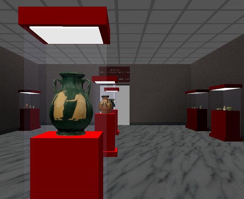
and not Alzheimer‟s disease (AD). Clinical cases that did not meet the “two cognitive
domains impaired” criteria required for an NINDS/ADRDA diagnosis of AD(McKhann et al.,
1984), were characterized by Petersen (R C Petersen et al., 1999) with the term “Mild
Cognitive Impairment”, a category in which these “single domain impaired” individuals were
assigned to. The term MCI however, lacked the precise definition and the clinical settings for
diagnosis. In the years that followed many criteria were debated and controversial discussions
were made regarding the clinical “image” of individuals with MCI. The end result was to
agree that MCI is somehow an intermediate stage between normal aging and
dementia(Gauthier et al., 2006)(R C Petersen, 2004)(Winblad et al., 2004).

1.1 MCI Subtypes

 Further research and meta-analysis, revealed the heterogeneity in the clinical description of
MCI leading to the classification of four subtypes. Based onthe number: single or multiple
and type: memory, non-memory or both, of impaired cognitive domains we have:

        1) Amnestic MCI – memory impairment only
        2) Multi-domain MCI-Amnestic (memory plus one or more non-memory domain)
        3) Multi-domain MCI-Non-Amnestic (more than one non-memory domain)
        4) Single Non-Memory MCI (one non-memory domain)(Winblad et al., 2004).

  Besides the four basic subgroups above, experts suggest that there are further cognitive,
functional and neuropsychiatric features that can distinguish individuals with MCI even
more(Hanfelt et al., 2011). This finding set the basis for improveddiagnosis because it
provided a common “language” among research centers forfuture research.However, a
common restriction has also been found. Approximately 16% of elderly subjects free of
dementia are affected by MCI with amnestic MCI being the most common type(R C Petersen
et al., 2010).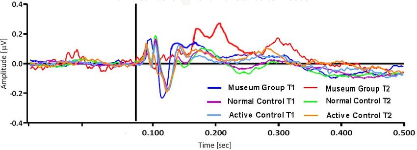
1.Complaint of cognitive deterioration from patient and/or
                                      informant

                        2.Objective deficit on neurocognitive testing

                                           YES                                     NO

                3. Persistent new disfunction in basic or instrumental       Probable Normal
                                        ADL

                                   NO                                              YES

                                  MCI                                           Probable
                                                                                Dementia

       Memory Affectd                                  Memory Non-Affected

       Amnestic MCI                                       Non-Amnestic
                                                              MCI

  Single           Multi-
 Domain            Domain                        Single Domain      Multi-Domain

                                        Image 2 MCI Subtypes

1.2 Diagnosis

 Recently, the criteria for the presence of MCI have been defined as (R C Petersen et al.,
1999; Ronald C Petersen et al., 2009):

        Subjective memory complaints, preferably validated by a third person
        Memory impairment, non-characteristic for given age and education level
        Preserved general cognitive function
        Intact activities of daily living
        Absence ofdementia.

  In addition to the cognitive deterioration, MCI may arise from vascular, neurogenerative,
traumatic, metabolic, psychiatric or other underlying medical disorders(Bennett, Schneider,
Bienias, Evans, & Wilson, 2005; Ronald C Petersen et al., 2009).In addition, impairment of
Activities of Daily Living (ADL) has been observed in MCI and therefore Instrumental ADL
( IADL) questionnaires or video assisted observation tools are more accurate as a diagnostic
marker(Gold, 2012; Perneczky et al., 2006).

 Of course, a thorough medical history of the patient is also taken into account. Low
education, generally impaired executive function, drug side-effects and general depression
may result in cognitive deficits, normally associated with MCI. A more precise clinical
evaluation should focus on the general psychosocial profile and it's impact on daily life(Patel
& Holland, 2012).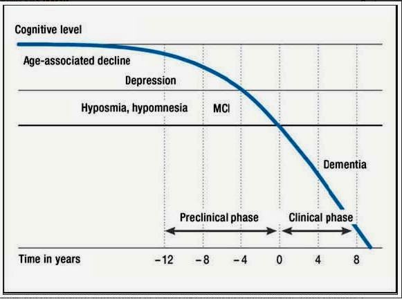
Baseline Education                                  Head Trauma
                Intellect                                           Sleep Disorders
                Learning Disabilities                               Substance Abuse
                Sensory Impairments                                 Polypharmacy
                Uncontrolled Pain                                   Medical     and     Psychosocial
                                                                    Illnesses   (Depression,Anxiety)

  In order to have a MCI diagnosis a variety of medical and neuropsychological examinations are required. A
thorough physical examination, blood sample studies, imaging (MRI,RiB-PET), genetic tests (APOE,
TREM2) as well as biomarkers in CSF (beta-amyloid, tau and phospho-tau protein)(Guerreiro et al., 2012).
Occasionally, a condition, such as vitamin B12 deficiency or thyroid disease, can be identified as a cause for
MCI(Patel & Holland, 2012).However, one general conclusion to be drawn is that none of the above
mentioned tools should be used alone, on the contrary the combination of different toolsresults in a more
precise diagnosis. Lastly, the most recent research findings showed that the pathophysiologic findings in
MCI may predict Alzheimer‟s Disease (and perhaps other diseases) and therefore the sooner the diagnosis
the more effective the intervention (Albert et al., 2011).

 1.3 Possible interventions

  Drug treatment of dementia and / or MCI is still a field of large on-going research.Instead promising results
have been shown by non-pharmacological treatments, such as increased physical activity that can be helpful
in the short term(Lautenschlager et al., 2008). Depressive mood and negative emotions is a risk factor and
better be addressed with psychological interventions. Some promising methods of cognitive rehabilitation
can compensate to some degree for deficits; e.g. computerized memory stimulations, memory games,
memory structuring techniques, personal digital assistants, cross word puzzles and mind games(Jean,
Bergeron, Thivierge, & Simard, 2010)(Tsolaki et al., 2011).

  It isimportant to note that MCI is not an one-way street to dementia. Several studies have shown that
patients with MCI have an annual rate of progression to dementia close to 5-15%(Farias, Mungas, Reed,
Harvey, & DeCarli, 2009; Ronald C Petersen, 2011). The usual annual percentage of dementia presence in
the general elderly population is 1-3%. In addition, some other studies show that a 15-40% of the MCI
patients, improve significantly and sometimes revert to a normal cognitive state(Larrieu et al., 2002; Ritchie,
Artero, & Touchon, 2001). For almost a decade, MCI is still a vast area for research. What's more, research
with the help of biomarkers shows that MCI is no longer a separate stage than AD. Recent scholars came to
the conclusion that mild cognitive impairment is a preclinical Alzheimer‟s disease. Early markers may
provide the necessary information for early diagnosis and also guide the intervention(Lazarczyk, Hof,
Bouras, & Giannakopoulos, 2012).

 Currently a global effort is under way in order to understand the nature of the problem by using the most
advancedtoolsin non– invasive intervention and computer based methods. One of the latest and
quitepromising efforts is the use of virtual environments for diagnostic and intervention purposes.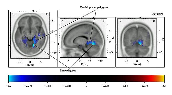
2. Virtual Environments and MCI

  Virtual Reality is a relatively new technology regarding its use for neuropsychological research.
Publications to date provide evidence of some cases where a virtual environment creates the desired
conditions and the necessary triggers for amnestic MCI patients to be classified and assessed. To be more
precise, applications of virtual reality in neuroscience can provide experiments in a controlled environment
where normal and impaired patient behavior, perception, control of movement, learning, memory and
emotional aspects can be observed(Rey & Alcañiz, 2010).VR creates interactive, multimodal sensory stimuli
that offer unique advantages over other approaches to neuroscientific research and applications. VR's
compatibility with imaging technologies such as functional MRI allows researchers to present multimodal
stimuli with a high degree of ecological validity and control while recording changes in brain activity.
Therapists, too, stand to gain from progress in VR technology, which provides a high degree of control over
the therapeutic experience.

  Normally, a real-time interaction is required in order to observe and analyze human reactions of any kind,
event or task. Otherwise, computer generated experimental tasks designed for specific variables and aspects
of human response are required. During the last decade and a half, research towards that directionprovided
information about the use of VR in Neuroscience(Cornwell, Johnson, Holroyd, Carver, & Grillon, 2008;
Harvey, Collman, Dombeck, & Tank, 2009; Plancher, Tirard, Gyselinck, Nicolas, & Piolino, 2012; Slater et
al., 2006; Waller & Richardson, 2008). For the purposes of this chapter we are going to analyze the way VR
can be used for evaluating spatial perception and memory aspects in individuals with MCI.

2.1 Spatial and visual memory

  Visual Memory is responsible for retaining visual shapes and colors whereas spatial memory is responsible
for information about locations and movement. It could be described as cognitive imaging and cognitive
mapping. This distinction is not always clear since part of visual memory involves spatial information and
vice versa(Klauer & Zhao, 2004). When it comes to MCI, impairment to both visual and spatial memory
could indicate memory deficits or may be by itself a sign of neurodegenerative disease(Mapstone,
Steffenella, & Duffy, 2003).

 Navigation can be argued that combines the two types of memory. Successful navigation requires a variety
of thoughts and actions; planning, selection of an appropriate strategy and possible alterations, prospective
memory and of course remembering previously visited locations. In particular, navigation seems to be
connected with the hippocampal function, a brain area already impaired in individuals with MCI(Lithfous,
Dufour, & Després, 2012). Thus, deficits on navigational skills and spatial memory could be a solid
cognitive indicator for MCI or early forms of Dementia.
Image 3 The neural network involved in spatial navigation(Lithfous et al., 2012)

2.2 Virtual Reality, Spatial Memory and MCI

  Immersive virtual reality environments can provide information and some times rehabilitate spatial
working memory(De Lillo & James, 2012). Ensuring that the desired conditions are ecologically valid, it is
possible to use VR as a tool to evaluate spatial memory in individuals with MCI by tracking their behavior
inside the virtual environment in real-time. As we discussed spatial memory can be impaired in amnestic
MCI. In one study, MCI participants were successively immersed in two virtual environments; the first, as
the driver of a virtual car (active exploration) and the second, as the passenger of that car (passive
exploration). Subjects were instructed to encode all elements of the environment as well as the associated
spatiotemporal contexts. Following each immersion, we assessed the patient's recall and recognition of
central information (i.e., the elements of the environment), contextual information (i.e., temporal, egocentric
and allocentric spatial information) and lastly, the quality of binding. The researchers found that the AD
patients' performances were inferior to that of the aMCI and even more to that of the healthy aged groups, in
line with the progression of hippocampal atrophy reported in the literature (Plancher et al, 2012). Spatial
allocentric memory assessments were found to be particularly useful for distinguishing aMCI patients from
healthy older adults. Active exploration yielded enhanced recall of central and allocentric spatial
information, as well as binding in all groups. This led aMCI patients to achieve better performance scores on
immediate temporal memory tasks. Finally, the patients' daily memory complaints were more highly
correlated with the performances on the virtual test than with their performances on the classical memory
test. Taken together, these results highlight specific cognitive differences found between these three
populations that may provide additional insight into the early diagnosis and rehabilitation of pathological
aging. In particular, neuropsychological studies would benefit to use virtual tests and a multi-component
approach to assess episodic memory, and encourage active encoding of information in patients suffering
from mild or severe age-related memory impairment. The beneficial effect of active encoding on episodic
memory in aMCI and early to moderate AD is discussed in the context of relatively preserved frontal and
motor brain functions implicated in self-referential effects and procedural abilities.
In another study, a virtual navigation based reorientation task (VReoT) was used (Caffo et al, 2012) and
again healthy subjects were compared with aMCI regarding their performance on the reorientation test. The
performance of the aMCI was significantly worse than the controls suggesting that VReoT detects spatial
memory deficits. A subsequent receiver-operating characteristics analysis showed a sensitivity of 80.4% and
a specificity of 94.3%.
3. The virtual reality museum
Virtual Reality (VR), Augmented Reality (AR) and Web3D technologies in conjunction with database
technology may facilitate the preservation, dissemination and presentation of cultural artifacts in museum‟s
collections and also educate the public in an innovative and attractive way. Virtual Reality signifies a
synthetic world, whereas Augmented Reality refers to computer generated 2D or 3D virtual worlds
superimposed on the real world. Web3D is used to represent the application of XML (eXtended Markup
Language) and VRML (Virtual Reality Markup Language) technologies to deliver interactive 3D virtual
objects in 3D virtual museums. Precedentsmade use of 3D multimedia tools in order to record, reconstruct
and visualize archaeological ruins using computer graphics and also provide interactive AR guides for the
visualization of cultural heritage sites (Liarokapiset al., 2010). These new emerging technologies are used
not only because of their popularity, but also because they provide an enhanced experience to the virtual
visitors. Additionally, these technologies offer an innovative, appealing and cost effective way of presenting
cultural information. Virtual museum exhibitions can present the digitized information, either in a museum
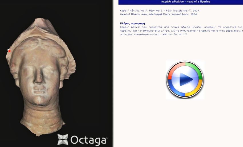
environment (e.g., in interactive kiosks), or through the World Wide Web.Image 4. Users can ‘walk’ freely in the virtual museum and interact with the artifacts. Once they select an
artifact they can choose to zoom in, rotate it at the X,Y,Z axis and read details in tags on the artifact itself
Our Virtual Museum system has been developed in XML and VRML and is described in detail(Tsatali et
al, 2012). The system allows museum curators to build, manage, archive and present virtual exhibitions
based on 3D models of artifacts. The innovation of our system is that it allows end-users to explore virtual
exhibitions implemented using very simple everyday interfaces (e.g. joystick, mouse) (Im.4).
The cultural artifacts are digitized by means of a custom built stereo photogrammetry system (Object
Modeler), mainly for digitizing small and medium-size objects and a custom modeling framework
(Interactive Model Refinement and Rendering tool) in order to refine the digitized artifact. The 3D models
are accompanied by images, texts, metadata information, sounds and movies (Im. 5). These virtual
reconstructions (3D models and accompanying data sets) are represented as eXtensible Markup Language
(XML) based data to allow interoperable exchange between the museum and external heritage systems.Image 5. The interface is ergonomically made so that it can tolerate errors. All icons, fonts and interactive
objects are large and understandable. The text is in Greek and reduced in size at this picture on intention.(?)
These virtual reconstructions are stored in a MySQL database system and managed through the use of a
specially designed Content Management Application, which also allows building and publishing virtual
exhibitions on the Internet or in a museum kiosk system. The system is a complete tool that enables
archiving of both content and context of museum objects. The described interactive techniques can transform
the museum visitors „from passive viewers and readers into active actors and players‟ (ibid).
3.1The Virtual Museum technical components
Two main components of the system are of interest for the evaluation: the Content Management
Application (CMA) and Augmented Reality Interface (ARIF). CMA allows publishing of virtual museums to
both Web (Im.4) and a specially designed application (ARIF) for switching between the Web and an AR
system (Im.5). The CMA application is implemented in Java and it includes the database of the
representations of cultural objects and their associated media objects, such as images, 3D models, texts,
movies, sounds and relevant metadata. It enables user-friendly management of different types of data stored
in the Virtual Museum database, through various managers, such as the Cultural Object Manager (deals with
virtual representations of cultural artifacts), the Presentation Manager (manages virtual exhibitions with the
help of templates) and the Template Manager (stores these visualization templates).
The ARIF component is a presentation or visualization framework that consists of three main sub-
components:
The ARIF Exhibition Server. Data stored in the Database is visualized on user interfaces via the
ARIF Exhibition Server.
The ARIF Presentation Domains with implemented web browser functionality, suited for web-based
presentations.The ARIF AR – Augmented reality functionality. This sub-component provides an AR based virtual
museum exhibition experience on a touch screen in the museum environment using table-top AR
learning experiences, e.g., AR quizzes and on-line museum exhibitions.
3.2 The Virtual Museum cognitive theory
According to the cognitive-enrichment hypothesis developed by Hertzog et al., (2009), the trajectory of
cognitive development across the life span is not fixed. Although the trajectory of cognitive development at
normal seniors is largely determined by a lifetime of experiences and environmental influences, there is
potential for discontinuity in the trajectory given a change in cognition-enriching behaviors. The cognitive-
enrichment hypothesis is corroborated by ample evidence for plasticity, i.e., the potential for improvement of
ability as a consequence of training (Denney, 1984) of everyday cognitive task-switching in the elderly
population. There are some improvements of updating (Baron and Mattila, 1989; Buschkuehl et al.,
2008; Dahlin et al., 2008), as well as shifting (Sammer et al., 2006; Bherer et al., 2008) and inhibition
(Davidson et al., 2003; Karbach and Kray, 2009) in the population of older adults, which have been reported.
In addition, domains such as selective attention (Ball et al., 2007) and inductive reasoning (Schmiedek et al.,
2010) can be improved in older adults.
We now know that the virtue of a cognitive-training technique depends on the generalization or transfer of
training to untrained tasks (Klingberg, 2010). Different degrees of transfer have to be distinguished. The
minimal degree of transfer that can occur, is improvement within the same cognitive domain as subjected to
training, assessed using different stimuli, and requiring a different response than the training task. This type
of transfer is referred to as near transfer. Improvement of abilities in other cognitive domains than the
cognitive domain subjected to training is referred to as far transfer.
Virtual reality museum exhibition and educational activities are considered to provide an ideal context for
cognitive enrichment (Achtman et al., 2008; Green and Bavelier, 2008). The unique characteristics of virtual
museums presumed to facilitate transfer are their motivating nature, frequent presentation of feedback,
precise reinforcement schedules, and stimulus variability (Gee, 2007). As a result of their entertainment
value, virtual museums maintain the motivation to engage in practice for much longer than monotonous
laboratory tasks or traditional training programs. Frequent feedback supports motivation and is also
important for conditioning the desired level of performance. When the difficulty level of the task is
continuously adapted to the performance, players will constantly be challenged at the limits of their ability. It
is in particular the phase of skill-acquisition that calls for cognitive control (CC), whereas continued
performance at a mastered level is associated with automatization and release of CC resources (e.g., Shiffrin
and Schneider, 1977; Logan, 1988). Furthermore, small increments of difficulty level maximize the
proportion of successful experiences with the task. The stimulus variability also plays an important role in
training CC, because it helps to generalize learnt cognitive skills to multiple stimulus contexts.
Transfer of virtual reality museum interventions to CC has, however, not been demonstrated
consistently. Owen et al. (2010), for instance, demonstrated that playing computerized cognitive training
games like Nintendo's® Dr. Kawashima's Brain Training™ was not more beneficial for CC functions than
answering general knowledge questions online. It is being assumed that because the sample of participants in
Owen et al.'s study was very heterogeneous and included both young and old adults, it is well possible that
improvements of cognitive test performance were attenuated in young adults due to ceiling performance at
pretest. This could have obscured possible transfer of training in the sub-sample of older adults. The notion
that sample heterogeneity can confound the observed effect of virtual reality training substantially is
corroborated by Feng et al. (2007). They found no effect of playing action virtual reality games on spatialattention in a sample of young adults. However, separate analysis of the effect in males and females revealed that females did actually benefit from playing. In addition, at the study Owen et al. the participant sample was very heterogeneous with respect to training adherence, so participants who completed only two training sessions could have had a negative impact on aggregated training outcomes. Another aspect of Owen et al.'s study that makes the observed absence of transfer difficult to interpret is that transfer was assessed using a test battery comprising only four cognitive tests, three of which were measures of working memory capacity. Ackerman et al. (2010) demonstrated that sample heterogeneity cannot account for Owen et al.'s (2010) findings. They found that playing cognitive training games (Nintendo® Wii™ Big Brain Academy™) does not benefit cognitive abilities to a greater extent than reading assignments do, in a homogeneous sample of healthy seniors on a relatively fixed and extensive training schedule. Moreover, a broader assessment of cognitive abilities of interest was made than in Owen et al.'s study. Still, Ackerman et al. focused predominantly on reasoning ability and perceptual processing speed, while a large share of the cognitive games under study taxed working memory updating and the large variety of the tasks probably stimulated participants' attention and task set shifting. Inclusion of transfer tasks gauging working memory updating and set shifting in Ackerman et al.'s study could have led to different conclusions regarding transfer of playing cognitive training games. Conversely, there is also some evidence against Owen et al.'s (2010) and Ackerman et al.'s (2010) pessimistic conclusions regarding the beneficial effects of playing virtual reality educational games on CC functions. Namely, Peretz et al. (2011) found a larger improvement of visuospatial working memory, visuospatial learning, and focused attention after playing Cognifit Personal Coach® cognitive training games than after playing conventional 3D videogames that were matched for intensity, in a sample of older adults. Even though there is some theoretical overlap in the cognitive functions assessed by Peretz et al. and Owen et al. and Ackerman et al., the specific cognitive tests used to assess transfer in these studies was different. It is conceivable that some cognitive tests are more sensitive to transfer effects than others, which might explain the discrepant results of these studies. Furthermore, playing 3D videogames not specifically designed for cognitive training can also improve CC functions in older adults. Basak et al. (2008) demonstrated that playing a particular complex 3-D real-time strategy game (Rise of Nations) was associated with greater improvements of shifting, updating, and inductive reasoning than observed in the control condition. It must be noted that the control group in this study was a no-contact control group, so it is not certain to what extent the observed improvements in the videogame group are attributable to placebo-effects. Nevertheless, the improvements of CC in this study were larger than practice effects due to repeated exposure to the same cognitive test. It has been argued that failures to demonstrate far transfer of playing cognitive training games in the population of older adults may be due to a general age-related decrease of the extent to which learning transfers to untrained abilities (Ackerman et al., 2010). This assertion is supported by Ball et al.'s (2002) finding that cognitive strategy training programs for improving memory, processing speed and reasoning, respectively, were associated with improvements within the trained cognitive domain but not with far transfer to untrained cognitive abilities of older adults. In contrast, however, far transfer of practicing basic cognitive tests has been reported repeatedly in the cognitive aging literature (Mahncke et al., 2006; Uchida and Kawashima, 2008; Karbach and Kray, 2009; Smith et al., 2009). Brain training games like Nintendo's® Dr. Kawashima's Brain Training™ share many task components of basic cognitive laboratory tasks and videogames have several additional characteristics facilitating transfer (Green and Bavelier, 2008). Therefore, it is reasonable to expect that transfer of computerized cognitive training games in the population of older adults is replicable.
3.3 TheVirtualMuseumcognitiveexercises

  It is difficult to reconcile inconsistent findings pertaining to the effect of playing cognitive training games
on cognition (Ackerman et al., 2010; Owen et al., 2010; Peretz et al., 2011), because the methodological
differences between these studies are substantial. More research is required to elucidate what aspects of brain
training games facilitate transfer to untrained cognitive abilities. Hence, the aim of our virtual museum was
to test whether playing some simple memory exercises inside an ecologically valid 3D environment does
transfer to different measures of CC in mild-cognitive impairment of the amnestic type (aMCI) older adults.

  The virtual reality museum is designed to speed up auditory processing, improve working memory,
improve the accuracy and the speed with which the brain processes speech information and reengage the
neuromodulatory systems that gate learning and memory. To reverse cognitive disuse and drive brain
plasticity, the program strongly engages the brain with demanding exercises and an adaptive and reward-
based daily training schedule. Cognitive exercises provided by it are divided into three interrelated
categories, that, in aggregate, span the cognitive functions of seniors, consistent with the recommendations
of (Tucker-Drob, 2011):

           Listen & Plan: Seniors follow instructions to locate and find items in an order. Instructions
           become more difficult (phonetically and syntactically) progressively (purpose: training on
           visuospatial abilities and planning following complex instructions with continuous processed
           speech).
           Storyteller: Seniors hear segments of museum items stories and are asked to answer a set of
           questions concerning the details of the respective segment (purpose: training on story
           comprehension and memory)
           Exer-gaming: Seniors are asked to actually represent the "scene" depicted at the archeological
           artifacts or multimedia description, e.g. movement, dance, wedding (purpose: training on
           executive function and orientation/praxis).

  This type of intervention was used in the recent study by Smith and colleagues (2009) which was the first
double-blind large-scale clinical trial that demonstrated marked improvement not only in the trained task, but
also in several generalized measures of memory and perception of cognitive performance in everyday life,
relative to an active control group that received a frequency and intensity-matched cognitive stimulation
program.

  Transfer was assessed by comparing performance on a battery of cognitive tests before and after the
intervention. Taking into account that some cognitive tests may be more sensitive to transfer effects than
others, several measures of updating, shifting, and inhibition were included in the test battery. Although it is
assumed that training interventions boost functional or even plastic changes to the brain, neuronal correlates
of the training induced changes in intervention studies were only examined in the last decade (Mozolic et al,
2010). Knowledge about the intervention related neuronal and functional changes is additionally useful in
order to understand the efficiency of the training and transfer effects to other tasks (Lustig et al, 2009).
Therefore, in the present study we used event-related brain potentials (ERPs) derived from the
electroencephalogram (EEG) in order to study more closely the neuronal processes which are affected by the
training intervention.
4. Research Methodology
4.1 Design
Single-site randomized controlled double-blind trial.
4.2 Participants
One hundred and fourteen patients with MCI according to the revised Petersen criteria (Petersen, 2006),
aged between 65 and 88 years, were recruited to participate in the experimental study which was conducted
in Alzheimer Hellas day clinic Agios Ioannis at Thessaloniki, Greece between May 2011 and October 2012.
The participants were randomly assigned to the training groups. We excluded subjects who met criteria for
dementia (DSM-IV), AD (NINCDS-ADRDA), depressive episode (IDC-10), subjects with significative
cerebrovascular disease (Hachinski scale score ≥4), and those with any other medical or psychiatric
identifiable cause accounting for their complaints.
The neuropsychological battery used for the pre- and post- testing included tests for the assessment of
memory (Rey Auditory Verbal Learning Test - RAVLT), language and semantic memory (15-items short-
form of the Boston Naming Test, category fluency), praxis and visuospatial skills (Rey complex figure
copy), attention and executive function (Symbol Digit Modalities Test, Trail Making part A and B, Stroop
interference Test and letter fluency). A cognitive domain was judged as impaired when subjects scored 1.5
SD below values for age and education matched controls in at least one test. According to the results of the
neuropsychological exploration, subjects were classified as pure amnestic MCI (a-MCI), patients fulfilling
Petersen's criteria for amnestic MCI, with memory being the only affected domain. (see Table 1 for details).
Group Cognitive training Active control Non-contact control
Mean age 70.5 years (4.3) 69.7 years (4.5) 70.9 (4.4) F(2, 102) = 1, P = .36
MMSE score 26.8 (3.6) 26.2 (3.6) 26.2 (3.1) F(2, 102) = 1.4, P = .24
Stroop-test (color repetition) 73.4 (34.24) 74.4(32.2) 70.6 (23.40)
RAVLT-immediate recall 15.4(4.3) 15.5 (4.6) 15.0(3.1)
RAVLT-delayed recall 1.6(1.5) 1.7 (1.5) 2.2(1.5)
RAVLT-recognition 5.6(2.2) 5.5 (2.2) 7.4(1.9)
BNT 10.42 (2.46) 10.60 (1.91) 11.22 (1.90)
Category fluency 10.6 (3.98) 11.3 (3.1) 11.2(4.3)
Letter fluency 7.4 (3.54) 7.1 (2.6) 6.0(3.4)
Ray figure copy 34.6(1.3) 32.7 (1.9) 28.9(8.5)
Ray figure immediate recall 11.9(9.2) 11.4 (9.2) 7.0(4.7)
Ray figure delayed-recall 11.6(9.4) 10.6(9.1) 7.2(4.6)
Ray figure recognition 6.6(2.9) 6.3 (2.5) 6.0(1.4)
Forward digit repetition 6.2(1.1) 6.1 (1.1) 5.8(1.1)
Backward digit repetition 3.8(0.8) 3.9 (0.7) 2.2(1.3)
Trail-Making Test B 193.9 sec (98.5) 179.0(83.7) 188.8 sec (55.1)
GDS 10.3 +/- 2.5 11.3 +/-3.1 13.3 +/- 2.5
Table 1.Demographic characteristics and cognitive status of the participant groups. Standard deviations are given in
parentheses behind the mean values. There were no significant group differences as is indicated by the statistical analysis
(last column).Image 6. Illustration of a possible Auditory ERP signal. On the X-axis the time is shown with 0 at the stimuli.
The Y-axis is the amplitude with 0 at the baseline. In the pre-stimuli window a baseline is visible from which a
horizontal average can be calculated
Participants also received an Auditory ERP-recording completed using a Nihon Kohden - Neuropack M1
MEB-9200 evoked potential/EMG measuring system. Event-related-potentials (ERPs) are being used as a
noninvasive clinical marker for brain function in human patients. Auditory ERPs are voltage changes
specified to a physical or mental occurrence that can be recorded by EEG (Papaliagkas et al, 2008). Different
ERPs were used in order to pinpoint the functional processes which would be improved by the cognitive
process training and which may be affected by retesting. The principal ERP components elicited after task-
relevant visual stimuli are among others the N1, the anterior N2, the P2, and the P3b. In Image 6, an
example of an Auditory ERP signal can be seen. The signal can be divided into two parts, a pre-stimuli
section consisting of a baseline with no clear potentials and a post-stimuli section consisting of various
potentials. The first positive potential is called P1, followed by a negative potential N1, then P2, N2, and so
forth. The latency of these potentials is measured from onset of stimuli to the peak of the potential.
Sometimes the peaks are named using the latency, e.g. if N1 occur at a latency of 40ms it is named N40 or if
P3 occur at a latency of 300ms it is named P300. The baseline amplitude is the difference between the peak
of a potential and the mean of the pre-stimulus baseline. The baseline measurement used to discriminate
between the MCI amnestic patients and the controls in our study is shown in Image 7 (Kimiskidis et al,
2012).
Image 7. Grand average baseline AERP waveforms for MCI amnestic patients at our study and comparison
to baseline for normal / controls.4.3 Procedures Thirty nine of the participants represented a virtual reality museum cognitive training group – experimental group (remaining N=32; 12 men, mean age: 70.5 years; range 65 to 82; seven drop-outs because of technical problems, illness, and tenancy changeover). The other participants formed an active control group (N=39; 16 men, mean age: 69.7 years; range: 65 to 88; no drop-outs) and a non-contact control group (remaining N=34; 13 men, mean age: 70.9 years; range: 65 to 87; two dropouts because of illness). The virtual reality museum cognitive training group was exposed to a multilayered cognitive training over a period of 5 month. At the same time, the active control group is a sample of the MCI amnestic population from the Agios Ioannis day clinic that received a learning-based memory training approach in which participants used computers to make cognitive exercises, viewed DVD-based educational programs on history, art and literature or participated at puzzle solving exercises. The active control group was required to have high face validity and match the experimental group for daily and total training time, interesting audiovisual content, and computer use. Thus the AC cognitive training program employed a learning-based memory training approach in which participants used computers to view DVD-based educational programs on history, art and literature. The participants in the virtual reality museum cognitive training and the active control group trained twice a week for 90 minutes across 5 months. The virtual reality museum cognitive training was conducted on an one-to-one basis while the active control trainings were conducted in small groups with not more than 12 participants by payed professional psychologists. Two extra sessions were offered at the end of the program for those participants who missed the regular sessions. The participants were not encouraged to train outside the training sessions. 4.4 Data recording and Analysis 4.4.1 Electrophysiologicalrecording The Electroencephalogram (EEG) was recorded from 32 active electrodes positioned according to the extended 10–20 system (the electrodes mounted directly on the scalp included the following positions: C3, C4, CP3, CP4, CPz, Cz, F3, F4, F7, F8, FC3, FC4, FCz, Fp1, Fp2, Fpz, Fz, O1, O2, Oz, P3, P4, P7, P8, PO3, PO4, POz, Pz, T7, and T8.). Electrodes A1 and A2 were placed at the left and right earlobes. The horizontal and vertical EOG was measured by electrodes placed at the outer canthi (LO1, LO2) and above and below both eyes (SO1, SO2, IO1, IO2). Electrode impedance was kept below 10 kOhm. The amplifier bandpass was 0.01–140 Hz. EEG and EOG were sampled continuously with a rate of 2048 Hz. Data were saved on a hard disc alongside with triggers marking significant events. Offline, the EEG was down-scaled to a sampling rate of 500 Hz and cut in stimulus locked epochs by using the software Neuroworkbench (Nihon-Kohden, Japan). The epochs were 1200 ms long ranging from 100 ms before and 1000 ms after stimulus onset. All epochs with EEG amplitudes of more than ±120 μV or with drifts of more than 150 μV within 300 ms were discarded. For all participants and conditions at mean 48 epochs (Min = 17; Max = 53; SD = 7.3) of the epochs remained for averaging after artefact rejection and correction. The epochs were averaged according to the stimulus conditions (target trials versus non-target trials) and referenced to linked earlobes (excluding the EOG electrodes). For stimulus locked averages only
correct epochs were used, excluding trials with false alarms or misses. A digital low-pass filter was set at
17 Hz.

4.4.2   Analysis

  Statistical analysis were performed by means of repeated measures ANOVAs with Greenhouse-Geisser
corrected degrees of freedom. In case of significant main effects (if the factor included more than two levels)
or interactions additional ANOVAs were applied for post hoc testing of contrasts and simple effects. For
response times (RTs; correct commission trials) the ANOVA included the within factor time(session one,
session two) and the between factor group (virtual reality museum cognitive training group, active control
group, no-contact control group). Separate ANOVAs were carried out for false alarms and for misses,
because they are different types of errors either demanding a response or not. Both analysis included the
factors time and group.

  The peak amplitude and latency of the N1 potential was measured at the two occipital electrodes O1 and
O2 were the potential showed its maximum. The N2 was quantified as the mean amplitude in the time
interval between 240 to 300 ms at the electrodes FCz, Cz and CPz were it showed the maximum amplitude.
A reliable measurement of the peak was not possible due to the overlapping P2, and P3b potentials. The P2
potential was quantified in amplitude and latency as the local maximum at the electrodes FCz, Cz and CPz in
the search interval between 200 and 400 ms where it showed the highest peaks. The peak amplitude and
latency of the P3b potential was measured as the local maximum at the electrodes Cz, CPz and Pz in the
search interval between 400 and 700 ms where it showed the highest amplitudes.

  Six separate ANOVAs were carried out for the peak amplitudes and latencies of the N1, P2 and the P3b,
respectively, including the between subject factor group and the within subject factors session (session one,
session two), stimulus type (target, nontarget) and electrodes (O1 and O2 for the N1; FCz, Cz, and CPz for
the P2 potential; Cz, CPz, and Pz for the P3b potential, resp.). An additional ANOVA was carried out for the
N2 mean amplitudes including the between subject factor group and the within subject
factors session, stimulus type, and electrodes (FCz, Cz, and CPz).

  We also used sLORETA (Pascual-Marqui, 2002) in order to closer examine the underlying neuronal
changes of the expected training effect of stimulus feature processing as reflected by the P2. We examined
only the target condition because the training gains may especially help to improve target detection. The
program sLORETA estimates the sources of activation on the basis of standardised current density at each of
6239 voxels in the grey matter of the MNI-reference brain with a spatial resolution of 5 mm. The calculation
is based upon a linear weighted sum of the scalp electric potentials with the assumption that neighbouring
voxels have a maximal similar electrical activity. The voxel-based sLORETA images were first computed
for each individual averaged ERP in the target condition in the interval from 170 to 190 ms surrounding the
P2 peak. Then, the differences of the sLORETA images between test sessions were statistically compared
between groups using the sLORETAvoxelwiserandomisation test (5000 permutations) which is based on
statistical nonparametric mapping (SnPM) and implemented in sLORETA. Two independent group tests
were carried out for comparison of the three groups (cognitive training group versus no-contact control
groups, and versus social control group). The tests were performed for an average of all time frames in the
interval with the null hypothesis that (T1groupA − T2groupA) = (T1groupB − T2groupB). The tests were
corrected for multiple comparisons (Holmes et al, 1996).
5. Results

5.1 Neuropsychological variables outcome

  In the virtual reality museum and active control aMCI group, there were significant differences between the
delayed-recall scores on the RAVLT at baseline and those at both the 5-month follow-up (1.6±1.5 vs.
4.4±1.5, p=0.04; 1.6±1.5 vs. 4.6±2.3, p=0.04) (Table 2). The immediate recall scores on the Rey Osterrieth
Complex Figure (11.9±9.2 vs. 15.8±9.4; p=0.04), the Trail-Making B (193.9±98.5 vs 104.1±28.7; p=0.04)
and the MMSE (26.8± 3.6 vs. 28.2±2.5; p=0.04) were significantly improved only at the 5-month follow-up
in the virtual reality museum aMCI group. There was a tendency toward improvement of the digit span
forward scores (6.2±1.1 vs. 7.8±1.3; p=0.07) at the follow-up of the virtual reality museum aMCI group and
a general training-induced BNT scores improvement (10.6±1.9 vs. 12.0±2.0; p=0.07) compared to the
baseline scores in the virtual reality and the active control aMCI group (Table 2). The GDS score was also
improved after cognitive training, but the difference did not reach statistical significance (10.3±2.5 vs.
8.9±1.7; p=0.23). There were no significant differences between the baseline and follow-up scores in other
outcome measures in the MCI wait-list control group.

                             Virtual Museum    Active Control aMCI
                             aMCI group        group                                            NormalControlaMCI group

                           Baseline              Follow-up           BaselineFollow-up      Baseline    After 20 weeks

  RAVLT, immediaterecall     15.4±4.3         16.6±5.1         15.5±4.6   15.6±4.1         15.0±3.1        12.8±5.9

  RAVLT, delayedrecall        1.6±1.5          4.4±1.5*         1.7±1.5   4.6±2.3*          2.2±1.5         2.4±2.6

  RAVLT, recognition          5.6±2.2          7.0±1.9          5.5±2.2    6.4±2.3          7.4±1.9         7.4±0.9

  ROCF copy                  34.6±1.3         36.0±0.0         32.7±1.9   34.2±1.6         28.9±8.5        26.2±8.8

  ROCF, immediaterecall      11.9±9.2         16.8±9.4*        11.4±9.2   11.0±3.4          7.0±4.7         9.3±5.4

  ROCF, delayedrecall        11.6±9.4         16.3±8.9         10.6±9.1   15.4±8.1          7.2±4.6         9.6±5.6

  ROCF, recognition           6.6±2.9          7.0±2.8          6.3±2.5    7.4±2.5          6.0±1.4         5.0±0.7

                                                        †
  Digitspanforward            6.2±1.1         7.8±1.3           6.1±1.1    7.2±1.1          5.8±1.1         6.4±1.5

  Digitspanbackward           3.8±0.8          4.0±1.6          3.9±0.7    3.6±0.9          2.2±1.3         2.6±0.5

  Stroop, colorreading     73.4±35.2          86.6±26.8       74.4±32.2 80.2±23.3          70.6±23.4       59.8±39.9

  Categoryfluency            10.6±2.2         13.4±5.7         11.3±3.1   13.2±4.4         11.2±4.3        11.6±4.8

                                                        †
  Letterfluency               7.4±3.6         8.6±4.8           7.1±2.6    7.6±2.9          6.0±3.4         6.0±5.0

  TRAIL-B                  193.9±98.5     104.10±28.7 *      179.0±83.7    210.0±62.6    188.8±55.1      228.8±75.0
† †
BNT score 10.4±1.9 15.4±2.4 10.6±1.9 12.0±2.0 11.2±1.9 10.0±2.2
MMSE score 26.8±3.6 28.2±2.5* 26.2±3.6 27.0±2.6 26.2±3.1 24.6±4.6
GDS score 10.3 +/-2.5 8.9 +/-1.7 11.3 +/-3.1 9.9 +/-2.7 13.3 +/-2.5 14.9 +/-2.2
†
*ptarget. Our findings however go further, because we used for a first time a 3D Virtual Museum environment and showed the neuronal correlates of the functional processes which likely were improved by the training: In the Virtual Museum cognitive training group the occipital N1 was enhanced after versus before the training for non-target stimuli. This suggests that the participants developed mechanisms for enhanced attention of arrays which were not immediately recognized as targets, that is, the non-targets (Im. 8). The frontal N2 to non-targets was also increased in amplitude for the cognitive training group after training. However, as this effect failed to reach significance, it can only be speculated that also the subsequent processing or even inhibition of the non-target stimuli improved after cognitive training. Based on the enhanced attention in non-target trials in the Virtual Museum cognitive training group as was reflected in the N1 amplitude, one may expect also a decrease in the false alarm rate (Im. 8). The N2 (see Image 8) showed a maximum at the electrodes FCz (1.2 μV) and Cz (1.4 μV) and was less negative at CPz (2,1 μV; main effect of f electrodes: F(2,204) = 30.7, P< .001). The tree-way-interaction of the factors session x stimulus type x group reached also a significance (F(2,102) = 3.01; P< .003). Image 8. Stimulus-locked event-related potentials at the occipital electrodes O1 and O2 separately for target and non-target trials, for the first (T1) and the second test session (T2) as well as for the Virtual Museum cognitive training group, the Active Control group and the normal control group. The increased amplitude of the P300 in target trials may suggest that feature based stimulus processing was significantly improved in our older participants after only the Virtual Museum cognitive training (Im. 9). Consequently, the improved discrimination of stimulus features in target-present trials should decrease the likelihood of missed targets and increase the likelihood of target detection. This effect on performance data was evident in our cognitive training group after the training compared to the pre-training session and also when compared to the control groups.
Image 9. Changes in P300 amplitudes in the Museum Group (Pz average) compared to the other groups pre- (T1) and post-
  (T2) training.

   The sLORETA analysis of the P300 amplitude differences between test sessions elucidates the neuronal
 basis of the training gain. Specifically, activation in the lingual and parahippocampalgyri was increased only
 in the cognitive training group and not in the two control groups. Most importantly, the increased P300
 amplitude together with the significant changes in brain activation show that the cognitive training caused a
 change in brain processes on a functional level in a near transfer task of visual search. Both regions are
 anatomically and functionally connected (Cant et al, 2007) and are discussed as being sensitive for global
 visual feature processing (Mechelh et al, 2000), as well as the global processing of spatial layout (Epstein,
 2008) and surface properties like color and texture of scenes and objects in visual arrays (Cant et al, 2007).
 For our training group this may mean that the cognitive process training improved the textual and spatial
 processing of visual arrays in general. Possibly, the use various kinds of visual material like pictures, objects,
 and text pages which were used in various tasks in the training sessions did improve one basic cognitive
 process of global processing of visual arrays. The present results also suggest the P300 potential of the ERP
 as a possible marker for the improvement of this cognitive process (Im. 10).

Image 10. Graphical representation of the sLORETA results comparing the differences of the target-P300. The blue colour
indicates local maxima of lower activation in the first compared to the second test session for the cognitive training group in
the right lingual and parahippocampal gyri, which may explain the amplitude difference of the P300 between sessions in the
tested interval surrounding the P300 peak.
5.3 Discussion

  In the present study we were able to distinguish the functional processes which were sensitive to the
training intervention from retest effects. In fact, the effect of test sessions on the topography of the P300
applies to all groups. We assume that the P300 may reflect memory-based stimulus processing. Thus,
whereas attentional processing of target-absent trials (N1 results) and feature-based stimulus processing of
target-present trials (P3 results) were only modulated by the cognitive training intervention, the improvement
of stimulus categorization, which is based on memory representations (P300), was sensitive to retesting. In
our study, the amplitude of the P300 component increased and latency shortened significantly following
training in both experimental groups. This adds to the evidence that the P3b contains a component related to
response selection or execution (Falkenstein, 1994a; 1994b). The idea of a functional compromise associated
with MCI is not new, and previous studies have reported a higher degree of functional impairment in MCI
subjects when compared with matched healthy subjects (Tam et al., 2007; Pereira et al., 2008; Ahn et al.,
2009; Burton et al., 2009; Schmitter-Edgecombe et al., 2009; Aretouli and Brandt, 2010; Bangen et al., 2010;
Teng et al., 2010a; 2010b).

  To a limited extent, the present findings support Basak et al.'s finding that inhibition can be improved by
playing videogames and Schmiedek et al.'s (2010) demonstration that functional impairment can be
improved by practicing basic cognitive tasks. The results from the present study suggest that modest
improvements of the functional ability, processing speed and memory can also be achieved by means of
playing virtual reality cognitive training games. A similar partially positive result of 3D games for cognition-
enriching everyday activities and processing speed was reported by Nouchi et al. (2012).

  Not all 3D virtual reality environments however are created equal (Achtman et al., 2008) and given an
individual's stage of cognitive development, one environment can be more beneficial for cognitive functions
than the other. For example, the cognitive training games used in the Virtual Reality Museum were very
similar to those used in an actual educational museum visit (Ball et al., 2002). Preliminary evidence for far
transfer of the cognitive training was found in the present study using the neural correlates. The different
extent of transfer in our study may be explained by the additional focus of the aMCI group to use specific
strategies to perform the training tasks.

  All our data support the a priori intuitive notion that highly cognitive-dependent skills are more likely to be
affected as a consequence of the virtual reality museum cognitive training, and that aMCI subjects show
significant improvement in these functional domains. On the other hand, it is noteworthy that differences
between groups were not restricted to the neuropsychological variables or the neural correlates, but also to
behavioral areas as well, such as depression and motivation, although this change was not significant. As
suggested by Green and Bavelier (2008), motivation is a key condition for transfer to occur. The engaging
nature of the virtual reality museum used in the present study could thus have facilitated transfer of training.
It is clear that more research in this direction is required. Nevertheless, it can be concluded that our findings
support the notion of plasticity in the neural system underlying virtual reality cognitive training and point to
a relationship between the more ecological validity of virtual reality and enhancement of specific cognitive
skills.
6. Conclusions

  The results from our study suggest that older adults do not need to be technologically savvy to benefit from
virtual reality training. Almost none of the aMCI participants in the reviewed studies had prior experience
with the technologies (i.e., video games, computers) used in the intervention study and yet they were still
able to benefit from these novel approaches. Previous research has shown participants‟ prior use of
computers was not significantly associated with acquisition of computer skills during training sessions,
suggesting older adults can benefit from novel technologies (Saczynski et al, 2004).

  Despite common misconceptions older adults do not enjoy learning to use new technology, perceptions of
the computerized training programs were positive for the older adults who completed computerized training
(Lee et al, 2011). In spite of many older adults reporting anxiety about using unfamiliar technology at the
beginning of training, most reported high levels of satisfaction after training was completed. Some patients
also stated they could use their new video game skills to connect more with their grandchildren, like we have
seen many times in the literature (e.g. Torres et al, 2008); whereas others were very willing to learn to use
video games and believed they could be a positive form of mental exercise (Belchior, 2008).

  In conclusion, the present study lends modest support to the notion that playing virtual reality cognitive
training games improves untrained cognitive functions in aMCI. Since these functions facilitate adaptive
behavior in various contexts, improved cognitive processing can be expected to help older adults to
overcome cognitive challenges in their daily routines. Virtual Reality provide an entertaining and thus
motivating tool for improving cognitive and executive functions and have other practical advantages as well.
The Virtual Reality Museum doesn't require physical well-being and mobility of the participant as much as
physical exercise interventions, although these seem to be more effective in buffering decline of executive
function (cf.Colcombe and Kramer, 2003). Additionally, the virtual reality museum is not expensive to
administer as compared to interventions supervised by a therapist. Virtual Reality comes in forms far more
complex than cognitive tests usually studied by cognitive psychologists. The present study suggests that the
virtual reality museum should not be dismissed as a cognitive training tool, but that we are just beginning to
understand how playing 3D videogames influences cognitive functions.

  Even within the homogeneous sample of older adults that participated in the present study, some
participants benefited more from playing the virtual reality museum than others. A variety of factors may be
responsible for individual differences in sensitivity to cognitive training. For instance, recent findings from
our lab indicate that inter-individual genetic variability modulates transfer of training to untrained tasks
(Colzato et al., 2011). Therefore, caution concerning the interpolation of aggregate data to individuals is
advised, and individual differences in cognitive training outcomes are an important topic to be addressed in
future studies.

  The artwork of the virtual reality museum we presented here was maybe not nearly as advanced and
capturing as commercial off-the-shelf games, and that applies to most studies of game training. Conversely,
commercial enhancement games are only seldom designed on the basis of cognitive insights, nor tested for
their effectiveness. Given that the creative industry and academic research are only just starting to inspire
each other's work, these first modest demonstrations of cognitive enhancement by games may only be
scratching the surface of its full potential.

 It is important to note that inconsistencies may be due to several factors not related to the actual training
program itself, including different cognitive outcome measures and modifications of the training program.
The electrophysiological data helped to elucidate the functional processes which were sensitive to the
You can also read