Assessment of Vestibulotoxicity of Henna Leaf (Lawsomnia Inermis) in A Rat Animal Model
←
→
Page content transcription
If your browser does not render page correctly, please read the page content below
Posted on 31 Mar 2023 — The copyright holder is the author/funder. All rights reserved. No reuse without permission. — https://doi.org/10.22541/au.168028569.95590410/v1 — This a preprint and has not been peer reviewed. Data may be preliminary.
Assessment of Vestibulotoxicity of Henna Leaf (Lawsomnia
Inermis) in A Rat Animal Model
Abdul Azim Al-Abrar Ahmad Kailani1 , Azliana Aziz2 , and Rosdan Salim2
1
Universiti Teknologi MARA Otorhinolaryngology Department
2
Universiti Sains Malaysia Jabatan Otorinolaringologi - Pembedahan Kepala dan Leher
March 31, 2023
Abstract
BACKGROUND: Otomycosis is complicated for patients and otologists since it always needs long-term care and follow-
up, but the recurrence rate remains high. Management is targeted toward intensive aural toileting with the administration
of topical antifungals. Classical antifungal agents, including azoles and polyenes, have shown increased resistance. This has
contributed to studies into medicinal plants as an alternative therapy for fungal infections. OBJECTIVE: This research aims
to examine the potential vestibulotoxic effects of the henna leaf (Lawsomnia inermis), an ancient folk medicine, on the inner ear
of a rat animal model. METHODS: Twenty-four healthy, adult male Wistar Kyoto rats were categorized into three groups
(A, B, C) with eight rats each. Baseline vestibular parameters were tested before endoscopic-guided transtympanic instillation
of aqueous and ethanol henna extract into the right middle ear of rats in Group A and B, respectively. At the same time,
normal saline was instilled in the right middle ear of rats in Group C. Vestibular parameter testing was conducted on rats
post-instillation of transtympanic Henna at intervals of 4 hours, 24 hours, 48 hours, 72 hours, one week, two weeks and three
weeks. RESULTS: All vestibular parameters documented were not affected after the instillation of henna extract into the
middle ear. CONCLUSION: Henna extract is a safe and potential alternative in treating otomycosis even with tympanic
membrane perforation, as it has no vestibulotoxic adverse effects.
Assessment of Vestibulotoxicity of Henna Leaf (Lawsomnia Inermis) in A Rat Animal Model
Abstract
BACKGROUND: Otomycosis is complicated for patients and otologists since it always needs long-term
care and follow-up, but the recurrence rate remains high. Management is targeted toward intensive aural
toileting with the administration of topical antifungals. Classical antifungal agents, including azoles and
polyenes, have shown increased resistance. This has contributed to studies into medicinal plants as an
alternative therapy for fungal infections.
OBJECTIVE: This research aims to examine the potential vestibulotoxic effects of the henna leaf (Law-
somnia inermis), an ancient folk medicine, on the inner ear of a rat animal model.
METHODS: Twenty-four healthy, adult male Wistar Kyoto rats were categorized into three groups (A, B,
C) with eight rats each. Baseline vestibular parameters were tested before endoscopic-guided transtympanic
instillation of aqueous and ethanol henna extract into the right middle ear of rats in Group A and B,
respectively. At the same time, normal saline was instilled in the right middle ear of rats in Group C.
Vestibular parameter testing was conducted on rats post-instillation of transtympanic Henna at intervals
of 4 hours, 24 hours, 48 hours, 72 hours, one week, two weeks and three weeks.
RESULTS: All vestibular parameters documented were not affected after the instillation of henna extract
into the middle ear.
1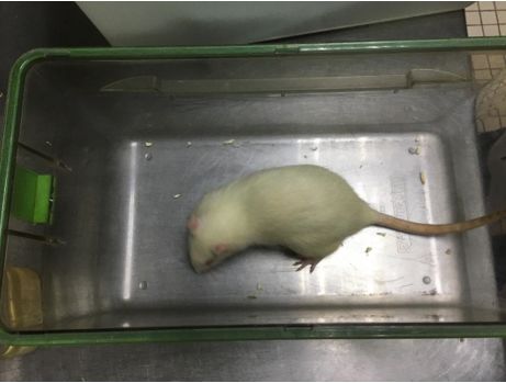
CONCLUSION: Henna extract is a safe and potential alternative in treating otomycosis even with tym-
panic membrane perforation, as it has no vestibulotoxic adverse effects.
5 succinct key points:
Posted on 31 Mar 2023 — The copyright holder is the author/funder. All rights reserved. No reuse without permission. — https://doi.org/10.22541/au.168028569.95590410/v1 — This a preprint and has not been peer reviewed. Data may be preliminary.
1. Animal phase trials using laboratory rats are widely used for otological studies as the middle and inner
ear anatomy is nearly identical to humans.
2. Henna leaf (Lawsomnia inermis) is widely available and has anti-fungal properties, but the safety profile
has never been studied to be used as an anti-fungal ear drops solution.
3. Henna extract can potentially be used as an alternative antifungal ear drops without any vestibulotoxic
effect in an animal study.
4. Henna extract is safe to be used even with tympanic membrane perforation without vestibulotoxic
effect in an animal study.
5. Henna extract has the potential to be commercialized as a new drug after thoroughly assessing the
cochleovestibulotoxicity effect in human trials.
Keywords :
Animal; Endoscopic; Henna; Ototoxicity; Vestibulotoxicity
Introduction
Otomycosis is an infection of the external auditory canal caused by a fungus commonly observed in the general
otology clinic. It has a global prevalence of 9% to 30% among patients with otalgia and otorrhea symptoms
[1]. The occurrence of otomycosis differs according to various climatic factors, with higher prevalence in
tropical and subtropical regions, where it is warm and humid. The most prevalent pathogenic fungi isolated
in otomycosis are Aspergillus niger andCandida albicans. There is currently no standardized medication
regimen for otomycosis that offers novel therapeutic choices, including herbal medication [2].
According to the World Health Organization, herbal plants are one of the ideal sources for obtaining a
range of medicines. Hence, these plants should be studied further to discover their benefits, safety, and
effectiveness. Henna leaf (Lawsonia inermis) has long been valued for its healing and medicinal properties
since ancient times. The therapeutic effects of various components of Henna are due to its biologically
active compound of 2-hydroxynapthoquinone (Lawsone) [3]. Studies have shown that henna leaf extracts in
Malaysia inhibit the development of pathogenic fungi [4].
For centuries, henna leaves have been used in Africa, the Middle East, and Asian countries due to their
healing attributes, such as antifungal, antibacterial, anti-amoebiasis, and antihemorrhagic effects [5]. This is
via the action of its phytoconstituents β-sitosterol glucoside, flavonoids, quinoids, naphthalene derivatives,
luteolin, betulin, lupeol galic acid, coumarins, xanthones, and phenolic glycosides [6]. Every part of the
henna tree has been studied and found to have antifungal and antitubercular activity [7], while the bark has
been found to show anti-inflammatory activity [8]. While its capacity to inhibit the growth of pathogenic
fungi causing otomycosis has been well established [4, 5, 7, 9], this medicinal plant’s ototoxicity status is
still unknown. Ototoxicity is defined as cellular and functional impairment of the inner ear by therapeutic
agents and can be classified into cochleotoxic and vestibulotoxic [10].
This study aims to determine the vestibulotoxic effect of the henna extract with the possibility of developing
it as an antifungal preparation in otomycosis. Balance disruptions as a drug-side effect are frequently
overlooked by doctors, whereas patients often see it as part of their condition and affecting their overall
health. Most drug-induced ototoxicity findings in patients are only restricted to the cochleotoxic effect.
However, studies of the vestibulotoxic effect are scarce. This is a pilot project to evaluate the ototoxic effects
of the henna extract by analyzing specifically the vestibulotoxicity in a rat animal model. To date, there is
only one study assessing the vestibular side effects of neem leaf extract in an animal study [10].
Methodology
Preparation of Henna Leaf Extract
2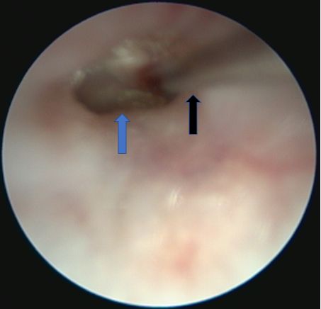
Six hundred grams of Malaysian henna leaves were collected from a single tree in Hospital Universiti Sains
Malaysia (USM) in the district of Kubang Kerian. The henna leaves were collected fresh and healthy from
the henna treetop without any fungal or bacterial contamination evidence. The leaves were green in colour;
Posted on 31 Mar 2023 — The copyright holder is the author/funder. All rights reserved. No reuse without permission. — https://doi.org/10.22541/au.168028569.95590410/v1 — This a preprint and has not been peer reviewed. Data may be preliminary.
neither brown nor yellow were used to avoid contamination.
The henna preparation was conducted at the Pharmacology Department, School of Medical Sciences, USM.
The leaves were cleaned in distilled water and dried at 50 degrees Celsius for two days until brittle. The dried
leaves were weighed on a scale balance before being pulverized into a fine powder and stored in a tightly
sealed glass jar.
Extraction by using a Soxhlet machine was used to prepare the sample. There were two types of solvents
used: aqueous and 70% ethanol. After extensive freeze-drying, 12 grams of ethanol henna extract and 15
grams of aqueous henna extract were produced. These powdered extracts were utilized to determine the
exact concentration of Henna required for this research, which is 25% aqueous extract and 25% ethanol
extract (Figure 1).
Preparation of the Rat Animal Model
Twenty-four healthy male, mature Wistar Kyoto rats weighing between 300g to 400g were used for this
study. They consisted of 24 males aged three months old. They were housed in a typical Macrolon cage
(280×520×145mm) with wood shavings as bedding, three to a cage. They were kept in a facility with 12
hours of on/off light cycles, replicating the regular day and night cycle at a constant temperature (20 ± 2 °C)
and humidity (55.5%). The rats were classified into three groups: Group A (8 animals), Group B (8 animals),
and Group C (8 animals).
The sample size of 24 rats was calculated via the ’resource equation’ method, a commonly used method
in animal studies. The inclusion criteria were Wistar male rats with normal vestibular parameters and
intact tympanic membrane. The exclusion criteria were Wistar rats with the diseased external ear canal, the
abnormal external ear canal, abnormal tympanic membrane, abnormal vestibular profile before perforation
of the tympanic membrane and female albino rats. The ARRIVE Guidelines 2.0 was the reporting guideline
that has been followed in this study.
The four baseline vestibular parameters were tested in all rats in each group. The parameters tested were dys-
kinetic head movements and circling behaviour, tail-hanging test, air-righting reflex, and contact inhibition
of the air-righting reflex. The animals were observed and rated for the presence or absence of the following
signs, while the observers were blinded to the treatments given. The scorings were rated 0, 1, and 2 for
every parameter tested. Zero indicates normal vestibular behaviour, while one indicates mild abnormalities,
and two indicate severe abnormalities observed. The signs of vestibulotoxicity were assessed using published
procedures as per the following [11]:
Dyskinetic head movements and circling
Each rat is placed in an observation chamber and monitored for dyskinetic head movements (head weaving)
and circling for 2 minutes (Figure 2).
Tail hanging
Each of the rats was raised by the tail, and the response was carefully examined and assessed in the fol-
lowing ways: 0-straight body posture with forelimbs extended toward the earth (normal), 1-slight bending
of the body ventrally (intermediate response), and 2-consistently bending the body, occasionally crawling
up towards its tail (severe response).
Air righting reflex
The rats were held supine and dropped onto a foam cushion from 30–40 cm height. The response was assessed
as follows: 0-successful in righting themselves and landing straight on their feet (normal), 1-inadequate in
3righting themselves or landing on the side (intermediate response), and 2-completely unsuccessful in righting
themselves and landing on their backs (severe response).
Contact inhibition of righting reflex
Posted on 31 Mar 2023 — The copyright holder is the author/funder. All rights reserved. No reuse without permission. — https://doi.org/10.22541/au.168028569.95590410/v1 — This a preprint and has not been peer reviewed. Data may be preliminary.
The rats were positioned supine on a horizontal surface, and another horizontal surface was placed slightly
in touch with the supine animal’s feet soles. The rating was done as follows: 0-the animal successfully righted
itself (normal), 1-partial righting, in which the animal exerts some effort (intermediate response), and 2-
complete loss of righting, in which the animal faces up its feet and walks on the upper surface (severe
response).
Ketamine (75-90 mg/kg, Ketalar, Pfizer, Istanbul, Turkey) and xylazine (5-8 mg/kg, Rompun, Bayer, Lever-
kusen, Germany) were used to anesthetize the rats intraperitoneally. The pedal reflex was used to determine
the depth of anaesthesia, and further anaesthesia was provided in half-dose increments as needed. Each rat’s
external ear canal and tympanic membrane were examined following general anaesthesia using a zero-degree
rigid Karl Storz surgical scope. Rats with abnormal external ear canals or a perforated tympanic membrane
were excluded from this study.
After the baseline vestibular parameters testing, an endoscopic-guided transtympanic instillation of aqueous
and ethanol henna extract was conducted into the right middle ear of the rats in Group A and B, respectively
(Figure 3). At the same time, normal saline was instilled in the right middle ear of rats in Group C. A total
of 0.1ml instillation of aqueous, ethanol extract, and saline for each rat was sufficient to fill the whole middle
ear cavity of the rat.
The rats were given time to recover from the anaesthesia. The four vestibular parameters testing were
assessed on the rat’s post-instillation of transtympanic Henna at intervals of 4 hours, 24 hours, 48 hours, 72
hours, one week, two weeks, and three weeks.
The rats would then be euthanized intraperitoneally with thiopental sodium (200 mg/kg Pentothal; Abboth,
Campoverde di Aprilla, Italy).
Results
In Group A (n=8), where the rats were subjected to transtympanic instillation of 25% ethanol henna extract,
no vestibular dysfunction was observed at 4 hours, 24 hours, 48 hours, 72 hours, one week, two weeks, and
three weeks post-instillation. Similar results were observed in Group B (n=8), where the rats were subjected
to transtympanic instillation of 25% aqueous henna extract, and Group C (n=8), where the control group
received transtympanic instillation of normal saline.
Provided the scores were 0 for the baseline and all post-test parameters (Table 1), statistical analysis could
not be performed for this research. In other words, there was no impairment of all vestibular parameters
documented post-instillation of both aqueous and ethanol henna extract in the middle ear, indicating that
Henna has no vestibulotoxic effects after it is administered into the middle ear mucosa.
This study used 24 Wistar Kyoto rats consisting of 8 male rats for Groups A, B, and C, with the latter
serving as a control group. The rats were three months old when this study was performed and weighed
between 300 to 400g each.
Discussion
Otomycosis refers to fungal infections of the external ear, including the auricle, auditory canal, eardrum,
and middle ear. The symptoms include otorrhea, otalgia, itchiness, and it is usually a recurrent disease
[12, 13]. Fortunately, this entity is not life-threatening, and complications are uncommon except in immu-
nocompromised patients as it is an opportunistic infection. However, this disease presents difficulties for
the otolaryngologist and the patient, as it has a high recurrence rate and residual disease. Additionally, it
necessitates long-term treatment and monitoring [13]. The mainstay of treatment for otomycosis is thorough
4removal and cleaning of the infected area with topical antifungal ear drops [14]. Topical medications are pre-
ferable to systemic medications because they have a low risk of systemic side effects and a high concentration
in the targeted infected area.
Posted on 31 Mar 2023 — The copyright holder is the author/funder. All rights reserved. No reuse without permission. — https://doi.org/10.22541/au.168028569.95590410/v1 — This a preprint and has not been peer reviewed. Data may be preliminary.
Aspergillus and Candida are the most prevalent fungal species isolated in otomycosis [13]. There has been a
report of growing resistance to classical antifungal agents that contain azoles, polyenes, and echinocandin,
which are usually effective againstAspergillus and Candida [15]. This is described as clinical resistance, which
is the inability to eliminate a fungal infection despite the antifungal agents having in vitro action against the
pathogen [15]. Currently, there is no FDA-approved antifungal otic solution for otomycosis treatment [16].
Physicians have struggled to discover the most efficient antimycotic medication to treat this ailment since
there are numerous medicines available with varying antimycotic properties. This opens up the research of
using herbal plants as an alternative drug to treat otomycosis.
According to the World Health Organization, herbal plants are one of the ideal sources for obtaining a
range of medicines [17]. Hence, these plants should be studied further to evaluate their benefits, safety, and
effectiveness. Henna leaf (Lawsonia inermis) has long been valued for its healing and medicinal properties
since ancient times. The therapeutic effects of various components of Henna are due to its biologically
active compound of 2-hydroxynapthoquinone (Lawsone) [3]. Studies have shown that henna leaf extracts in
Malaysia inhibit the development of pathogenic fungi [4]. Although commonly used as a natural remedy to
treat skin, outer ear, and hair lesions, the safety status and possible ototoxic effects of henna leaves are still
to be reported. Ototoxicity is identified as functional disability and cellular degradation of the inner hair cells
by therapeutic drugs and chemical compounds and is categorized into cochleotoxicity and vestibulotoxicity
[18].
Two Malaysian henna extracts were used, which are aqueous extract and ethanol extract, which, their fun-
gitoxic activity has been established [4]. Yaroko et al. tested the antifungal activity of Malaysian henna
leaf extract against two prevalent otomycotic pathogenic fungi, which were Aspergillus niger and Candida
albicans . He discovered that the aqueous extract of Malaysian Henna is superior in inhibiting the growth of
A. niger . In contrast, ethanol extract is more effective in suppressing the growth of C. Albicans [4]. Three
concentrations of aqueous and ethanol henna extracts were used in this study; 25mg/ml, 50mg/ml, and
75mg/ml. Statistical analysis revealed significant inhibition of A. niger with aqueous extract concentrations
of 75mg/ml and 50mg/ml, while ethanol extract was significant with a concentration of 75mg/ml only. Mean-
while, henna ethanol extract demonstrated near-significant inhibition againstC. Albicans with a 75mg/ml
concentration. The least inhibition was recorded with a 25mg/ml concentration for both extracts [4]. Howe-
ver, to ensure that the substance is administered into the ear canal and absorbed by the inner ear and middle
ear mucosa, aqueous and ethanol henna at a concentration of 25% were used and instilled transtympanically
[4]. Nadjib et al. also studied using the same concentration of 25% by mixing 25g of powdered henna plant
with 100 ml of different solvents to test against C.albicans and various microbes [9]. Meanwhile, Zakaria et
al. recommended 20mg/ml as the best concentration to inhibit many pathogenic fungi in aqueous or etha-
nol extract [19]. This concentration has been shown to have a sufficient antifungal effect in a thin diluted
solution. It is suitable for physicians and patients to administer in the form of an ear drop.
Wistar rats are widely used in otological studies and were chosen in our study [20, 21]. The middle and inner
ear anatomy is nearly identical to humans. The rat cochlear comprises two and a half turns, making it a
good model for cochleotoxicity and vestibulotoxicity research [22]. The rats were all three months old when
used in the experiment, equivalent to a 20-26-year-old human [23]. This is due to the widespread recognition
of the age connection between mice and humans, particularly regarding neurological development.
The four established vestibular parameters observed in rats after transtympanic instillation of ototoxic agents
were dyskinetic head movements and circling, tail hanging, air righting reflex, and contact inhibition of
righting reflex [11]. A normal baseline vestibular parameters were ensured in all rats following the inclusion
criteria. The henna extract was injected into the right middle ear of each rat for constant observational
behaviour. Vestibular parameters were recorded at four hours, twenty-four hours, seventy-two hours, one
week, two weeks, and three weeks after transtympanic instillation. Acute vestibular changes can be observed
5in rats as early as 4 hours and as late as three weeks after transtympanic instillation. This is because rats can
reestablish normal postural locomotor function following a temporary vestibular insult on one side [23]. The
findings recorded by blinded observers were zero or normal responses in all rats at every vestibular interval
Posted on 31 Mar 2023 — The copyright holder is the author/funder. All rights reserved. No reuse without permission. — https://doi.org/10.22541/au.168028569.95590410/v1 — This a preprint and has not been peer reviewed. Data may be preliminary.
tested. Hence, statistical analysis could not be implemented in this study.
To conclude, henna extract in aqueous and ethanol extract can safely be used in otomycosis without vesti-
bulotoxic effect in an animal model, even with a tympanic membrane perforation. Malaysian Henna showed
potential to be used as an alternative topical antifungal eardrop in treating otomycosis. We recommend
conducting a histological study of the cochlear in an animal model to assess the effect on inner hair cells to
fully establish the safety efficacy of Henna before human clinical trials.
References
[1] Agarwal P, Devi LS. Otomycosis in a rural community attending a tertiary care hospital: Assessment of
risk factors and identification of fungal and bacterial agents. J Clin Diagn Res 2017; 11:Dc14.
[2] Saniasiaya J, Salim R, Mohamad I, Harun A. Antifungal effect of Malaysian aloe vera leaf extract on
selected fungal species of pathogenic otomycosis species in in vitro culture medium. Oman Med J 2017; 32:41.
[3] Tripathi RD, Srivastava HS, Dixit SN. A fungitoxic principle from the leaves of Lawsonia inermis Lam.
Experientia 1978; 34:51-2.
[4] Yaroko AA, Salim R, Mohamed Z, Mohamad I. Antifungal activity of Malaysian Henna leaves extracts
on pathogenic fungi of otomycosis. Int Med J 2015; 22:389-91.
[5] Khattak SG, Gilani SN, Ikram M. Antipyretic studies on some indigenous Pakistani medicinal plants. J
Ethnopharmacol 1985; 14:45-51.
[6] Lal JB, Dutta SB. Constitution of the colouring matter of Lawsonia alba or Indian Mehndi. J Indian
Chem Soc 1933; 10:577-9.
[7] Bhatnagar SS, Santapau H, Desa JD, Maniar AC, Ghadially NC, Solomon MJ et al. Biological activity of
Indian medicinal plants. Part I. Antibacterial, antitubercular and antifungal action. Indian J Med Res 1961;
49:799-813.
[8] Singh VK, Pandey DK. Fungitoxic studies on bark extract of Lawsonia inermis against ringworm fungi.
Hindustan Antibiot Bull 1989; 31:32-5.
[9] Rahmoun N, Boucherit-Otmani Z, Boucherit K, Benabdallah M, Choukchou-Braham N. Antifungal ac-
tivity of the Algerian Lawsonia inermis (Henna). Pharm Biol 2013; 51:131-5.
[10] Salim R, Daud Mk, Narayanan Ms, Ab Rani A. Assessment of vestibulotoxicity of Neem leaf (Azadirachta
indica) in a rat animal model. Indian J Otol 2020; 26:159.
[11] Al Deeb S, Al Moutaery K, Khan HA, Tariq M. Exacerbation of iminodipropionitrileinduced behavioral
toxicity, oxidative stress, and vestibular hair cell degeneration by gentamicin in rats. Neurotoxicol Teratol
2000; 22:213-20.
[12] Jadhav VJ, Pal M, Mishra GS. Etiological significance of Candida albicans in otitis externa. Mycopa-
thologia 2003; 156:313-15.
[13] Ho T, Vrabec JT, Yoo D, Coker NJ. Otomycosis: clinical features and treatment implications. Otolaryngol
Head Neck Surg 2006; 135:787-91.
[14] Vennewald I, Klemm E. Otomycosis: diagnosis and treatment. Clin Dermatol 2010; 28:202-11.
[15] Kanafani ZA, Perfect JR. Resistance to antifungal agents: mechanisms and clinical impact. Clin Infect
Dis 2008; 46:120-8.
6[16] Nemati S, Hassanzadeh R, Jahromi SK, Abadi AD. Otomycosis in the north of Iran: common pathogens
and resistance to antifungal agents. Eur Arch Otorhinolaryngol 2014; 271:953-7.
[17] Rates SM. Plants as source of drugs. Toxicon 2001; 39:603-13.
Posted on 31 Mar 2023 — The copyright holder is the author/funder. All rights reserved. No reuse without permission. — https://doi.org/10.22541/au.168028569.95590410/v1 — This a preprint and has not been peer reviewed. Data may be preliminary.
[18] Hawkins JE, Keidel WD, Neff WD. Handbook of sensory physiology. Drug ototoxicity 1976; 5:707.
[19] Almola Z. The inhibitory effect of Henna Lawsonia inermis leaves on some fungi. Iraq Acad Sci J 2010;
10:501-10.
[20] Fetoni AR, Quaranta N, Marchese R, Cadoni G, Paludetti G, Sergi B. The protective role of tiopronin
in cisplatin ototoxicity in Wistar rats. Int J Audiol 2004; 43:465-70.
[21] Rybak LP, Somani S. Ototoxicity. Amelioration by protective agents. Ann N Y Acad Sci 1999; 884:143-
51.
[22] Albuquerque AA, Rossato M, Oliveira JA, Hyppolito MA. Understanding the anatomy of ears from
guinea pigs and rats and its use in basic otologic research. Braz J Otorhinolaryngol 2009; 75:43-49.
[23] Dutta S, Sengupta P. Men and mice: relating their ages. Life sci 2016; 152:244-8.
Figures legend:
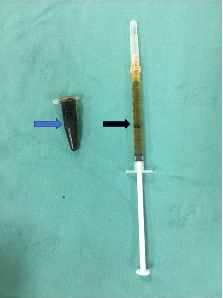
1. Figure 1: The final extract in paste form (blue arrow) with 100% concentration. One cc syringe was
used for intratympanic instillation after serial dilution into the exact concentration of 25% aqueous
and ethanol Henna extract each (black arrow).
2. Figure 2: Observation and scoring of the rat vestibular parameter (dyskinetic head movements and
circling) in a glass cube.
3. Figure 3: Endoscopic-guided intra-tympanic instillation of henna extract. The blue arrow shows the
rat’s right tympanic membrane and the black arrow shows the 25-gauge needle attached to the 1cc
syringe for intratympanic instillation.
7Posted on 31 Mar 2023 — The copyright holder is the author/funder. All rights reserved. No reuse without permission. — https://doi.org/10.22541/au.168028569.95590410/v1 — This a preprint and has not been peer reviewed. Data may be preliminary. 8
Posted on 31 Mar 2023 — The copyright holder is the author/funder. All rights reserved. No reuse without permission. — https://doi.org/10.22541/au.168028569.95590410/v1 — This a preprint and has not been peer reviewed. Data may be preliminary.

         Hosted file

9
Posted on 31 Mar 2023 — The copyright holder is the author/funder. All rights reserved. No reuse without permission. — https://doi.org/10.22541/au.168028569.95590410/v1 — This a preprint and has not been peer reviewed. Data may be preliminary.
10
vestibulotoxicity-of-henna-leaf-lawsomnia-inermis-in-a-rat-animal-model
Table 1.docx available at https://authorea.com/users/601970/articles/632928-assessment-of-You can also read