Case Report Hantavirus Cardiopulmonary Syndrome and Diffuse Alveolar Hemorrhage in the Era of COVID-19
←
→
Page content transcription
If your browser does not render page correctly, please read the page content below

Hindawi
Case Reports in Infectious Diseases
Volume 2021, Article ID 8800500, 4 pages
https://doi.org/10.1155/2021/8800500

Case Report
Hantavirus Cardiopulmonary Syndrome and Diffuse Alveolar
Hemorrhage in the Era of COVID-19

          Khizar Hamid ,1 Swaminathan Perinkulam Sathyanarayanan,1 Touba Naim,1
          Muhammad Hamza,1 Mirza Omer Mahmood Baig,2 and Emad Abu Sitta2
          1
              Internal Medicine, University of South Dakota Sanford School of Medicine, Sioux Falls, SD, USA
          2
              Infectious Disease, Sanford USD Medical Center, Sioux Falla, SD, USA

          Correspondence should be addressed to Khizar Hamid; khizar.hamid@usd.edu

          Received 30 April 2021; Accepted 11 September 2021; Published 24 September 2021

          Academic Editor: Dawei Cui

          Copyright © 2021 Khizar Hamid et al. This is an open access article distributed under the Creative Commons Attribution License,
          which permits unrestricted use, distribution, and reproduction in any medium, provided the original work is properly cited.
          Hantavirus Cardiopulmonary Syndrome (HCPS) can occur after infection with Hantavirus which can occur by inhaling
          aerosolized rodent urine, feces, and saliva contaminated with the virus. It presents with the rapid development of pulmonary
          edema, respiratory failure, and cardiogenic shock with the hallmark being microvascular leakage. We report a patient with a
          history of alcohol abuse and recent exposure to mice and sick kittens who presented with cough with sputum production,
          shortness of breath, orthopnea, and new-onset lower extremity edema. Imaging revealed bilateral infiltrates more common on the
          left with an unremarkable echocardiogram. Testing for COVID-19, Human Immunodeficiency Virus (HIV), influenza, bacterial
          pneumonia including tuberculosis and methicillin-resistant Staphylococcus aureus (MRSA), aspergillosis, histoplasmosis,
          Blastomyces, and Coccidiodes was negative. Bronchoscopy and bronchoalveolar lavage revealed diffuse alveolar hemorrhage
          (DAH) and were negative for acid-fast bacilli and Nocardia cultures. He was further tested for Hantavirus, Q fever, leptospirosis,
          toxoplasmosis, and empiric treatment with doxycycline initiated. His Hantavirus IgM antibody came back positive. Human
          Hantavirus infection occurs after inhalation of infected rodent excreta; fortunately, human-to-human transmission has not been
          documented. HCPS most commonly occurs due to the Sin Nombre virus (SNV), has a case fatality rate of 50%, and is a notifiable
          disease in the United States. It has 3 distinct phases, prodromal, cardiopulmonary, and convalescent/recovery. The cardio-
          pulmonary phase occurs from increased permeability of pulmonary capillaries and in severe cases can progress to cardiogenic
          shock. Diagnosis is based on the presence of IgM and IgG Hantavirus antibodies. Treatment is mainly supportive; however,
          patients are usually treated with broad-spectrum antibiotics while workup is underway. In animal models, ribavirin and favipiravir
          are only effective when administered in the prodromal phase. If suspicion of Hantavirus infection exists, early mobilization to the
          intensive care unit for treatment is recommended. Extracorporeal membrane oxygenation (ECMO) has been suggested to
          improve outcomes in severe HCPS with refractory shock.

1. Introduction                                                         [1, 2]. HCPS, also known as Hantavirus pulmonary syn-
                                                                        drome, is a severe illness denoted by the rapid development
Hantaviruses are RNA viruses belonging to the Bunyaviridae              of pulmonary edema, respiratory failure, and cardiogenic
family whose transmission in humans is caused by inhaling               shock. The hallmark of HCPS is microvascular leakage [3].
aerosolized rodent urine, feces, and saliva contaminated by             Here, we report a 33-year-old man with HCPS presenting
the virus [1]. They mainly attack vascular endothelial cells            with diffuse alveolar hemorrhage (DAH).
along with alveolar macrophages and follicular dendritic
cells. Another potential infection site is the epithelium of            2. Case Presentation
renal tubules [2]. Hemorrhagic fever with renal syndrome
(HFRS) and Hantavirus Cardiopulmonary Syndrome                          A 33-year-old Native American man, a welder by profession,
(HCPS) are two syndromes caused by Hantavirus in humans                 with a past medical history significant for alcohol use,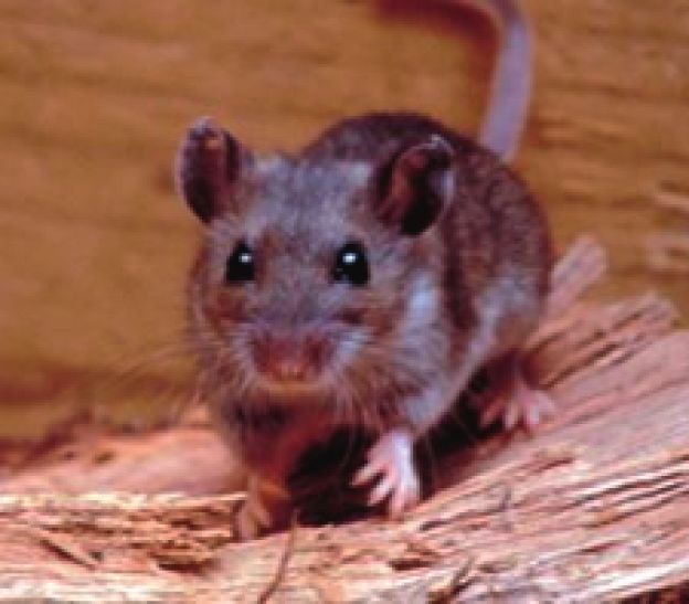
2                                                                                            Case Reports in Infectious Diseases

hypertension, and asthma presented to the hospital with
complaints of cough with occasional bloody sputum pro-
duction. His cough started four days before presentation,
was initially dry but progressively worsened, and became
productive. It was also associated with dyspnea and
orthopnea along with new-onset lower extremity edema. He
further endorsed preceding nausea, vomiting, and diarrhea
without abdominal pain, melena, or hematemesis. He re-
ported an extensive history of alcohol consumption and had
quit smoking 6 years ago.
    On physical examination, he appeared restless and his
vitals comprised a temperature of 98.5°F, blood pressure
158/91 mm of Hg, respiratory rate 30 breaths/minute, pulse          Figure 1: Computed tomographic angiogram of the chest showing
116 beats/minute, oxygen saturation 96% on 2 liters of              bilateral infiltrates more extensive on the left.
supplemental oxygen via a nasal cannula, weight 290
pounds, and a body mass index of 39.32. His pertinent
laboratory findings included a white blood cell count of                 While on mechanical ventilation, aggressive diuresis was
16.3 K/uL, hemoglobin 8.6 g/dL, platelets 124,000 K/uL,             initiated for pulmonary edema. Thereafter, he was weaned
potassium 3.0 mEq/L, sodium 125 mEq/L, magnesium                    off the ventilator and extubated within a week. His he-
1.6 mg/dL, folate level 5.7 ng/mL, creatinine kinase 455 U/L,       moptysis resolved, but he continued to cough with infre-
lactate dehydrogenase 310 U/L, albumin 2.9 g/dL, CRP                quent mucoid phlegm. Antibiotics were discontinued after
16.9 mg/L, lactic acid 3.1 mmol/L, procalcitonin 0.14 ng/mL,        ten days when he was clinically better, and repeat CXR
and D-dimer 10.56 ug/mL FEU. His ethanol level was                  exhibited slight improvement in left-sided infiltrates along
96.7 mg/dL, while liver function tests showed aspartate             with the resolution of his right lung consolidation.
transaminase 132 U/L, alanine transaminase 33 U/L, and                  Steroids were transitioned from IV to oral prednisolone
alkaline phosphatase 219 U/L. Testing for SARS-CoV-2,               40 mg daily for 26 days followed by a taper to cover for
Human Immunodeficiency Virus (HIV), influenza, and                    suspected alcoholic hepatitis. His mental function improved
methicillin-resistant Staphylococcus aureus (MRSA) nasal            with alcohol withdrawal treatment and he became more alert
screen was negative.                                                and oriented. On further questioning, he recalled having
    His computed tomographic angiogram of the chest                 exposure to sick kittens and mice few days before admission.
(Figure 1) revealed bilateral infiltrates more extensive on the      He was tested for Hantavirus, Q fever, leptospirosis, and
left as compared to the right and excluded pulmonary                toxoplasmosis, and empiric treatment with doxycycline
embolism. Ultrasound abdomen revealed hepatomegaly. An              100 mg twice a day was initiated for 3 weeks. TB testing
echocardiogram showed an ejection fraction of 65–70% with           (QuantiFERON, sputum acid-fast bacilli, and PPD) and
moderate left ventricular hypertrophy, left atrial enlarge-         antibodies for leptospirosis and Q fever were all negative.
ment, and right atrial dilation. He was started on empiric              On day 18, the patient was hemodynamically stable with
treatment with ceftriaxone 2 gm daily and doxycycline               complete resolution of respiratory failure and was deemed
100 mg twice a day for suspected bacterial pneumonia along          stable for discharge. Four days after discharge, Hantavirus
with management of possible alcoholic hepatitis with thi-           IgM returned positive with equivocal IgG. Retrospectively,
amine and folic acid and Clinical Institute Withdrawal              his presentation correlated with HCPS. Upon follow-up in
Assessment for Alcohol (CIWA) protocol.                             the clinic 10 days after discharge from the hospital, he re-
    On day 2 of hospitalization, his hemoptysis and breathing       ported improvement in cough and dyspnea. Outpatient CXR
deteriorated, and his white cell count rose further to 18.1 K/uL.   (Figure 2(b)) revealed mild improvement of left upper lobe
His supplemental oxygen requirements increased, and chest           consolidation.
X-ray (CXR) (Figure 2(a)) showed worsening left-sided infil-
trates. Following this, the antibiotic regimen was broadened to     3. Discussion
cefepime 2 gm three times a day for 8 days and vancomycin
dosed by the pharmacy for 5 days. Urine Legionella antigen,         Hantaviruses are enveloped, segmented negative-strand
Streptococcus pneumoniae direct antigen, Aspergillus, Blasto-       RNA viruses that belong to the Bunyaviridae family. Human
myces, Coccidiodes, histoplasma antibodies, and repeat SARS-        spread occurs by inhalation of aerosolized excreta from
CoV-2 testing were all negative. Blood cultures and sputum          infected rodents [4]. In the US, rodents that carry hantavirus
cultures were obtained and were found to have no growth.            include cotton rat, deer mouse, rice rat, and white-footed
Bronchoscopy was performed revealing DAH thought sec-               mouse (Figure 3) (transmission in the Midwest is pre-
ondary to pulmonary edema. Intravenous (IV) steroids were           dominantly from deer mouse and white-footed mouse). Sin
started, and further infectious and autoimmune workup was           Nombre virus (SNV) is most prevalent in North America,
undertaken. Bronchoalveolar lavage sampling was negative for        while in Central and South America, the most common one
acid-fast bacilli, Nocardia cultures, and CMV DNA. With the         is Andes (AND) virus. Person-to-person transmission has
continued decline in respiratory function, he underwent en-         not been documented in North America, Europe, or Asia
dotracheal intubation.                                              [5].
Case Reports in Infectious Diseases 3
(a) (b)
Figure 2: (a) Chest X-ray showing worsening left-sided infiltrates. (b) Chest X-ray showing mild improvement of left upper lobe
consolidation.
(a) (b)
Figure 3: (a) Deer mouse [6]. (b) White-footed mouse [6].
Close to 40 different species of Hantaviruses have been abdominal pain lasting anywhere from 1 to 5 days like our
identified, and 22 are considered to cause infections in patient’s presentation. There are usually no upper respi-
humans [7]. The hantaviruses that circulate in Europe and ratory tract symptoms in this phase. Another characteristic
Asia called the “old world Hantaviruses” cause HFRS which feature of the prodrome is thrombocytopenia. About 80 to
is characterized by hemorrhagic manifestations such as skin 95% of people will have a platelet count of less than 150,000
petechiae and ecchymoses, epistaxis, hematuria, hematem- units [4].
esis, melena, fatal intracranial hemorrhages, and renal failure The cardiopulmonary phase consists of cough, dyspnea,
[8]. Hantaviruses in the Americas called the “new world and hypoxia with the development of noncardiogenic pul-
Hantaviruses” cause the HCPS [5, 8]. HCPS was first de- monary edema from increased permeability of pulmonary
scribed in the US in 1993. SNV is the most common cause of capillaries referred to as “pulmonary leak.” In severe cases, it
HCPS in North America [8]. HCPS has been listed as a progresses to myocardial dysfunction resulting in cardio-
notifiable disease since 1995 in the US. From 1993 to 2017, a genic shock. Complete blood count and a peripheral smear
total of 728 cases of HCPS have been reported in the US (17 aid in the diagnosis of HCPS. Thrombocytopenia (platelets
in South Dakota) [9, 10]. 50% in men and >48% in
women, lack of toxic granulations in polymorphs, left shift of
myeloid series, and >10% immunoblasts are some typical
3.1. Hantavirus Cardiopulmonary Syndrome. HCPS has a features of HCPS. The presence of four out of the above-
severe disease course with a high case fatality rate ranging mentioned five findings has a 96% sensitivity for Hantavirus
from 30 to 50%. The disease course progresses through 3 infection. Other laboratory findings include high levels of
distinct phases: prodromal, cardiopulmonary, and con- CK, LDH, and transaminases along with low albumin [4].
valescent/recovery [8]. The prodromal phase is charac- Our patient had similar findings on testing. Chest radio-
terized by nonspecific complaints including fevers, graph often shows bilateral interstitial markings, and some
headaches, malaise, myalgias, nausea, vomiting, and patients may have unilateral opacities in the beginning, later4 Case Reports in Infectious Diseases
progressing to bilateral infiltrates [4, 5]. This phase usually Conflicts of Interest
lasts about a week, with a subsequent diuretic period and
resolving pulmonary edema. Convalescence may take up to 6 There are no conflicts of interest.
months [8].
The definitive diagnosis is always based on serologies, the References
presence of IgM and IgG antibodies in the serum. The
[1] P. A. Vial, F. Valdivieso, G. Mertz et al., “Incubation period of
enzyme-linked immunosorbent assay (ELISA) provided by hantavirus cardiopulmonary syndrome,” Emerging Infectious
the CDC and the strip provided by TriCore Reference Diseases, vol. 12, no. 8, pp. 1271–1273, 2006.
Laboratories at Albuquerque, New Mexico, are commonly [2] S. Q. Simpson, L. Spikes, S. Patel, and I. Faruqi, “Hantavirus
used assays [4]. Reverse transcription-polymerase chain pulmonary syndrome,” Infectious Disease Clinics of North
reaction (RT-PCR) is another method that detects viral RNA America, vol. 24, no. 1, pp. 159–173, 2010.
during the viremic phase of infection [4, 5]. [3] A. Macneil, A. MacNeil, S. T. Nichol, and C. F. Spiropoulou,
Currently, there are no FDA-approved treatments for “Hantavirus pulmonary syndrome,” Virus Research, vol. 162,
Hantavirus diseases except for supportive management for no. 1-2, pp. 138–147, 2011.
respiratory failure and cardiogenic shock [11]. People are [4] S. T. Llah, S. Mir, S. Sharif, S. Khan, and M. A. Mir, “Han-
usually treated with broad-spectrum antibiotics while tavirus induced cardiopulmonary syndrome: a public health
concern,” Journal of Medical Virology, vol. 90, no. 6,
awaiting results like in our case. Administration of fluids
pp. 1003–1009, 2018.
should be carried out very cautiously, and if suspicion for [5] B. Chang, M. Crowley, M. Campen, and F. Koster, “Hanta-
HCPS is high, patients should be transferred to the Intensive virus cardiopulmonary syndrome,” Seminars in Respiratory
Care Unit (ICU) early in the course [12]. Some antiviral and Critical Care Medicine, vol. 28, no. 2, pp. 193–200, 2007.
medications studied in animal models such as ribavirin and [6] Centers for Disease Control and Prevention, “Rodents in the
favipiravir show effectiveness only when administered in the United States that carry Hantavirus,” 2021, https://www.cdc.
prodromal phase before the onset of viremia [11]. Patients gov/hantavirus/rodents/index.html.
with HCPS who progress to severe cardiopulmonary failure [7] N. Munir, M. Jahangeer, S. Hussain et al., “Hantavirus dis-
and refractory shock have very high mortality, and early eases pathophysiology, their diagnostic strategies and thera-
extracorporeal membrane oxygen (ECMO) has been sug- peutic approaches: a review,” Clinical and Experimental
Pharmacology and Physiology, vol. 48, no. 1, pp. 20–34, 2020.
gested to improve outcomes in such patients [13]. However,
[8] T. Avšič-Županc, A. Saksida, and M. Korva, “Hantavirus
according to a case report, a good response to continuous infections,” Clinical Microbiology and Infection, vol. 21,
high-volume hemofiltration for a limited time duration may pp. e6–e16, 2019.
assist in identifying patients who may improve with con- [9] H. Jiang, X. Zheng, L. Wang, H. Du, P. Wang, and X. Bai,
ventional ICU management decreasing the need for ECMO “Hantavirus infection: a global zoonotic challenge,” Virologica
and transfer to ECMO-capable facilities [4]. Sinica, vol. 32, no. 1, pp. 32–43, 2017.
[10] Centers for Disease Control and Prevention, “National center
for emerging and zoonotic infectious diseases (NCEZID), di-
4. Conclusions vision of high-consequence pathogens and pathology
(DHCPP),” 2017, https://www.cdc.gov/hantavirus/surveillance/
HCPS has high mortality, and no antiviral treatments or reporting-state.html.
vaccines have proven to be efficacious to this date. It can [11] R. L. Brocato and J. W. Hooper, “Progress on the prevention
present with DAH, and an extensive negative infectious and treatment of hantavirus disease,” Viruses, vol. 11, no. 7,
workup in the case of pulmonary infiltrates should raise the p. 610, 2019.
suspicion for this diagnosis, especially if exposure to rodents [12] A. B. Kuenzli, J. Marschall, J. C. Schefold et al., “Hantavirus
has been documented. Our patient had multiple tests cardiopulmonary syndrome due to imported andes hantavi-
conducted for COVID-19 which were negative. Not all rus infection in Switzerland: a multidisciplinary challenge, two
unexplained cases of shortness of breath and pulmonary cases and a literature review,” Clinical Infectious Diseases,
infiltrates should be attributed to COVID-19 during this vol. 67, no. 11, pp. 1788–1795, 2018.
[13] G. Bugedo, J. Florez, M. Ferres, E. Roessler, and A. Bruhn,
pandemic, and effective history taking can help identify
“Hantavirus cardiopulmonary syndrome successfully treated
other rare causes of infection. More research needs to be with high-volume hemofiltration,” Revista Brasileira de Ter-
undertaken to prevent the fatal outcomes of HCPS. apia Intensiva, vol. 28, no. 2, pp. 190–194, 2016 Jun.
Data Availability
Previously reported published data which can be found at
Google Scholar and PubMed were used to support this study
and are available and reported in the manuscript. These prior
studies (and datasets) are cited at relevant places within the
text as references. Pictures were extracted from https://www.
cdc.gov/hantavirus/rodents/index.htm.You can also read