Case Report Hemorrhagic Shock after Neonatal Circumcision: Severe Congenital Factor XIII Deficiency
←
→
Page content transcription
If your browser does not render page correctly, please read the page content below
Hindawi
Case Reports in Pediatrics
Volume 2021, Article ID 5550199, 6 pages
https://doi.org/10.1155/2021/5550199

Case Report
Hemorrhagic Shock after Neonatal Circumcision: Severe
Congenital Factor XIII Deficiency

          Erin L. Cohen ,1 Samantha E. Millikan ,1 Perry C. Morocco ,2 and Jill L. O. de Jong                                          2

          1
              Department of Pediatrics, Comer Children’s Hospital, Pritzker School of Medicine, University of Chicago, Chicago, IL, USA
          2
              Department of Pediatrics, Section of Pediatric Hematology, Oncology, Stem Cell Transplant, Comer Children’s Hospital,
              Pritzker School of Medicine, University of Chicago, Chicago, IL, USA

          Correspondence should be addressed to Perry C. Morocco; perry.morocco@uchospitals.edu

          Received 26 January 2021; Revised 22 April 2021; Accepted 22 April 2021; Published 3 May 2021

          Academic Editor: Anselm Chi wai Lee

          Copyright © 2021 Erin L. Cohen et al. This is an open access article distributed under the Creative Commons Attribution License,
          which permits unrestricted use, distribution, and reproduction in any medium, provided the original work is properly cited.
          A Caucasian male infant born full term via normal spontaneous vaginal delivery was given vitamin K after birth, circumcised on
          day of life (DOL) 1, and discharged from the nursery on DOL 2. At the time of circumcision, oozing from the surgical site was
          noted and initially resolved with silver nitrate. Over the next two days, he presented to local emergency rooms multiple times for
          recurrent bleeding, eventually developing hemorrhagic shock resulting in admission to the neonatal intensive care unit. After
          extensive work up, he was ultimately diagnosed with severe congenital factor XIII deficiency. Congenital factor XIII deficiency is a
          rare bleeding disorder characterized by normal prothrombin time (PT) and activated partial thromboplastin time (aPTT)
          coagulation labs on routine screening, and has a high risk of complications, such as spontaneous intracranial hemorrhage.
          Although uncommon, when caring for a child with bleeding, physicians must have a high index of suspicion to make this
          diagnosis in order to initiate proper treatment and start prophylaxis given the risk of morbidity and mortality in
          untreated patients.

1. Introduction                                                          2. Case Presentation
Factor XIII deficiency is a rare bleeding disorder with an                The patient was a full-term infant boy born via an uncom-
incidence of approximately one in two million births. Unlike             plicated spontaneous vaginal delivery after an uncomplicated
other bleeding disorders, clots are formed normally but are              pregnancy. He was circumcised on DOL 1 after receiving
unstable due to a defect in fibrin cross-linking which leads to           vitamin K. There was oozing noted at the circumcision site
excessive fibrinolysis. As such, patients may present with                after the procedure that resolved with application of silver
delayed bleeding after a procedure or injury, or recurrence of           nitrate, and he was discharged home on DOL 2. (See Table 1
bleeding. Characterized by normal PT, INR, and aPTT                      for detailed chronology of the case.) Laboratory studies drawn
coagulation values, the diagnosis of factor XIII deficiency is            in response to the episode of bleeding at the time of nursery
often delayed, which can result in high morbidity and                    discharge revealed a normal hemoglobin level (Table 2). Of
mortality. Here, we present the case of an infant who ex-                note, coagulation studies (PT and aPTT) were not done prior
perienced hemorrhagic shock after prolonged bleeding due                 to hospital discharge.
to congenital factor XIII deficiency, which was diagnosed                     After arriving home, his parents took him to a local
after multiple medical visits. A review of the scientific lit-            emergency room (ER) three times within 24 hours for re-
erature related to the clinical course, pathophysiology, and             current bleeding of the circumcision site. At the initial ER
treatment options for factor XIII deficiency is also presented.           visit on the night of hospital discharge, application of a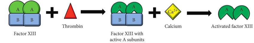
2                                                                                                                    Case Reports in Pediatrics

                                                          Table 1: Chronology of care.
Event                                                                          Day of life (DOL) and time
Birth                                                                              DOL 1 at 1:16 am
Vitamin K administered                                                             DOL 1 at 8:26 am
Circumcision performed                                                             DOL 1 at 11:52 am
Bleeding noted from circumcision                    DOL 1 at 2:55 pm, 3:10 pm and 7:00 pm; unknown timing of silver nitrate application
Labs checked (Hgb 15.2 g/dL)                                                       DOL 2 at 3:55 am
Discharged to home                                                                 DOL 2 at 4:56 pm
First visit to ER                                                                  DOL 3 at 1:11 am
Second visit to ER                                                                 DOL 3 at 4:33 am
Third visit to ER (Hgb 12.5 g/dL)                                                  DOL 3 at 2:25 pm
Fourth visit to ER (Hgb 3.4 g/dL)                                                  DOL 4 at 12:35 pm

                                                           Table 2: Laboratory values.
                                   Reference range         Newborn nursery (DOL 2)             3rd ER visit (DOL 3)          4th ER visit (DOL 4)
          ∗
WBC (10 3/uL)                            6–30                       15.12                              10.9                           8.0
Hemoglobin (g/dL)                       12–20                        15.2                              12.5                           3.4
Hematocrit (%)                          36–66                        46.1                              36.9                          12.7
Platelets (10∗ 3/uL)                   90–475                        330                               312                           138
RBC (10∗ 6/uL)                          3.3–6                        4.80                              3.87                           —
MCV (fL)                               85–125                        96.0                              95.4                         123.0
RDW (%)                               11.6–14.2                      16.9                              15.3                          17.5
nRBCs abs (10∗ 3/uL) [%]                0–0.1                     0.22 [1.5]                            —                             [1]
MPV (fL)                               9.0–12.6                       9.4                               —                             —
Total bilirubin (mg/dL)                  0–12                         2.4                               —                             0.9
aPTT (seconds)                          25–36                         —                                 —                             92
PT (seconds)                          10.1–13.1                       —                                 —                            27.1
INR                                       —                           —                                 —                             2.3
ER � emergency room; DOL � day of life; WBC � white blood cell count; RBC � red blood cell count; MCV � mean corpuscular volume; RDW � red cell
distribution width; nRBCs � nucleated red blood cells; MPV � mean platelet volume; aPTT �activated partial thromboplastin time; PT �prothrombin time;
INR � international normalized ratio.

Surgicel dressing controlled the bleeding. After this dressing               90 mL/kg), and 1 unit of fresh frozen plasma (FFP). He was
fell off, he was brought back to the hospital a few hours later               started on ampicillin and gentamicin due to concern for
where a stitch was placed and another Surgicel dressing was                  possible infection, given hydrocortisone and tranexamic
applied. Later that same day, the bleeding restarted within 10               acid (antifibrinolytic), and placed on a dopamine infusion
hours, leading to another ER visit for a third Surgicel                      for blood pressure control. With these interventions, his
dressing. Labs at this time (Tables 1 and 2) demonstrated a                  clinical condition stabilized, although he had continuous
loss of about 2.7 g/dL of hemoglobin. There was no report of                 oozing from his circumcision site. Surgicel was again placed
bleeding from other sites, including no bleeding from the                    on the site, with a thrombin soaked gauze applied over the
umbilical cord stump. The bleeding appeared to be con-                       Surgicel.
trolled, so he was discharged home with plans to follow up                       At this point, he was transferred to a level IV neonatal
with his pediatrician the next day.                                          intensive care unit at a tertiary care center for escalation of
     When he arrived to his pediatrician appointment on                      care. Upon arrival, he was immediately evaluated by pedi-
DOL 4, he appeared pale with weak pulses. He was trans-                      atric surgery and urology and taken to the operating room
ported immediately by ambulance to the nearest ER. Vital                     where diffuse oozing was noted from the circumcision site.
signs were notable for a heart rate of 92 bpm, respiratory rate              Hemostasis was obtained, and the patient did well post-
of 20, and blood pressure of 72/59 upon arrival to the ER. He                operatively with no bleeding recurrence. Head and ab-
became lethargic with weak respiratory effort requiring                       dominal ultrasounds were performed and did not reveal
bagging, intubation, chest compressions, emergent umbilical                  other sites of bleeding. Over the next several postoperative
artery and venous catheter placement, bicarbonate, and                       days, he was extubated and his hemoglobin stabilized.
epinephrine. Initial ER labs (Table 2) revealed severe anemia                    Further workup demonstrated an elevated factor VIII
(Hgb 3.4 g/dL) and hemorrhagic shock causing severe                          with normal factors II, V, VII, IX, X, XI, and XII for age
metabolic acidosis (pH 6.415, lactate 25 mmol/L), mild                       (Table 3) [1]. Given that his PT, INR, and PTT also nor-
thrombocytopenia (platelets 138 10∗ 3/uL), and a con-                        malized without additional FFP, workup was sent to evaluate
sumptive coagulopathy (PT 27.1 sec, INR 2.3, aPTT 92 sec).                   for bleeding disorders with normal PT and aPTT values,
Fluid resuscitation was performed with 20 mL/kg of normal                    including von Willebrand disease, plasminogen activator
saline, 290 mL of packed red blood cells (approximately                      inhibitor-1 (PAI-1) deficiency, and factor XIII deficiency.
Case Reports in Pediatrics 3
Table 3: Coagulation factor levels tested.
Newborn reference range
Lab reference range Initial results (DOL 4) Repeat results (DOL 6)
(mean ± standard Deviation)∗
Fibrinogen
163–463 — 153 297
(mg/dL)
Factor II (%) 77–138 58 ± 14 49 57
Factor V (%) 75–137 90 ± 25 31 86
Factor VII (%) 71–147 85 ± 26 81
Factor VIII (%) 57–152 89 ± 33 212 202
Factor IX (%) 80–147 49 ± 17 47 73
Factor X (%) 76–146 46 ± 14 42 49
Factor XI (%) 78–133 57 ± 16 — 63
Factor XII (%) 47–173 44 ± 17 — 46
Factor XIII (%) 69–143 89 ± 24 — 20
∗
Source: reference [1].
PAI-1 antigen level and von Willebrand disease panel were A 2019 report by the World Federation of Hemophilia
normal. described 1537 patients with factor XIII deficiency registered
Ultimately, the quantitative factor XIII activity, a chro- from 70 countries, with 55% males and 39% females, in-
mogenic assay performed by ARUP laboratories, was found to cluding 108 patients in the United States [9]. Factor XIII
be low at 20% (age-adjusted mean and standard deviation deficiency is categorized as a rare bleeding disorder that is
89 ± 24) [1], raising concern for a congenital factor XIII most commonly congenital but can be acquired [10]. When
deficiency. Given recent FFP administration, this number was congenital, it is inherited in an autosomal recessive pattern,
thought to be artificially elevated, so repeat testing was distributing evenly amongst males and females, in contrast
performed 2 months after discharge which confirmed the to hemophilia A and B, which are inherited in an X-linked
diagnosis with an undetectable ( A, p.Arg261His in exon 6) and was with decreased protein synthesis, or qualitative type 2 de-
heterozygous for a different missense variant in the other copy ficiency with a functionally defective A subunit [11]. B
of the F13A1 gene (c.610G > A, p.Glu204Lys in exon 5), subunit deficiencies are much less common, estimated to be
confirming a deficiency due to compound heterozygous less than 5% of cases [3]. Acquired factor XIII deficiency can
factor XIII subunit A mutations. He is currently receiving be seen in many medical conditions, including leukemia,
monthly recombinant factor XIII prophylaxis and has had no inflammatory bowel disease, cirrhosis, and thromboembolic
further episodes of bleeding. events, and can result from increased consumption or de-
creased production of factor XIII or associated with anti-
3. Discussion factor XIII antibodies [11]. If due to antibody formation,
systemic lupus erythematosus is a common cause, but an-
3.1. Pathophysiology. Factor XIII is a protransglutaminase tibodies can develop spontaneously in geriatric patients [11].
composed of two catalytic A subunits and two carrier B Acquired factor XIII deficiency is even rarer than the
subunits (Figure 1). When activated by thrombin and cal- congenital form, with a 2017 review finding only 93 cases
cium, factor XIII plays an important role in the final stage of reported in the literature in adult patients [12] and only one
the coagulation pathway, with its primary function being to report in a pediatric patient [13].
stabilize newly formed clots by cross-linking the fibrin
strands [2]. In addition to protecting the fibrin clot against
fibrinolysis, factor XIII plays an important role in wound 3.3. Presentation. The presentation of factor XIII deficiency
healing and maintenance of pregnancy [2, 3]. Factor XIII can be quite variable but usually involves pathologic
deficiency is thus a disorder of secondary hemostasis, de- bleeding and often recurrence of bleeding over days. Our
fined as involving the processes of coagulation and clot patient presented with prolonged bleeding from a circum-
stabilization. The cutoffs for severity are controversial, with cision site, with intermittent hemostasis, eventually leading
some authors defining mild deficiency when factor XIII to profound blood loss and hemorrhagic shock. When factor
levels are greater than 10%, moderate when 5–10%, and XIII deficiency is congenital, it usually presents in neonatal
severe when under 5% [4], while others consider mild de- life, with umbilical cord bleeding being the most common
ficiency when over 30% and severe under 1% [5]. presentation [14]. Other common presentations include
intracranial hemorrhage, mucosal tract bleeding, sponta-
neous hematoma and hemarthrosis, and postsurgical
3.2. Epidemiology. Factor XIII deficiency has an incidence of bleeding [15]. Consistent with our patient’s presentation of
approximately 1 in 2 million people, with higher prevalence intermittent hemostasis, clots in patients with factor XIII
in areas with higher rates of consanguineous marriage [6–8]. deficiency can form normally but then undergo premature4 Case Reports in Pediatrics
(a)
(b)
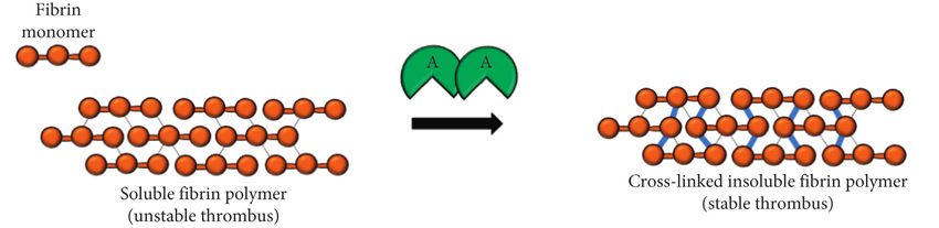
Figure 1: Diagram of factor XIII activation and function. (a) Factor XIII is a protransglutaminase zymogen heterotetramer composed of two
catalytic A subunits and two carrier B subunits. Thrombin cleaves the A subunits to make them catalytically active, followed by calcium-
dependent dissociation of the B subunit to expose the active enzymatic site on the A subunit. (b) Activated factor XIIIa is then able to
crosslink the fibrin strands, by catalyzing the formation of covalent bonds between glutamine and lysine residues on the fibrin a and g chains,
resulting in a stable thrombus that is more resistant to degradation and less soluble.
degradation after approximately 24–48 hours from poor diagnosis includes von Willebrand disease, thrombocyto-
fibrin cross-linking (Figure 1) [3]. Intracranial hemorrhage penia, a platelet function disorder, PAI-1 deficiency, alpha II
occurred in 18% of patients in one case series of 190 Iranian antiplasmin deficiency, and factor XIII deficiency [19, 20].
patients [14] and often is the initial bleed that leads to a For patients with recurrent bleeding symptoms or intra-
diagnosis of factor XIII deficiency [15]. Intracranial hem- cranial hemorrhage, providers need to have a high index of
orrhage is reported more frequently in factor XIII deficiency, suspicion for factor XIII deficiency and laboratory testing for
as compared to hemophilia A and B, and is often the cause of factor XIII activity should be prioritized. Traditionally, this
death in patients with factor XIII deficiency [3, 11]. Another was done using the urea clot solubility test, which is a
case series of 10 patients in Karachi reported prolonged nonquantitative test of factor XIII activity still in use today in
bleeding and bruising in 80% of their patients, and 2 patients developing countries due to its cost-effectiveness and ease of
with prolonged bleeding from the umbilical stump [16]. Two use [21]. In developed countries, the clot solubility test is no
patients had intracranial hemorrhage as the presentation longer recommended as it has low sensitivity for mild to
[16]. Another 20-year case review from Saudi Arabia looked moderate deficiency, including patients who are heterozy-
at 16 patients’ initial presentations and found ecchymoses gous carriers [21]. The first-line screening test for factor XIII
and recurrent hematomas in the majority of patients (71%), deficiency is a quantitative factor XIII assay. Once a low
bleeding after circumcision as the initial presentation in 6 factor XIII activity level is noted, the next step in diagnosis is
male patients (55%), bleeding from the umbilical stump in 7 to establish the subtype of factor XIII deficiency by mea-
patients (41%), poor wound healing in 3 patients (18%), and suring factor XIII-A2B2 antigen concentration in plasma,
intracranial hemorrhage in 3 patients (18%) [6]. A final case followed by factor XIII-A and factor XIII-B antigens, or
series of 6 patients with factor XIII deficiency found that all through genetic sequencing of the F13A1 and F13B genes
presented with umbilical cord bleeding, and a significant [11].
delay in final diagnosis was noted for half of the patients,
resulting in further bleeding complications [17]. Other less
3.5. Treatment. In order to prevent bleeding complications,
frequent presentations included cephalohematoma, spon-
particularly spontaneous intracranial hemorrhage, patients
taneous abortion, abruption placenta, and intraperitoneal
with severe factor XIII deficiency should be started on
bleeding [6]. Compared to other coagulation factors, factor
prophylactic factor infusion [18]. The half-life of factor XIII
XIII does not increase in pregnancy, making spontaneous
is long at 11–14 days, and levels greater than 3% are typically
abortion in the first trimester a common complication of
sufficient to prevent bleeding [4], so only monthly pro-
women with severe factor XIII deficiency who did not re-
phylaxis is needed. In the event a patient with factor XIII
ceive prophylaxis [18].
deficiency presents with bleeding and factor XIII is not
available, cryoprecipitate or FFP can be given, with cry-
3.4. Diagnosis. Any patient who presents with bleeding oprecipitate having a higher concentration of factor XIII
requires a laboratory workup that includes PT and aPTT. If (mean 2.81 factor XIII activity per mL in cryoprecipitate
these coagulation studies are normal, the differential versus mean 1.18 factor XIII activity per mL in FFP) [22]. AsCase Reports in Pediatrics 5
of 2021, there are currently two products approved for use in Authors’ Contributions
the US for factor XIII deficiency: factor XIII concentrate
Erin L. Cohen, Samantha E. Millikan, and Perry C. Morocco
®
(human) (Corifact by CSL Behring) and coagulation factor
contributed equally to the conception, design, writing, and
®
XIII A subunit (recombinant) (Tretten by Novo Nordisk
Inc.). Both these options have been found to be efficacious in revision of this manuscript. Jill L. O. de Jong contributed to
treating factor XIII deficiency, and are overall well tolerated. the design of the figure, and conception, editing and revi-
sions of the manuscript.
®
An analysis of Corifact postmarketing safety reports from
1993–2013 by CSL Behring found 75 cases of adverse events,
for an estimated 1 in 15,700 standard doses [23]. Of these Acknowledgments
adverse events, 20 experienced a possible pathogen trans-
mission, 12 a hypersensitivity reaction, 7 a possible The authors would like to thank the patient’s family for
thromboembolic event, and 5 developed a possible inhibitor allowing them to use their child’s presentation for this
®
[23]. For Tretten , an international phase 3b trial exploring
its safety and efficacy in pediatric patients with congenital
manuscript.
factor XIII deficiency found that prophylaxis over 1.8–3.5 References
years dropped the annualized bleeding rate to zero with
mostly mild adverse events [24]. Over the 16.6 patient years [1] M. Andrew, B. Paes, R. Milner et al., “Development of the
of the study, there were no thromboembolic events or hy- human coagulation system in the full-term infant,” Blood,
persensitivity reactions observed [24]. Of note, during the vol. 70, no. 1, pp. 165–172, 1987.
study, two patients experienced a head injury and neither [2] L. Muszbek, Z. Bereczky, Z. Bagoly, I. Komáromi, and
developed an intracranial hemorrhage [24]. É. Katona, “Factor XIII: a coagulation factor with multiple
plasmatic and cellular functions,” Physiological Reviews,
vol. 91, no. 3, pp. 931–972, 2011.
4. Conclusion [3] L. Hsieh and D. Nugent, “Factor XIII deficiency,” Haemo-
philia, vol. 14, no. 6, pp. 1190–1200, 2008.
Bleeding is an uncommon but potentially life-threatening [4] A. Tahlan and J. Ahluwalia, “Factor XIII: congenital deficiency
complication after circumcision, particularly in the setting of factor XIII, acquired deficiency, factor XIII A-subunit, and
an undiagnosed congenital bleeding disorder such as factor factor XIII B-subunit,” Archives of Pathology & Laboratory
XIII deficiency. As coagulation labs are typically not checked Medicine, vol. 138, no. 2, pp. 278–281, 2014.
prior to circumcision, parents should be counseled on the [5] F. Peyvandi, R. Palla, M. Menegatti et al., “Coagulation factor
risk of bleeding complications from the procedure and the activity and clinical bleeding severity in rare bleeding dis-
possibility (albeit rare) that the procedure itself could un- orders: results from the European network of rare bleeding
mask a congenital bleeding disorder. Our patient presented disorders,” Journal of Thrombosis and Haemostasis, vol. 10,
no. 4, pp. 615–621, 2012.
on multiple occasions with refractory bleeding after cir-
[6] F. Z. Al-Sharif, M. D. Aljurf, A. M. Al-Momen et al., “Clinical
cumcision and ultimately had excessive blood loss resulting and laboratory features of congenital factor XIII deficiency,”
in life-threatening hemorrhagic shock. Hemostasis was Saudi Medical Journal, vol. 23, no. 5, pp. 552–554, 2002.
obtained after administration of FFP, but the diagnosis of [7] A. Dorgalaleh and J. Rashidpanah, “Blood coagulation factor
factor XIII deficiency was delayed. Physicians must remain XIII and factor XIII deficiency,” Blood Reviews, vol. 30, no. 6,
vigilant to diagnose congenital factor XIII deficiency in pp. 461–475, 2016.
children with recurrent bleeding, particularly in the setting [8] C. J. Killick, C. J. Barton, S. Aslam, and G. Standen, “Prenatal
of the classic laboratory findings of a normal PT, INR, and diagnosis in factor XIII-A deficiency,” Archives of Disease in
aPTT. Childhood - Fetal and Neonatal Edition, vol. 80, no. 3,
pp. F238–F239, 1999.
[9] World Federation of Hemophilia, Report on the Annual
Abbreviations Global Survey 2019, World Federation of Hemophilia,
Montréal, Canada H3G 1T7, 2021, http://www1.wfh.org/
PT: Prothrombin time publications/files/pdf-1806.pdf.
aPTT: Activated partial thromboplastin time [10] S. S. Acharya, “Rare bleeding disorders in children: identi-
DOL: Day of life fication and primary care management,” Pediatrics, vol. 132,
ER: Emergency room no. 5, pp. 882–892, 2013.
FFP: Fresh frozen plasma [11] H. P. Kohler, A. Ichinose, R. Seitz, R. A. S. Ariens, and
INR: International normalized ratio. L. Muszbek, “Diagnosis and classification of factor XIII de-
ficiencies,” Journal of Thrombosis and Haemostasis, vol. 9,
no. 7, pp. 1404–1406, 2011.
Data Availability [12] A. Ichinose and Japanese Collaborative Research Group on
AH13, “Autoimmune acquired factor XIII deficiency due to
The data used to support the findings of this case report are anti-factor XIII/13 antibodies: a summary of 93 patients,”
included within the article. Blood Reviews, vol. 31, no. 1, pp. 37–45, 2017.
[13] R. Kessel, C. Hu, L. Shore-Lesserson, J. Rand, and
Conflicts of Interest D. Manwani, “A child with acquired factor XIII deficiency:
case report and literature review,” Haemophilia, vol. 19, no. 6,
The authors declare they have no conflicts of interest. pp. 814–826, 2013.6 Case Reports in Pediatrics
[14] M. Naderi, A. Dorgalaleh, S. Alizadeh et al., “Clinical man-
ifestations and management of life-threatening bleeding in the
largest group of patients with severe factor XIII deficiency,”
International Journal of Hematology, vol. 100, no. 5,
pp. 443–449, 2014.
[15] M. Lak, F. Peyvandi, A. Ali Sharifian, K. Karimi, and
P. M. Mannucci, “Pattern of symptoms in 93 Iranian patients
with severe factor XIII deficiency,” Journal of Thrombosis and
Haemostasis, vol. 1, no. 8, pp. 1852-1853, 2003.
[16] Z. Fadoo and A. F. Saleem, “Factor XIII deficiency in chil-
dren--clinical presentation and outcome,” Journal of the
College of Physicians and Surgeons--Pakistan: JCPSP, vol. 18,
no. 9, pp. 565–568, 2008.
[17] R. Anwar, A. Minford, L. Gallivan, C. H. Trinh, and
A. F. Markham, “Delayed umbilical bleeding—a presenting
feature for factor XIII deficiency: clinical features, genetics,
and management,” Pediatrics, vol. 109, no. 2, p. e32, 2002.
[18] S. Bouttefroy, S. Meunier, V. Milien et al., “Congenital factor
XIII deficiency: comprehensive overview of the FranceCoag
cohort,” British Journal of Haematology, vol. 188, no. 2,
pp. 317–320, 2020.
[19] R. Mehta and A. D. Shapiro, “Plasminogen activator inhibitor
type 1 deficiency,” Haemophilia, vol. 14, no. 6, pp. 1255–1260,
2008.
[20] S. L. Carpenter and P. Mathew, “α2-antiplasmin and its de-
ficiency: fibrinolysis out of balance,” Haemophilia, vol. 14,
no. 6, pp. 1250–1254, 2008.
[21] M. Karimi, F. Peyvandi, M. Naderi, and A. Shapiro, “Factor
XIII deficiency diagnosis: challenges and tools,” International
Journal of Laboratory Hematology, vol. 40, no. 1, pp. 3–11,
2018.
[22] J. S. C. Caudill, W. L. Nichols, E. A. Plumhoff et al.,
“Comparison of coagulation factor XIII content and con-
centration in cryoprecipitate and fresh-frozen plasma,”
Transfusion, vol. 49, no. 4, pp. 765–770, 2009.
[23] C. Solomon, W. Korte, D. Fries et al., “Safety of factor XIII
concentrate: analysis of more than 20 Years of pharmacovi-
gilance data,” Transfusion Medicine and Hemotherapy, vol. 43,
no. 5, pp. 365–373, 2016.
[24] B. A. Kerlin, A. Inbal, A. Will et al., “Recombinant factor XIII
prophylaxis is safe and effective in young children with
congenital factor XIII-A deficiency: international phase 3b
trial results,” Journal of Thrombosis and Haemostasis, vol. 15,
no. 8, pp. 1601–1606, 2017.You can also read