CHIP-NEXUS (VERSION 2019) - STOWERS INSTITUTE RESEARCH WEBSITES
←
→
Page content transcription
If your browser does not render page correctly, please read the page content below
Last updated: 06/13/2019

ChIP-nexus (version 2019)
Sabrina Krueger, Wanqing Shao, Sergio Garcia-Moreno Alcantara, Vivek Ramalingam,
Cindi Staber, Melanie Weilert, and Julia Zeitlinger
Stowers Institute for Medical Research

This protocol describes the updated, simplified version of the ChIP-nexus approach
introduced by He Q, Johnston J, and Zeitlinger J, in Nature Biotechnology, 2015:
http://www.nature.com/nbt/journal/v33/n4/full/nbt.3121.html

In ChIP-nexus (Chromatin-ImmunoPrecipitation with nucleotide resolution using
exonuclease digestion, unique barcode and single ligation) is a ChIP-exo protocol that
makes use of a unique barcode to identify duplicate reads and a library preparation
strategy that involves self-circularization by circLigase.

Overview of updates
  • Overall hands-on-time was reduced to ~8 h (when working with 8 samples) by
    streamlining the protocol and removal of several steps without compromising the
    quality of the prepared libraries (see below for details).
  • Part C: ChIP-exo treatment
      o Strand extension after Nexus adapter ligation is now performed with
         phi29 DNA polymerase to circumvent excessive 3’ end trimming by T4 DNA
         polymerase.
      o The RecJ digestion and RNase A digestion steps were removed (RNase A
         digestion is only important when using columns or beads for subsequent
         DNA purification).
      o All Tris washes are performed with a Tris buffer of the same pH (pH 8.0).
  • Part E: ChIP-nexus library preparation
      o The circularized ssDNA fragments are directly used as template for the PCR
         library amplification. (Enzymatic digestion with the help of cut oligos prior to
         PCR is not necessary since the polymerase does not have strand
         displacement activity to produce rolling circles)
  • The protocol has been extensively tested and optimized for application on
    mammalian cells (details for library preparations are included).

An official updated protocol can be found at: http://research.stowers.org/zeitlingerlab

                                            1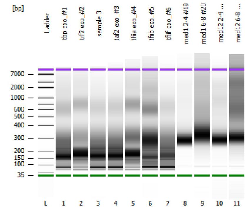
Last updated: 06/13/2019

A. Preparation of cross-linked cells
Cross-linking of cultured cells and Drosophila embryos should be performed in a timely
manner, without interruption of the procedures detailed below.

Option 1: Culture cells
Drosophila cells
Harvest up to ~50 million cells and transfer to a 15 ml conical tube. Adjust volume to
10 ml using standard media.
Add 270 µl of 37% formaldehyde and fix for 10 min at room temperature (rotating).
Add 1 ml of 2.5 M glycine in PBS and quench for 1 min at room temperature (rotating).
Pellet fixed cells at 200 x g for 3 min at 4 °C.
Remove media and wash cells twice with 10 ml cold PBS. Pellet cells at 200 x g for 3 min
at 4 °C. Remove supernatant.
Optional: freeze the cross-linked cells in liquid nitrogen and store at -80 °C.

Mammalian cells
Protocol for up to ~50 million cells in 150 mm plate.
Wash cells twice with 10 ml PBS.
Add 15 ml PBS and 400 µl of 37% formaldehyde to plate. Fix cells for 10 min at room
temperature (with movement, e.g. on orbital shaker).
Add 1.5 ml of 2.5 M glycine in PBS and quench for 5 min at room temperature (with
movement, e.g. on orbital shaker).
Wash cells twice with 10 ml cold PBS (with protease inhibitors).
Add 15 ml PBS (with protease inhibitors) and scrape cells of the plate. Transfer to 15 ml
conical tube.
Pellet fixed cells at 1000 x g for 3 min at 4 °C. Remove supernatant.
Optional: freeze the cross-linked cells in liquid nitrogen and store at -80 °C.

Option 2: Drosophila embryos
Add Clorox bleach (50-100%) to apple plates with embryos for up to 3 min. Use a soft
paintbrush to help removing the embryos by scraping the plate surface and pour into a
sieve. Use dH2O to rinse embryos from plate into sieve.

                                                   2
Last updated: 06/13/2019

Wash embryos well with lots of dH2O. Remove mesh containing embryos from sieve,
transfer embryos to 15 ml conical tube by washing mesh with PBT (PBS with
0.1% Triton X-100).
Let embryos settle down without centrifugation. Discard supernatant and add fixation
solution. Fix up to ~1 g embryos in:
       2.3 ml Fix Buffer (50 mM Hepes, 1 mM EDTA, 0.5 mM, EGTA, 100 mM NaCl;
              total volume with embryos should be ≤ 3 ml)
       130 µl 37% formaldehyde (final concentration in water phase: 1.8%)
       7.5 ml n-heptane
Shake vigorously (e.g. on vortexer) for 15 min at room temperature.
Spin for 1 min at 500 x g and 4 °C to pellet embryos and discard supernatant by carefully
pipetting off as much fixation solution as possible.
Add ~15 ml PBT-glycine (PBT with 250 mM glycine) to quench fixation. Shake embryos
vigorously for 1 min at room temperature and spin for 1 min at 500 x g and 4 °C.
Carefully decant the supernatant and remove remaining liquids with a pipette.
Wash twice with ~15 ml PBT. Add PBT, shake 1 min by hand and spin for 1 min at 500 x g
and 4 °C. Decant supernatant very carefully (pellet is not as stable as in the step above).
Resuspend embryos in ~2 ml PBT.
Optional for staging embryos: Transfer ~50 µl embryos with a cut pipette tip into a
microcentrifuge tube (continue with DAPI staining after freezing embryos, described
below).
Transfer the remaining embryos into 1.5 ml pre-weighed microcentrifuge tube.
Spin for 30 s at 500 x g and 4 °C. Remove all excess PBT by pipetting. Re-weigh tube and
determine weights of embryos.
Optional: freeze the cross-linked embryos in liquid nitrogen and store at -80 °C.

DAPI staining for embryo staging
Incubate embryos in 1 ml PBT with 10 µl 100x DAPI for 10 min at room temperature
(note: DAPI is light sensitive, cover tubes during incubation).
Wash twice with 500 µl PBT for 10 min.
Remove PBT and resuspend embryos in 120 µl 70% glycerol/ 30% PBS. Transfer ~100 µl
embryos with a cut pipette tip onto a glass slide. Putting on the coverslip gently and at
an angle will help to avoid bubbles. Seal the edges of the coverslip with nail polish to
prevent from drying out.

                                             3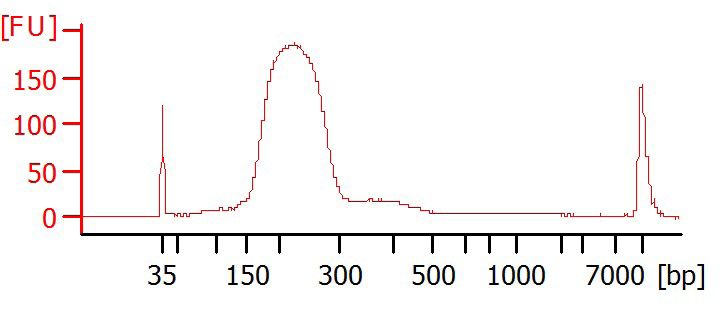
Last updated: 06/13/2019

B. ChIP set up
Bead preparation
Use 50 µl beads (e.g. Dynabeads Protein A and/or G from Invitrogen) per ChIP. Prepare
beads in microcentrifuge tubes (e.g. in Eppendorf Safe-Lock to prevent evaporation).
Wash beads 3 times with 1 ml Standard ChIP buffer or ChIP Buffer A2.
Per ChIP, resuspend beads in 500 µl Standard ChIP buffer or ChIP Buffer A2 and add
~10 µg antibody.
Incubate on rotator at 4 °C for at least 2 h to overnight.
Note: antibodies with background benefit from pre-incubation with materials that do
not contain the epitope (e.g. fixed tissue from mutants or different stage, Western blot
without the epitope band).

Option 1: Preparation of chromatin extracts for tissue culture cells
Use Standard ChIP buffer (with protease inhibitors) for chromatin extraction from
Drosophila cells. Use ChIP Buffer A2 (with protease inhibitors) for mammalian cells.
Place buffer on ice.
Resuspend cross-linked cells in Standard ChIP buffer or ChIP Buffer A2 (use 300 µl per
10 million cells). Incubate for 10 min on ice.
Sonicate in Bioruptor on HIGH, 30 sec on/30 sec off for 4-6 cycles. Cycle number and
duration depends on the type of bioruptor and may need to be optimized. Desired size
distribution of fragments is typically 100-500 bp.
Spin for 20-30 min at max. speed and 4 °C. Transfer and combine supernatants in a clean
tube. This is the input for the ChIP.

Option 2: Preparation of chromatin extract for Drosophila embryos
This protocol is for up to ~1 g embryos. The amount of embryos per ChIP depends on
the embryonic stage: 100-300 mg for early stages or 10-15 mg for later stages
(> stage 6).
Place cross-linked embryos, Lysis Buffers A1 (with protease inhibitors) and ChIP
buffer A2 (with protease inhibitors) on ice.
Resuspend cross-linked embryos in 5 ml Lysis Buffer A1 and transfer to 7 ml Dounce
homogenizer. Work with embryos on ice from now on.
Dounce with each pestle until homogenized (this may take 5-40 times for each pestle
dependent on stage and amount of embryos).

                                              4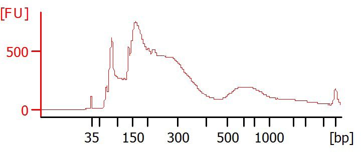
Last updated: 06/13/2019

Transfer homogenate to 15 ml tube. and spin for 3 min at 650 x g at 4 °C.
Discard supernatant and add 5 ml Lysis Buffer A1. Resuspend pellet by gently pipetting
with a wide bore or cut pipette tip. Do not vortex. Spin for 3 min at 650 x g at 4 °C.
Decant supernatant. Wash pellet two more times with 5 ml Lysis Buffer A1 (three
washes total).
Wash sample with 5 ml ChIP Buffer A2 and remove supernatant. Resuspend the sample
in ChIP Buffer A2 for sonication (optimized for our conditions: 300 µl per ChIP). Incubate
for 10 min on ice.
Sonicate in Bioruptor on HIGH, 30 sec on/30 sec off for 5 cycles. Cycle number and
duration depends on the type of bioruptor and may need to be optimized. Desired size
distribution of fragments is typically 100-500 bp.
Spin for 20-30 min at max. speed and 4 °C. Transfer supernatant to a clean tube. This is
the input for the ChIP.

Incubate ChIP
If not already done, aliquot beads for each ChIP into a separate microcentrifuge tube
(e.g. Eppendorf Safe-Lock).
Wash beads 3 times with 1 ml ChIP Buffer A2 or Standard ChIP buffer (with protease
inhibitors). Remove supernatant.
Add 300 µl input to each microcentrifuge tube. Add 300 µl ChIP Buffer A2 or Standard
ChIP buffer (with protease inhibitors) to each tube to increase total volume to a suitable
volume for tube rotation (at least 500 µl in 1.5 ml tube).
Incubate samples over night at 4 °C with rotation.
Note: These conditions work for a wide variety of antibodies and epitopes but some
ChIPs benefit from optimization. For example, high background may be reduced by
decreasing the amount of antibody-coated beads (e.g. in case of rare epitopes).
Changing the concentration of the input may also improve the signal-to-noise ratio.

                                             5
Last updated: 06/13/2019
C. ChIP-exo treatment
Note: protocol is optimized for 50 µl Dynabeads, which have a ~5 µl volume after
removing liquids. If a different volume is used, water volumes need to be adjusted
accordingly.
For all wash steps:
• Keep wash buffers A-D and 10 mM Tris (pH 8.0) cold (in fridge, on ice or work in
cold room).
• Wash each sample with 1 ml buffer fast and fairly vigorously, i.e. shake with a 90°
wrist motion around 10 times within 5 s. All beads should be resuspended after
each wash.
• To remove liquid, place samples on a magnetic rack that retains the beads. Let the
liquid settle for about 1 min, then remove the beads that are stuck in the lid using
2-5 wrist shakes (repeat if necessary to avoid losing beads). Wait another 1 min
and completely remove the liquid, preferably by aspiration (if decanting is used,
remove residual liquid by centrifuging and pipetting prior to washing with buffer A
and after washing with buffer D and Tris).
• After the last wash in each round of washes, briefly spin tubes to collect the beads
at the bottom of the tube so that they can be resuspended in master mix of the
next enzyme reaction.
Wash ChIP samples with buffers A, B, C, D, then 10 mM Tris, pH 8.0.
End repair
Add master mix: 1x
NEBNext End Repair reaction buffer (10x) 5 µl
NEBNext End Repair enzyme mix 1 µl
dH20 39 µl
45 µl
Incubate at 25 °C for 30 min.
Wash with buffers A-D, then 10 mM Tris, pH 8.0.
6Last updated: 06/13/2019
dA-tailing
Add master mix: 1x
NEBNext dA-tailing reaction buffer (10x) 5 µl
Klenow Fragment (3’ > 5’ exo-) 1 µl
dH20 39 µl
45 µl
Incubate at 37 °C for 30 min.
Wash with buffers A-D, then 10 mM Tris, pH 8.0.
Nexus adapter ligation
Add master mix: 1x
Quick Ligase buffer (2x) 25 µl
Nexus adapters (1 µM working stock) 1 µl
Quick Ligase 3 µl
dH20 16 µl
45 µl
Incubate at 25 °C for 30 min.
Note: the adapters can be diluted further to reduce adapter dimers.
Wash with buffers A-D, then 10 mM Tris, pH 8.0.
Barcode extension
Add master mix: 1x
Phi29 reaction buffer (10x) 5 µl
dNTPs (10 mM working stock) 1 µl
BSA (20 mg/ml) 0.5 µl
Phi29 DNA polymerase 1 µl
dH20 37.5 µl
45 µl
Incubate at 30 °C for 30 min.
Wash with buffers A-D, then 10 mM Tris, pH 8.0.
7Last updated: 06/13/2019
Lambda exonuclease digestion
Add master mix: 1x
Lambda exonuclease buffer (10x) 10 µl
Triton-X100 (10% working stock) 1 µl
DMSO 5 µl
Lambda exonuclease 4 µl
dH20 75 µl
95 µl
Incubate at 37 °C for 60 min in thermomixer at 1000 rpm.
Wash 3 times with RIPA buffer.
D. Standard DNA purification
Reverse cross-linking and DNA purification
To each sample, add 300 μl elution buffer and 3 µl Proteinase K (20 mg/ml). Incubate for
6 h to overnight at 65 °C in thermomixer at 1000 rpm.
Add 300 µl Phenol:Chloroform:IAA (25:24:1, v/v) to each sample and mix thoroughly (at
least 10-times if done by hand inversion). Spin at max. speed for 5 min and room
temperature. Transfer upper aqueous phase to a new microcentrifuge tube. Add 3 µl
glycogen (20 mg/ml), 12 µl 5 M NaCl and 750 µl cold 100% EtOH. Incubate for 30 min
at -80 °C.
Spin for 30 min at max. speed and 4 °C. Remove supernatant, and wash sample with
500 µl cold 70% EtOH. Spin for at least 5 min at max. speed at 4 °C. Air dry and
resuspend pellet in 12 µl dH20. Transfer sample into PCR tubes.
8Last updated: 06/13/2019
E. ChIP-nexus library preparation
Single-stranded DNA circularization
Denature sample for 5 min at 95 °C, then chill on ice.
Add master mix: 1x
CircLigase Buffer (10x) 1.5 µl
MnCl2 (50 mM) 0.75 µl
ATP (1 mM) 0.75 µl
3.0 µl
Add individually per sample:
CircLigase ssDNA Ligase 0.5 µl
Incubate for 1 h at 60 °C.
After circularization, chill samples on ice and proceed to PCR amplification.
PCR library amplification
To each sample, add master mix: 1x
Q5 Master Mix (2x) 25 µl
Universal primer (Nex_primer_U, 10 µM) 1 µl
dH20 8 µl
34 µl
Add individually per sample:
Index primer (e.g. Nex_primer_01, 10 µM) 1 µl
PCR program
1x 98 °C 2 min
98 °C 10 s
18x 65 °C 30 s
72 °C 30 s
1x 72 °C 5 min
Hold at 4 °C.
9Last updated: 06/13/2019
Library DNA extraction with agarose gel
Prepare a 2% agarose gel (with dye) in 1x TAE buffer. Use ultrapure agarose for gel
preparation.
Load 50 bp or 100 bp DNA ladder according to manufacturer’s instructions.
Add 8 μl of 6x loading dye to 50 μl PCR library DNA and load each sample in two lanes of
the gel. Leave at least one empty lane between samples.
Run gel for ~50 min (or until samples reach near end of gel) at 80 V.
View and document gel on a transilluminator (minimize UV exposure). Use a clean,
sharp razor blade to precisely excise the band containing the library, avoiding the primer
and adaptor dimers. Place gel slice into a new microcentrifuge tube.
Example of agarose gel (before excision)
Purification of DNA from gel slice
Purify the DNA from agarose slices with e.g. Monarch DNA Gel Extraction Kit (NEB)
according to manufacturer’s instructions.
Weigh the gel slices in tube. Add 4 volumes of Gel Dissolving Buffer to 1 volume of gel
(100 mg gel = 400 μl buffer). Incubate at room temperature to 32 °C in thermomixer
(with mixing at 500-1000 rpm) until the gel slices are completely dissolved.
Before proceeding, vortex each sample and spin briefly.
Load sample onto column and spin for 1 min. Discard flow-through and place the
column back into the same collection tube.
Wash with 200 μl Wash Buffer and spin for 1 min. Repeat washing step.
10Last updated: 06/13/2019
Discard flow-through and spin for 2 min to remove residual Wash Buffer.
To elute, place each column into a clean 1.5 ml microcentrifuge tube. Add 10-20 μl
water to the center of the column membrane. Let the column stand for 2 min, and spin
for 1 min (elution step can be repeated which might improve DNA yield).
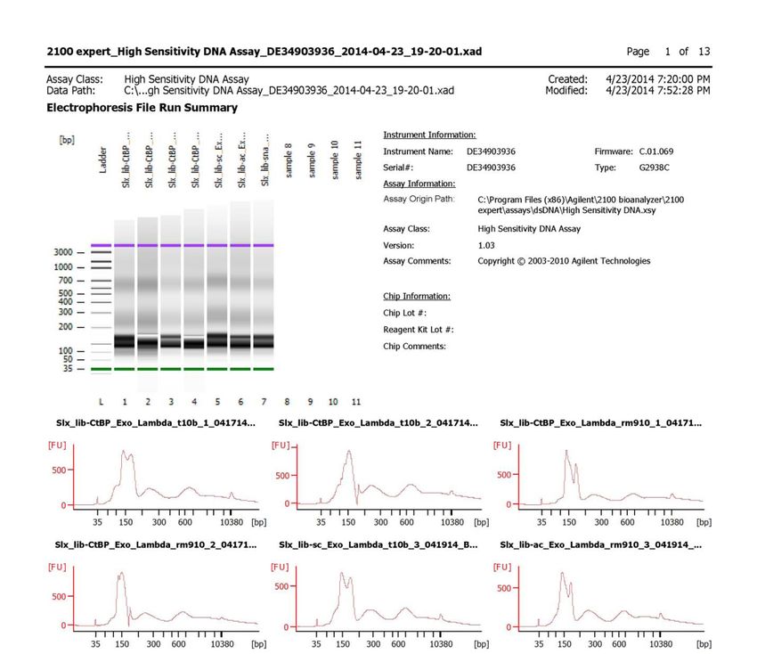
Optional: Bioanalyzer
Run samples on Bioanalyzer (e.g. Agilent, DNA HS Chip) to determine purity and
fragment size distribution.
Examples of Bioanalyzer results
1
clean size selection
150-300 bp
Good result
1
1
Undesired results
contamination from agarose
contamination from adapter dimer
11Last updated: 06/13/2019
Sequencing
Sequence DNA samples on an Illumina NextSeq platform with the single-end sequencing
primer over 50 cycles of extension according to manufacturer’s instructions. Paired-end
sequencing is also possible, but not necessary since each mapped read’s “start” site will
be the only position kept.
For a typical transcription factor in Drosophila melanogaster (and organisms with
similarly sized genomes), we recommend targeting at least 15 million uniquely alignable
reads (~25 million raw sequencing reads). For mammalian cells, we recommend
targeting at least 60 million uniquely alignable reads (~100 million raw sequencing
reads).
Data processing
There are two sets of tools recommended for the (1) preprocessing, (2) alignment, and
(3) deduplication of aligned ChIP-nexus reads. Each toolset performs the same set of
tasks, with varying customizability and compatibilities.
• Original scripts provided in R (Johnston and Weilert)
https://github.com/mlweilert/chipnexus-processing-scripts
o Scripts are intended to be run in the command-line and provide option parsing.
o Scripts are intended to be run individually - good starting point if analysis/script
customization is required.
o Includes paired-end sequencing preprocessing scripts.
• C/C++ executable CLI using Nim (Avsec)
https://github.com/Avsecz/nimnexus
o Highly parallelizable - each job has low memory requirements.
o Easy to install and use.
12Last updated: 06/13/2019
Below is a summary of the steps documented in the above links:
Format sequencing to FASTQ files (BCL à FASTQ)
If your sequencing returns raw .bcl files, apply bcl2fastq to demultiplex raw .bcl files to
.FASTQ files. Keep reads passing the default Illumina quality filter (CASAVA v1.8.2).
Formatting ChIP-nexus reads (FASTQ à FASTQ)
ChIP-nexus fragment reads should now have the format:
In order to align ChIP-nexus reads, a few preprocessing steps need to occur:
1. For each read in the .FASTQ file, check for the presence of the fixed barcodes (AGCT,
CAGT, GTCA, TCAG) at read position 6. If you use different sets of fixed barcodes, make
sure to adjust your scan accordingly when using the tools provided above.
2. Remove the fixed barcode sequence (read positions 6-9) and reassign the random
barcode sequence (read positions 1-5) and fixed barcode sequence (read positions 6-9)
to the FASTQ read name (format: “[random]_[fixed]”). A graphical representation of an
example FASTQ read in a preprocessed .FASTQ file is below:
3. Trim any remaining adapter sequences from the right end of the fragment using
cutadapt.
4. Filter all remaining reads to be at least 22 bp in length.
Trimming adapters (FASTQ à FASTQ)
Using cutadapt (-m 22 -O 4 -e 0.2), trim the remaining ChIP-nexus adapters of the
3’ end of the preprocessed fragment in the .FASTQ files. The -m 22 parameter removes
reads that would be less than 22 bp after this trimming step.
Aligning ChIP-nexus (FASTQ à BAM)
Align to appropriate reference genome using Bowtie/BWA/etc. and keep uniquely
aligning reads. We suggest a maximum of 2 mismatches.
13Last updated: 06/13/2019
Deduplication of aligned reads (BAM à BAM/GRANGES/BIGWIG)
To remove ChIP-nexus duplicates, remove reads with identical alignment coordinates
(chromosome, start position and strand) and identical random barcode (which was
assigned to the name of the FASTQ read in the preprocessing step). Tools for doing this
can be found in the GitHub links above.
Conversion of aligned fragments to Nexus coverage (BAM/GRANGES/BIGWIG à
BAM/GRANGES/BIGWIG)
Split reads by strand orientation and calculate the genome-wide counts of the start
positions (Lambda exonuclease’s stop position) for each strand. This will result in a near
single-bp resolution of ChIP-nexus mapping coverage on the forward and reverse strand,
with read pileups found at TF binding sites.
Application of exo-specific peak callers (BAM/BIGWIG à BED/IGV/TSV)
Due to the strand-specific nature of ChIP-nexus data, canonical peak callers often have
difficulties accurately assigning peaks to ChIP-nexus coverage. However, there have
been exo-specific peak callers published in recent years:
• PeakXus: Hartonen et al., Bioinformatics, 2016
https://www.ncbi.nlm.nih.gov/pubmed/27587683
• Q-nexus: Hansen et al., BMC Genomics, 2016
https://www.ncbi.nlm.nih.gov/pubmed/27814676
Some already-existing peak callers have published suggestions for application of their
peak calling tools towards ChIP-exo/nexus data, such as GEM:
http://groups.csail.mit.edu/cgs/gem/
Depending on the needs of the investigator, the selection of a peak caller and associated
parameter selection within the tools will vary.
14Last updated: 06/13/2019
F. Reagents
Lysis Buffer for Drosophila embryos
Lysis Buffer A1
15 mM HEPES, pH 7.5
15 mM NaCl
60 mM KCl
4 mM MgCl2
0.5% Triton X-100
0.5 mM DTT (add fresh)
ChIP Buffer A2
15 mM HEPES, pH 7.5
140 mM NaCl
1 mM EDTA
0.5 mM EGTA
1% Triton X-100
0.5% N-lauroylsarcosine
0.1% sodium deoxycholate
0.1% SDS
Add protease inhibitors freshly to an aliquot of each buffer before starting the protocol.
Lysis buffers for tissue culture cells
Standard ChIP Buffer
10 mM Tris, pH 8.0
140 mM NaCl
1% Triton X-100
0.1% sodium deoxycholate
0.5% N-lauroylsarcosine
0.1% SDS
Add protease inhibitors freshly to an aliquot of each buffer before starting the protocol.
15Last updated: 06/13/2019
ChIP-nexus buffers
Wash Buffer A
10 mM Tris, pH 8.0
1 mM EDTA
0.1% Triton X-100
Wash Buffer B
20 mM Tris, pH 8.0
150 mM NaCl
5 mM EDTA
5.2% sucrose
1% Triton X-100
0.2% SDS
Wash Buffer C
5 mM Tris, pH 8.0
25 mM HEPES
250 mM NaCl
0.5 mM EDTA
0.5% Triton X-100
0.05% sodium deoxycholate
Wash Buffer D
10 mM Tris, pH 8.0
250 mM LiCl
10 mM EDTA
0.5% IGEPAL CA-630
0.5% sodium deoxycholate
Tris buffer
10 mM Tris, pH 8.0
RIPA Buffer
50 mM HEPES, pH 7.5
500 mM LiCl
1 mM EDTA
0.7% sodium deoxycholate
1% IGEPAL CA-630
Elution buffer
25 mM Tris, pH 8.0
5 mM EDTA
0.5% SDS
16Last updated: 06/13/2019
Reagents to order
Vendor Reagent Catalog #
NEBNext End Repair Module E6050S
NEBNext dA-Tailing Module E6053S
Quick Ligation Kit M2200S
Phi29 DNA polymerase M0269S
New England Biolabs
Lambda exonuclease M0262S
Q5 High-Fidelity 2x Master Mix M0492S
10 mM dNTPs N0447S
Monarch DNA Gel Extraction Kit T1020
Invitrogen Proteinase K (20 mg/ml) 25530049
Roche Glycogen (20 mg/ml) 10901393001
Lucigen CircLigase ssDNA Ligase CL4111K
17Last updated: 06/13/2019
Nexus oligos to order
Name Identity Modification Barcode Sequence
Adaptor: /5Phos/GATCGGAAGAGCACACGTCTACGACG
Nex_adapter_U 5' phosphate /
universal CTCTTCC
Adaptor: /5Phos/AGCTNNNNNAGATCGGAAGAGCGTC
Nex_adapter_1 5' phosphate AGCTNNNNN
barcoded GTAGACGTGTGCTCTTCCGATCT
Adaptor: /5Phos/CAGTNNNNNAGATCGGAAGAGCGTC
Nex_adapter_2 5' phosphate CAGTNNNNN
barcoded GTAGACGTGTGCTCTTCCGATCT
Adaptor: /5Phos/GTCANNNNNAGATCGGAAGAGCGTC
Nex_adapter_3 5' phosphate GTCANNNNN
barcoded GTAGACGTGTGCTCTTCCGATCT
Adaptor: /5Phos/TCAGNNNNNAGATCGGAAGAGCGTC
Nex_adapter_4 5' phosphate TCAGNNNNN
barcoded GTAGACGTGTGCTCTTCCGATCT
Primer: 3' phosphoro- AATGATACGGCGACCACCGAGATCTACACTCTT
Nex_primer_U /
universal thioate bond TCCCTACACGACGCTCTTCCGATC*T
Primer: 3' phosphoro- CAAGCAGAAGACGGCATACGAGATCGTGATGT
Nex_primer_01 ATCACG
indexed thioate bond GACTGGAGTTCAGACGTGTGCTCTTCCGATC*T
Primer: 3' phosphoro- CAAGCAGAAGACGGCATACGAGATACATCGGT
Nex_primer_02 CGATGT
indexed thioate bond GACTGGAGTTCAGACGTGTGCTCTTCCGATC*T
Primer: 3' phosphoro- CAAGCAGAAGACGGCATACGAGATGCCTAAGT
Nex_primer_03 TTAGGC
indexed thioate bond GACTGGAGTTCAGACGTGTGCTCTTCCGATC*T
Primer: 3' phosphoro- CAAGCAGAAGACGGCATACGAGATTGGTCAGT
Nex_primer_04 TGACCA
indexed thioate bond GACTGGAGTTCAGACGTGTGCTCTTCCGATC*T
Primer: 3' phosphoro- CAAGCAGAAGACGGCATACGAGATCACTGTGT
Nex_primer_05 ACAGTG
indexed thioate bond GACTGGAGTTCAGACGTGTGCTCTTCCGATC*T
Primer: 3' phosphoro- CAAGCAGAAGACGGCATACGAGATATTGGCGT
Nex_primer_06 GCCAAT
indexed thioate bond GACTGGAGTTCAGACGTGTGCTCTTCCGATC*T
Primer: 3' phosphoro- CAAGCAGAAGACGGCATACGAGATGATCTGGT
Nex_primer_07 CAGATC
indexed thioate bond GACTGGAGTTCAGACGTGTGCTCTTCCGATC*T
Primer: 3' phosphoro- CAAGCAGAAGACGGCATACGAGATTCAAGTGT
Nex_primer_08 ACTTGA
indexed thioate bond GACTGGAGTTCAGACGTGTGCTCTTCCGATC*T
18Last updated: 06/13/2019
Preparation of ChIP-nexus oligos
Nexus adapter: Oligos for the universal adapter (Nex_adapter_U) and the barcoded
adapters (Nex_adapter_1 to _4) are annealed to become the Nexus adapter. Each
barcoded adapter contains a random barcode (NNNNN) followed by one of four fixed
barcodes (AGCT, CAGT, GCTA, or TCAG). The random barcode is used to detect
overamplification in the PCR and to determine the complexity of the final library.
To prepare 50 µM Nexus adapter mix, anneal:
Nex_adapter_U (200 µM) 50 µl
Nex_adapter_1 (AGCT, 200 µM) 12.5 µl
Nex_adapter_2 (CAGT, 200 µM) 12.5 µl
Nex_adapter_3 (GTCA, 200 µM) 12.5 µl
Nex_adapter_4 (TCAG, 200 µM) 12.5 µl
10x TE 20 µl
5 M NaCl 2 µl
dH20 78 µl
total 200 µl
Create the following adapter annealing program on a thermocycler:
95 °C 5 min
cool to 25 °C 1 min with 1% ramp
25 °C 30 min
hold at 4 °C
Dilute this stock 1:50 to obtain a working stock of 1 µM Nexus adapter mix (aliquot to
avoid repeated freeze-thaw-cycles).
PCR library amplification primers: Universal primer (Nex_primer_U) and index primers
(Nex_primer_01 to _08) are used during PCR amplification of the libraries. Prepare
working stocks of 10 µM of all primer.
19Last updated: 06/13/2019
Other reagents and equipment
Beads (e.g. Dynabeads Protein A or Protein G from Invitrogen)
Protease inhibitor (e.g. Roche)
DMSO
Phenol:chloroform:IAA
Formaldehyde
100% Ethanol
70% Ethanol
10% Triton X-100
3 M NaOAC
5 M NaCl
10x TE buffer
TAE buffer
Agarose (ultrapure)
Agarose gel dye (e.g GelRed)
6x DNA gel loading dye
DNA ladder for agarose gel
Microcentrifuge tubes (e.g. Eppendorf Safe-Lock)
15 ml tubes (e.g. Falcon)
50 ml tubes (e.g. Falcon)
PCR tubes
Disposable scalpels or razor blades
7 ml Dounce homogenizer (Wheaton)
Magnetic racks (DynaMag-2 Magnet, Invitrogen, #12321D) *expensive, but works well
Bioruptor/ sonicator (e.g. Diagenode)
Benchtop centrifuges (both room temperature and 4 °C; e.g Eppendorf)
Thermomixer (e.g. Eppendorf)
Rotator for 1.5 ml microcentrifuge tubes
Thermocycler (e.g Eppendorf)
Equipment for agarose gel electrophoresis
Agilent 2100 Bioanalyzer (optional)
20You can also read