CHOROIDAL NEVUS: GROWTH WITHOUT TRANSFORMATION
←
→
Page content transcription
If your browser does not render page correctly, please read the page content below
s OCULAR ONCOLOGY
CHOROIDAL NEVUS: GROWTH
WITHOUT TRANSFORMATION
One case illustrates how slow enlargement over time doesn’t necessarily mean the
patient has melanoma.
BY ANNIKA G. SAMUELSON, BS, AND CAROL L. SHIELDS, MD
I
n the United States, choroidal A B C
nevus—a stable, melanocytic
tumor—is found in up to 6.5% of the
White population, 0.6% of the Black
population, and 2.7% of the Hispanic
population.1,2 Choroidal nevi can grow
into melanoma, or they can enlarge
slowly over a long period of time with-
out melanoma transformation.3
Although choroidal nevi can affect
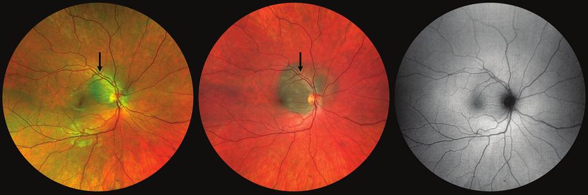
Figure 1. Wide-angle photography of the right eye shows a pigmented juxtapapillary choroidal nevus (arrow, A). Four years
vision, most are asymptomatic with little later, wide-angle photography reveals enlargement of the choroidal nevus (arrow denoting original margin, B) and fundus
impact on visual function or refractive autofluorescence demonstrates absence of orange pigment or subretinal fluid (C).
error. Shields et al evaluated a cohort
of 3,422 consecutive eyes with choroidal nevi, categorized as nevus has been considered a key determining feature sug-
either subfoveal or extrafoveal, and found that the median gestive of melanoma transformation.8 However, recent lit-
VA at presentation was 20/20 in both cohorts. However, at erature shows that some choroidal nevi can enlarge slowly
the 15-year follow-up, vision loss of ≥ 3 logMAR lines of vision during a patient’s younger years and thereafter remain
was observed in 26% of eyes with subfoveal tumors compared stable.9 Here we describe a case of slow enlargement of a
4
with only 2% of eyes with extrafoveal tumors. Vision loss due benign choroidal nevus. Importantly, this case emphasizes
to a subfoveal choroidal nevus is most often related to tumor- that slow nevus growth in the absence of risk factors can
induced retinal pigment epithelial (RPE) alterations (especially represent benign enlargement, especially in young patients.
RPE detachment), lipofuscin pigment, and foveal edema.4
Regarding progression to melanoma, Qiu and Shields CASE REPORT
used the US National Health and Nutrition Examination A 28-year-old White woman was diagnosed, using wide-
Survey to identify 5,575 participants 40 years or older angle imaging, with a choroidal nevus 4 years prior to pre-
and found no association between choroidal nevus and sentation to our clinic (Figure 1A). The nevus was monitored
skin melanoma; however, there was a relationship with annually and remained stable for 3 years, according to the
uveal melanoma.2 Singh et al retrospectively estimated referring physician. However, in year 4, enlargement was
that one in 8,845 choroidal nevi demonstrated evolution noted, and the patient was referred for our opinion. Medical
into choroidal melanoma, presuming that all melanoma and ocular history were noncontributory. Family history
arises from a nevus.5 Shields et al longitudinally studied the revealed cutaneous melanoma in a paternal grandparent.
growth of choroidal nevi into melanoma and found that On examination, BCVA was 20/20 OU. The pupils, IOP,
growth occurred in 2% at 1 year, 9% at 5 years, and 13% at and anterior segment findings were within normal limits
10 years.6 Shields and colleagues subsequently identified in each eye. The left fundus was unremarkable. The right
objective criteria, based on multimodal imaging, to identify fundus revealed a juxtapapillary pigmented choroidal mass
at-risk nevi for early treatment.7 measuring 7 mm in basal diameter, appearing approximately
Choroidal nevi that slowly enlarge without progressing 1 mm larger than was documented 4 years prior (Figure 1B).
to melanoma are poorly understood. Growth of a choroidal Fundus autofluorescence (FAF) showed no areas of orange
44 RETINA TODAY | SEPTEMBER 2021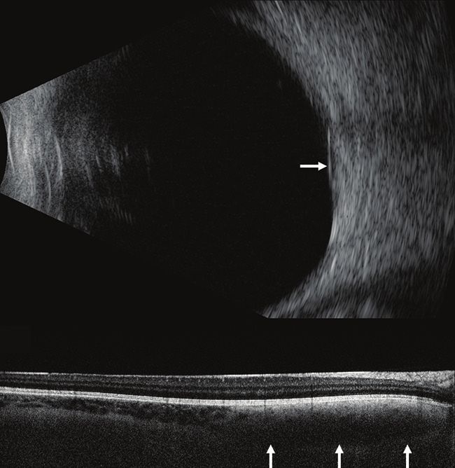
OCULAR ONCOLOGY

                                                                                                                                                                                s
pigment or subretinal fluid (Figure 1C). Ultrasonography                          A
demonstrated a flat, dense choroidal mass with a thickness
of 1.83 mm (Figure 2A). OCT showed an intact retina with
no subretinal fluid (Figure 2B). Multimodal imaging revealed
only one risk factor: diameter > 5 mm. A diagnosis of benign,
slow enlargement of choroidal nevus was made, and observa-
tion was recommended.

DISCUSSION
   Evaluation and imaging are important steps to determine if a
choroidal nevus is at risk for progression into melanoma. There
are six important risk factors related to the transformation of a
choroidal nevus into melanoma, remembered by the mnemonic
to find small ocular melanoma doing imaging (TFSOM-DIM),
which represents Thickness > 2.0 mm on ultrasonography, Fluid                     B
(subretinal) on OCT, Symptoms (VA ≤ 20/50) on Snellen acu-
ity, Orange pigment on FAF, Melanoma acoustic hollowness on
ultrasonography, and DIaMeter > 5.0 mm on fundus photogra-
phy (Table 1).7 Each of these risk factors is identified by imaging
or visual acuity testing using objective criteria.
   In this patient, all imaging risk factors were absent except
for nevus diameter > 5.0 mm. Based on the mean 5-year                            Figure 2. Ultrasonography of the right eye shows a flat, dense choroidal mass with
estimates, patients with one risk factor have an overall                         thickness of 1.83 mm (arrow, A). OCT shows an intact retina with no subretinal fluid and the
11% rate of growth into melanoma.7 Furthermore, tumor                            deep choroidal mass with loss of vascular markings (arrows, B).
diameter > 5.0 mm was found to be the weakest risk factor
(P = .0275; hazard ratio, 1.84).7 Thus, cautious observation                     and acoustic hollowness (30%).7 Benign choroidal nevus
was advised for our patient with the intent to treat if further                  enlargement, however, is a relatively slow process with a
growth or development of other factors was observed.                             mean diameter increase of only 0.06 mm/year.9 In a study of
   Choroidal nevus with growth into melanoma tends to occur                      284 choroidal nevi, researchers observed 31% of the nevi with
with a mean 1.0 mm/year diameter growth rate and 0.5 mm/                         very slow enlargement on follow-up over a mean 15 years.9
year increase in thickness, often with development of other                      Enlargement was inversely related to age, with 54% of nevus
features such as subretinal fluid (63%), orange pigment (40%),                   growth observed in patients < 40 years, 34% in patients

                                TABLE. CHOROIDAL NEVUS TRANSFORMATION INTO MELANOMA IN 2,355 CASES
Variable                                                    Letter(s) Mnemonic        Representation                             Hazard ratio (95% Cl) by P value
                                                                                                                                 multivariable analysis
Tumor thickness:                                            T          To             Thickness > 2 mm by                        3.80 (2.22–6.51)                 < .0001
> 2 mm vs ≤ 2 mm                                                                      ultrasonography
Fluid subretinal: Cap vs none                               F          Find           Subretinal fluid by OCT                    3.00 (1.77–5.09)                 < .0001
≤ 3 mm from nevus vs none                                                                                                        3.56 (1.78–7.12)                   .0003
Symptoms: visual acuity loss 20/50 or worse vs better       S          Small          Symptoms, vision loss by Snellen 2.28 (1.28–4.04)                           .0050
Orange pigment: present vs absent                           O          Ocular         Orange pigment by fundus                   3.07 (1.65–5.74)                 .0004
                                                                                      autofluorescence
Melanoma acoustic density: hollow vs solid                  M          Melanoma       Melanoma hollow by                         2.10 (1.31–3.37)                 .0020
                                                                                      ultrasonography
Tumor diameter: > 5 mm vs ≤ 5 mm                            DIM        Doing          Diameter by photography                    1.84 (1.07–3.17)                 .0275
                                                                       IMaging
Adapted from: Shields CL, Dalvin LA, Ancona-Lezama D, et al. Choroidal nevus imaging features in 3,806 cases and risk factors for transformation into
melanoma in 2,355 cases: The 2020 Taylor R. Smith and Victor T. Curtin Lecture. Retina. 2019;39(10):1840-1851.

                                                                                                                                          SEPTEMBER 2021 | RETINA TODAY 45
s   OCULAR ONCOLOGY

            between 41 and 60 years, and 19% in patients > 60 years.9
               We speculate that benign nevus enlargement may be
            more common in young adults. Most notably, patients with
            slow enlargement of choroidal nevus demonstrate further
            stability without the development of melanoma features
            over a mean follow-up of 15 years.9
               In this case, the patient had only one risk factor, a basal
            diameter of 7 mm, with slow nevus enlargement of approxi-
            mately 0.25 mm/year. Although this is faster than most nevi
            enlargement, it is slower than melanoma growth. Thus, we
            recommended cautious observation with long-term follow-
            up. This case highlights that slow growth of choroidal nevus,
            especially in young patients, is not a definitive sign of mela-
            noma transformation.
               Clinicians must assess all six risk factors of choroidal nevus
            when making a judgement regarding the potential for future
            growth, keeping in mind that a subset of patients might
            show slow enlargement of nevus without risk factors and
            without transformation into melanoma. For those patients,
            observation may be a suitable management option. n
            1. Sumich P, Mitchell P, Wang JJ. Choroidal nevi in a white population: The Blue Mountains Eye Study. Arch Ophthalmol.
            1998;116(5):645-650.
            2. Qiu M, Shields CL. Choroidal nevus in the United States adult population: racial disparities and associated factors in the
            national health and nutrition examination survey. Ophthalmology. 2015;122(10):2071-2083.
            3. Shields CL, Furuta M, Mashayekhi A, et al. Clinical spectrum of choroidal nevi based on age at presentation in 3422
            consecutive eyes. Ophthalmology. 2008;115(3):546-552.
            4. Shields CL, Furuta M, Mashayekhi A, et al. Visual acuity in 3422 consecutive eyes with choroidal nevus. Arch Ophthalmol.
            2007;125(11):1501-1507.
            5. Singh AD, Kalyani P, Topham A. Estimating the risk of malignant transformation of a choroidal nevus. Ophthalmology.
            2005;112(10):1784-1789.
            6. Shields CL, Furuta M, Berman EL, et al. Choroidal nevus transformation into melanoma: Analysis of 2514 consecutive cases.
            Arch Ophthalmol. 2009;127(8):981-987.
            7. Shields CL, Dalvin LA, Ancona-Lezama D, et al. Choroidal nevus imaging features in 3,806 cases and risk factors for trans-
            formation into melanoma in 2,355 cases: The 2020 Taylor R. Smith and Victor T. Curtin Lecture. Retina. 2019;39(10):1840-1851.
            8. Char DH, Heilbron DC, Juster RP, et al. Choroidal melanoma growth patterns. Br J Ophthalmol. 1983;67(9):575-578.
            9. Mashayekhi A, Siu S, Shields CL, et al. Slow enlargement of choroidal nevi: a long-term follow-up study. Ophthalmology.
            2011;118(2):382-388.

            ANNIKA G. SAMUELSON, BS
            n  MD Candidate, Sidney Kimmel Medical College at Thomas Jefferson
                University, Philadelphia
            n a nnika.g.samuelson@gmail.com

            n F inancial disclosure: None

            CAROL L. SHIELDS, MD | SECTION EDITOR
            n  D irector of the Ocular Oncology Service, Wills Eye Hospital, Thomas
                Jefferson University, Philadelphia
            n E ditorial Advisory Board Member, Retina Today

            n c arolshields@gmail.com

            n F inancial disclosure: None

            Support provided in part by the Eye Tumor Research Foundation,
            Philadelphia (CLS). The funders had no role in the design and conduct of the
            study, in the collection, analysis and interpretation of the data, and in the
            preparation, review or approval of the manuscript. Carol L. Shields, MD, has
            had full access to all the data in the study and takes responsibility for the
            integrity of the data.

    46 RETINA TODAY | SEPTEMBER 2021
You can also read