Chyle leak post laparoscopic cholecystectomy: a case report, literature review and management options
←
→
Page content transcription
If your browser does not render page correctly, please read the page content below
Case Report
                                                                                                                                             Page 1 of 7

Chyle leak post laparoscopic cholecystectomy: a case report,
literature review and management options
Ferdinand Ong1, Amitabha Das1, Kheman Rajkomar2
1
    Department of Upper Gastrointestinal Surgery, Liverpool Hospital, Sydney, NSW, Australia; 2Department of Upper Gastrointestinal Surgery,
Bankstown-Lidcombe Hospital, Sydney, NSW, Australia
Correspondence to: Dr. Kheman Rajkomar. Eldridge Road, Bankstown-Lidcombe Hospital, Bankstown NSW 2200, Sydney, Australia.
Email: kheman205@yahoo.com.

                  Abstract: Chyle leak after a laparoscopic cholecystectomy (LC) is very rarely reported. However, it is
                  needs to be recognised promptly and managed as otherwise it can lead to further metabolic and infective
                  complications. We present the case of a 48 years old man who was admitted with ultrasound proven acute
                  calculous cholecystitis. His vital signs were within normal range but his murphy’s sign was positive. His white
                  cell count (WCC) and liver function tests were within normal limit. He underwent an uneventful standard
                  LC with cholangiography during the same admission with no anomalous biliary or hepatic arterial anatomy
                  noted during the procedure. Post operatively he was noted to have 125 mL of white fluid in his drain. The
                  fluid triglyceride was 23.2 mmol/L, cholesterol level was 2.8 mmol/L, and drain/serum triglyceride of 15.5,
                  hence confirming it to be chyle. He was clinically otherwise very well. He was managed conservatively
                  as a low volume chyle leak with a fat free diet. The triglyceride content in the drain effluent decreased to
                  1.3mmol/L by day 6 and the fluid turned straw coloured in that interval. The drain was removed and the
                  patient discharged home without any further issues. This is the 6th reported case of chyle leak after a LC
                  for calculous disease in the English literature. The main ‘take away’ message is that although rare chyle
                  leak should be considered even in uncomplicated LC. We include a review of previous cases, propose
                  pathogenesis, diagnostic workup and a review of the options of management of this rare complication. We
                  suggest a step-up approach to management of such leaks depending on severity.

                  Keywords: Chyle leak; laparoscopic cholecystectomy (LC); calculous cholecystitis; complications; case report

                  Received: 04 July 2020; Accepted: 12 October 2020; Published: 20 April 2021.
                  doi: 10.21037/ales-20-99
                  View this article at: http://dx.doi.org/10.21037/ales-20-99

Introduction                                                                    significant morbidity due to the loss of lymph fluid rich
                                                                                in triglycerides, lymphocytes, immunoglobulins and
Gallstone disease is a highly prevalent problem worldwide.
                                                                                electrolytes. A prompt diagnosis and initiation of treatment
Laparoscopic cholecystectomy (LC) is currently a well-
                                                                                is necessary. Our current understanding and management of
accepted operation to deal with symptomatic gallstones,                         abdominal chyle leak is derived from cases that occur in the
with more than 300,000 such operations being performed                          setting of oncological resections in the retroperitoneum. It
in USA each year (1). Its postoperative complications have                      is most unusual in the setting of an uncomplicated LC.
been well described in the literature, including bile leak                          We report the sixth case of chyle leak after a LC performed
and bile duct injury, although this procedure is usually                        for acute cholecystitis. In an attempt to understand this
associated with low morbidity and mortality.                                    phenomenon we undertake a literature review, propose
   Chylous ascites is a rare complication of many abdominal                     pathogenesis, diagnostic workup and management options
and thoracic operations, especially where retroperitoneal                       for this rare complication. We suggest a step-up approach to
and mediastinal dissection has been undertaken. It carries                      the management of such leaks.

© Annals of Laparoscopic and Endoscopic Surgery. All rights reserved.           Ann Laparosc Endosc Surg 2021;6:25 | http://dx.doi.org/10.21037/ales-20-99
Page 2 of 7                                                                      Annals of Laparoscopic and Endoscopic Surgery, 2021

                                                                        no palpable masses. Murphy’s sign was positive. Laboratory
                                                                        investigations showed a normal white cell count (WCC),
                                                                        C reactive protein (CRP), liver function tests and lipase
                                                                        levels. A computed tomography (CT) scan showed a
                                                                        distended gallbladder with pericholecystic fluid (Figure 1).
                                                                        An ultrasound again revealed a thickened and oedematous
                                                                        gallbladder wall up to 1.2 cm (Figure 2), with two mobile
                                                                        gallstones and no bile duct dilatation. He was diagnosed
                                                                        with acute cholecystitis and commenced on intravenous
                                                                        antibiotics.
                                                                           The patient underwent a LC later that day. Intraoperatively
                                                                        he had an acutely inflamed gallbladder. The hepatocystic
                                                                        triangle was dissected, with clear exposure of the cystic
                                                                        duct. The cystic artery was exposed after the cystic node
                                                                        was reflected cranially and its attachments taken down
                                                                        with monopolar diathermy. The cystic artery was clipped
                                                                        before being cut. An intraoperative cholangiogram was
Figure 1 Ultrasound showing acute calculous cholecystitis with
                                                                        performed showing no abnormal findings before the cystic
1.2 cm gallbladder wall.
                                                                        duct was doubly clipped. The gallbladder was dissected
                                                                        off the cystic plate and exteriorised via a retrieval bag. A
                                                                        closed-suction silicone drain was left in the subhepatic
                                                                        space as per the surgeon’s practice in the setting of acute
                                                                        cholecystitis. Postoperative pathology would later confirm
                                                                        acute cholecystitis with cholelithiasis.
                                                                           He was commenced on an unrestricted diet immediately
                                                                        after the surgery and a slightly milky-white drain effluent
                                                                        was noted on the first postoperative day. The patient was
                                                                        clinically asymptomatic with no pain, fever or signs of
                                                                        peritonitis. The drain fluid was sent off for analysis which
                                                                        showed a raised triglyceride concentration (23.2 mmol/L)
                                                                        and normal bilirubin and amylase, with a serum
Figure 2 Axial CT of abdomen showing thick walled gallbladder           triglyceride of 1.5 mmol/L, i.e., drain/serum TG ratio
with some pericholecystic fluid.                                        15.5. Microbiological analysis showed polymorphs in the
                                                                        drain fluid with no organism being cultured. Hence, he was
                                                                        diagnosed with a chyle leak, rather than bile leak or infected
   We present the following case in accordance with the                 collection. His WCC and liver function tests remained
CARE reporting checklist (available at http://dx.doi.                   normal throughout the postoperative period. The CRP
org/10.21037/ales-20-99).                                               levels peaked at 42 mg/L on the first postoperative day and
                                                                        gradually improved.
                                                                           The drain volume was 125 mL on the first day (Figure 3A).
Case presentation
                                                                        He was started on a fat-free diet which he tolerated. The
A 48-year-old male presented acutely with a day history of              subsequent drainage volume improved (Figure 3B). The
right upper quadrant pain associated with nausea. He has a              content of the drain fluid gradually became serous on
history of ankylosing spondylitis managed with etanercept               postoperative day 4 (Figure 3C). Resolution of the chyle
injections every 2 weeks.                                               leak was confirmed by repeated testing of the drain fluid
   On examination, his vitals were within normal limits.                which demonstrated normal triglyceride concentration
His abdomen was tender in the right upper quadrant with                 (1.3 mmol/L) (Table 1), with drain/serum triglyceride ratio

© Annals of Laparoscopic and Endoscopic Surgery. All rights reserved.   Ann Laparosc Endosc Surg 2021;6:25 | http://dx.doi.org/10.21037/ales-20-99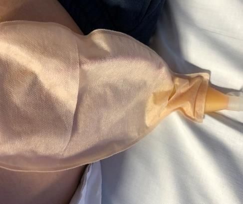
Annals of Laparoscopic and Endoscopic Surgery, 2021                                                                                  Page 3 of 7

        A

        B

        C

Figure 3 Timeline since operation. (A) Number of days post operation; (B) volume in drain (mL) since operation; (C) colour of drain
effluent on day 2, 4, 6 post op.

 Table 1 Biochemical analysis of serum and drain effluent
                                                  Serum values           Drain effluent values Day 2           Drain effluent values Day 6

 Cholesterol (mmol/L)                                   3.7                           2.8                                   2.5

 Triglycerides (mmol/L)                                 1.5                         23.2                                    1.3

of 0.9. The drain was removed and he was discharged                     identified only 5 reported cases of chyle leak after LC for
home on day 6 on a low-fat diet for another week. He had                calculous disease making it an exceedingly rare complication
no further complications during his hospital stay or after              (Table 2).
discharge.                                                                 Chyle leaks are usually of low volume and are self-
   All procedures performed in studies involving human                  limiting. However high-volume leaks can cause significant
participants were in accordance with the ethical standards of           morbidity. Those patients are prone to infections and sepsis
the institutional and/or national research committee(s) and             as the lymph fluid is rich in lymphocytes. Deficiencies
with the Helsinki Declaration (as revised in 2013). Written             in protein, vitamins, and calories can occur, along with
informed consent was obtained from the patient.                         electrolyte imbalances (6). In some cases mortality rates up
                                                                        to 70% have been associated with this complication (7).
                                                                           The mechanism of a chyle leak following a cholecystectomy
Discussion
                                                                        is possibly explained by the lymph drainage pattern of the
Whilst complications related to bile duct and hepatic                   gallbladder. Lymph from the latter drains into the first-
artery injuries after LC are uncommon, they have been                   order nodes, either the cystic or the pericholedochal
well documented in the literature. In comparison, we have               node(s) (8). From here there are 3 pathways described:

© Annals of Laparoscopic and Endoscopic Surgery. All rights reserved.   Ann Laparosc Endosc Surg 2021;6:25 | http://dx.doi.org/10.21037/ales-20-99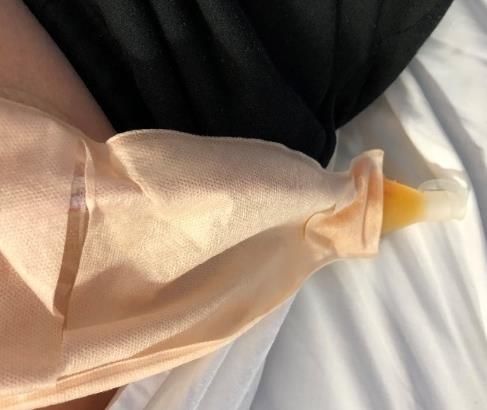
Page 4 of 7                                                                                 Annals of Laparoscopic and Endoscopic Surgery, 2021

 Table 2 Cases of chyle leak post laparoscopic cholecystectomy in literature
                                              Presentation post
 Authors/Year               Indication                                                                    Management
                                                 operatively

 Jensen 2006 (1)           Biliary colic           2 weeks              Percutaneously placed drain output was up to 1 L per day. TPN started
                                                                        with no resolution of leak. Lymphoscintigraphy localised leak in gallbladder
                                                                        fossa. Underwent laparoscopic oversew of leak (timing unreported) and
                                                                        used Tisseel fibrin glue (Baxter, Illinois, USA). Leak resolved after ‘several’
                                                                        days. Patient discharged home

 Huang 2009 (2)              Biliary                3 days              Conservative, with low fat diet and medium-chain triglyceride
                           pancreatitis                                 supplementation. Patient discharged home

 Gogalniceanu                Biliary                 1 day              Initial drain output was 340 mL/day. Fat free diet was started. Output went
 2010 (3)                  pancreatitis                                 to zero over next 7 days. Patient discharged home and was asymptomatic
                                                                        at 3 months

 Yao 2018 (4)                 Acute                 2 days              Drain output 500 mL/day. TPN started on day 3. Output increased to
                           cholecystitis                                8 L/day at day 7. Trial of somatostatin infusion/diuretics ineffective. Open
                                                                        exploration on day 10. Oversew of leak in liver bed with 3-0 prolene and
                                                                        fibrin glue placed. Drain output reduced to zero after 7 months. No issues
                                                                        noted at 1 year follow up

 Bansal 2016 (5)          Not reported              3 days              MRI showed free fluid. Re laparoscopy performed (timing unreported).
                                                                        1.4 L of chyle aspirated, no leak site identified, drain placed. Intolerant of
                                                                        sandostatin and TPN. Kept nil orally, then fat free diet introduced on day
                                                                        5. Drain output was 400 mL/day by day 4, stopped on day 15. Discharged
                                                                        home on day 19

(I) right (more predominant) route which descends                                 report non-specific symptoms, including abdominal
along the common bile duct to join the posterosuperior                            distension, discomfort, or nausea. It may also present as a
pancreaticoduodenal node(s); (II) left route which runs                           milky discharge from the wound or the drain (13). Analysis
medial to the hepatoduodenal ligament following the                               of the fluid samples from the abdomen will help confirm the
common hepatic artery before reaching the nodes around                            diagnosis. The presence of a milky fluid in the peritoneal
the coeliac trunk; (III) mesenteric route which flows along                       cavity with a triglyceride count greater than 110 mg/dL
the portal vein to the nodes around the superior mesenteric                       (1.2 mmol/L) is characteristic of chylous ascites (1).
artery (9,10).                                                                    The fluid is typically odourless, alkaline, sterile, rich
   However, during an uncomplicated LC, damage to                                 in lymphocytes but poor in bilirubin and amylase.
minor lymphatics annexed to the cystic node can potentially                       Furthermore, drain fluid/serum ratio for triglycerides is
happen and would explain minor postoperative chyle                                usually greater than 1.0. In our case, the characteristic
leaks, most of which are likely to remain subclinical. A                          appearance of the post-operative drain fluid, with its
cholecystectomy done for an impacted stone causing                                triglyceride content and drain/serum triglyceride ratio
chronic cholecystitis whereby the dissection is undertaken                        allowed us to make the diagnosis. Alternatively, the
more centrally, i.e., in close proximity to the bile duct or an                   diagnosis could be confirmed with chylomicron testing,
anomalous hepatic artery may lead to inadvertent injury to                        which unfortunately cannot be processed by our laboratory.
the lymph outflow along the right or left routes mentioned                           Different imaging modalities can aid in the diagnosis. On
above. Alternatively it has been suggested that an anatomical                     CT, it will appear as a low attenuated fluid with a possible
lymphatic variant may exist which can predispose some                             fat-fluid level (13). Lymphangiography is traditionally the
patients to a chyle leak (11). Others have suggested transient                    method employed to locate leaks, but has fallen out of
mechanical compression of lymphatics by an inflamed                               favour due to its invasive nature, technical difficulty and
pancreas (12). Interestingly, as with our patient, none of the                    adverse effects associated with the oil-based contrast used.
previously reported cases were technically difficult (Table 2).                   Some studies have shown its usefulness in the treatment
   Diagnosis of a chyle leak can be difficult. Often patients                     of refractory leaks due to the inflammatory reaction of

© Annals of Laparoscopic and Endoscopic Surgery. All rights reserved.              Ann Laparosc Endosc Surg 2021;6:25 | http://dx.doi.org/10.21037/ales-20-99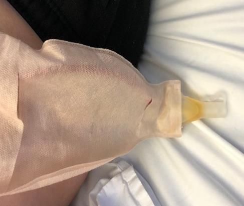
Annals of Laparoscopic and Endoscopic Surgery, 2021                                                                                  Page 5 of 7

Lipiodol when extravasated (14). Lymphoscintigraphy is                  supplementation. MCTs bind to albumin and enter the
able to provide the initial diagnosis of a chyle leak but has           portal system directly bypassing the lymphatic system,
had varying success in the exact localisation (7). It involves          whereas long-chain triglycerides are absorbed directly into
injecting technetium (99Tc) labelled colloid into the dermis            the intestinal lymphatics (17). However, the literature has
of the interdigital web spaces which gets taken up into                 reported lower success rates compared to TPN (16). There
the lymphatic channels, and therefore may be visualised                 are no randomised trials to compare these. In the acute
as collecting at the site of the leak. It has the advantage of          inpatient setting or in leaks with significant volumes, it may
being less invasive with less associated adverse reactions.             be suitable to place the patient on TPN for rapid resolution,
Some studies advocate for its use over lymphangiography,                while reserving MCT diets for low volume leaks less than
particularly in selecting patients for surgical repair (2,13).          100 mL/day (11,18). On discharge, the continuation of
Imaging should be reserved for cases where the diagnosis is             a low-fat diet with medium-chain triglycerides for a few
unclear or as a localisation tool whilst intervention is being          months has been suggested to prevent recurrence.
planned. Interestingly, in our literature review (Table 2)                 In our case we started a normal diet as per our practice
among 2 cases that did not settle with conservative means               after an uneventful cholecystectomy. This certainly
only the one who underwent lymphoscintigraphy before                    compounded the chyle leak. However, a prompt diagnosis
surgical intervention had a more favourable postoperative               of the complication and institution of a fat free diet helped
outcome (1).                                                            with rapid resolution of the low volume leak. The risk of
    In our case we did not perform any postoperative imaging            a chyle leak after LC for calculous disease is very low, as
as the leak settled and there were no signs of undrained                evidenced by 5 cases in the literature search in Table 2.
infected collection. Otherwise we would have organised a                There might be a role for an upfront restrictive diet in high
CT before contemplating percutaneous drainage in the first              risk patients for example those having a complex biliary
instance before moving to lymphoscintigraphy in case of                 dissection and especially if there is intraoperative chyle
persistent leakage.                                                     leakage noted. Those patients should be nil by mouth with
    The management of chyle leaks after LC draws upon                   slow progression to fat free/high protein/MCT diet while
our experiences with chylous ascites and chylothoraces.                 observing for chyle leakage.
However, there is no strong consensus with regards to                      Pharmacological agents may be added to the treatment
treatment due to a lack of evidence from controlled trials.             regimen if dietary modification alone is not effective in
It is agreed that therapy should be initiated swiftly in cases          reducing the chylous ascites. The reported usefulness is
of high-volume leaks. Management can be broadly divided                 variable. Pancreatic lipase inhibitors (e.g., orlistat) and
into 2 categories: conservative and surgical. Conservative              synthetic somatostatin analogues (e.g., octreotide) may help
treatment should be started first, with surgical intervention           decrease triglyceride absorption and enteric chyle flow (19).
reserved for more severe or persistent cases.                           Some studies have shown a significant improvement in output
    Paracentesis is useful in symptomatic chylous ascites with          after a few days of subcutaneous administration or continuous
no drain in situ. Repeated paracentesis should be avoided               infusion, whereas others have had less success (13). The
due to the risk of infection, increased nutritional losses and          addition of octreotide to TPN or a medium-chain triglyceride
possibility of prolonging the leak (13,15).                             diet has been shown to resolve leaks significantly earlier
    Conservative management revolves around reducing                    in hepatopancreaticobiliary surgeries and nephrectomies
enteric lymph flow and replacing any fluid, protein or                  (16,18). Somatostatin therapy can be started at 100 µg 3 time
electrolyte losses (13). Lymph normally flows through the               a day. Additionally, antibiotic prophylaxis is also advocated to
thoracic duct at an average 1 mL/kg/h but increases up to               prevent superimposed infection (1).
200 mL/kg/h after ingestion of a fatty meal (2). The use                   Surgical intervention is another cornerstone of
of short term total parenteral nutrition (TPN) is the most              management, especially since conservative approaches are
reliable and immediate way to decrease intestinal lymph                 not always effective. As expected, there is a large paucity
flow allowing the damaged lymph channels to heal. Studies               of evidence in the timing of reoperation for chyle leaks
have shown TPN alone can resolve cases in 77% to 100%                   post LC. Important clinical factors to consider in each
of cases (16). It is advantageous to maintain the benefits              case would be the current clinical status of the patient, the
of enteral feeding by placing the patient on a low fat, high            severity of the chyle leak (especially if >500 mL per day)
protein-based diet with medium-chain triglycerides (MCT)                and the original surgical procedure. Also chyle leaks that

© Annals of Laparoscopic and Endoscopic Surgery. All rights reserved.   Ann Laparosc Endosc Surg 2021;6:25 | http://dx.doi.org/10.21037/ales-20-99
Page 6 of 7 Annals of Laparoscopic and Endoscopic Surgery, 2021
are clearly seen on lymphoscintigraphy may be unlikely dose as needed. Imaging should be done early in these cases
to resolve with non-operative management (2). The aims to localise of the leak. Surgical intervention and ligation of
of surgical intervention are drainage of the leaked chyle the leaking lymphatic should then be attempted early before
followed by ligation of the culprit lymphatic. A laparoscopic inflammation sets in and makes reoperation hazardous.
approach can be safe and feasible in experienced hands,
with resolution achieved if the leak is clearly visualised
Acknowledgments
at the time of reoperation. Fibrin glue can also be used
as an adjunct (1,13). Nonetheless, the timing of surgical Funding: None.
intervention is not defined, especially since some leaks can
take several weeks to resolve on conservative treatment.
Footnote
Currently, the decision to return to theatre is strongly based
on surgeon discretion. Reporting Checklist: The authors have completed the CARE
Various other treatments for chylous ascites has reporting checklist. Available at http://dx.doi.org/10.21037/
been reported in the literature. However, these are not ales-20-99
commonly used either due to adverse effects or lack of
evidence for success. Etilefrine, a sympathomimetic used in Peer Review File: Available at http://dx.doi.org/10.21037/
postural hypotension, has been shown to reduce chyle leaks ales-20-99
by acting on smooth muscles to constrict the lymphatics (20).
Peritoneovenous shunts can reduce the nutritional, fluid and Conflicts of Interest: All authors have completed the ICMJE
immunological losses from lymph leaks, but predispose the uniform disclosure form (available at http://dx.doi.
patient to disseminated intravascular coagulopathy, sepsis, org/10.21037/ales-20-99). The authors have no conflicts of
and fat emboli (13). As such, shunts have fallen largely out interest to declare.
of favour.
Our review suggests that the volume of leak may predict Ethical Statement: The authors are accountable for all
likelihood of resolution with conservative treatment alone aspects of the work in ensuring that questions related
(Table 2). Of the 5 cases, 3 (2,3,5) settled simply with to the accuracy or integrity of any part of the work are
low fat diet, without TPN or ongoing octreotide use. appropriately investigated and resolved. All procedures
Interestingly they all had less than 1 L of leak per day after performed in studies involving human participants were in
initial drainage. The other cases (1,4) had >1 L/day output accordance with the ethical standards of the institutional
and did not settle with TPN or somatostatin infusion and and/or national research committee(s) and with the Helsinki
eventually required surgical intervention. Resolution of our Declaration (as revised in 2013). Written informed consent
low volume leak without the need for TPN or intervention was obtained from the patient.
in line with the review.
This report only describes the case of a low volume Open Access Statement: This is an Open Access article
chyle leak that settled with conservative means. However, distributed in accordance with the Creative Commons
we highlight it in the context of a literature review Attribution-NonCommercial-NoDerivs 4.0 International
with a detailed summary of similar cases. We include a License (CC BY-NC-ND 4.0), which permits the non-
pathogenesis of this rare complication and provide an up commercial replication and distribution of the article with
to date evidence-based approach to the investigative and the strict proviso that no changes or edits are made and the
management options available. original work is properly cited (including links to both the
This case shows that chyle leaks, although very formal publication through the relevant DOI and the license).
rare, can occur even in technically uncomplicated LC. See: https://creativecommons.org/licenses/by-nc-nd/4.0/.
Our recommended management protocol would be to
commence the patient on a low fat, high protein, medium
References
chain triglyceride diet and assess its efficacy as we did for
our case. If the output is >500 mL per day despite those 1. Jensen EH, Weiss CA 3rd. Management of chylous ascites
measures, then we suggest the commencement of TPN after laparoscopic cholecystectomy using minimally
with the inclusion of somatostatin therapy, escalating its invasive techniques: a case report and literature review. Am
© Annals of Laparoscopic and Endoscopic Surgery. All rights reserved. Ann Laparosc Endosc Surg 2021;6:25 | http://dx.doi.org/10.21037/ales-20-99Annals of Laparoscopic and Endoscopic Surgery, 2021 Page 7 of 7
Surg 2006;72:60-3. 11. Cheung CX, Kelly ME, El Tayeb O, et al. Chylous ascites
2. Huang YM, Chen JH, Liu SH, Lin MT. Chyle leakage post open cholecystectomy after severe pancreatitis. JOP
after laparoscopic cholecystectomy for acute biliary 2012;13:278-81.
pancreatitis: a case report. Hepatogastroenterology 12. Ben-Ami H, Nagachandran P, Assalia A, et al. Acute
2009;56:39-42. transient chylous ascites associated with acute biliary
3. Gogalniceanu P, Purkayastha S, Spalding D, et al. Chyle pancreatitis. Am J Med Sci 1999;318:122-3.
leak following laparoscopic cholecystectomy: a rare 13. Leibovitch I, Mor Y, Golomb J, et al. The diagnosis and
complication. Ann R Coll Surg Engl 2010;92:W12-4. management of postoperative chylous ascites. J Urol
4. Yao BZ, Li L, Jiang M, et al. Refractory chyle leakage 2002;167:449-57.
after laparoscopic cholecystectomy for gallstone disease: 14. Kawasaki R, Sugimoto K, Fujii M, et al. Therapeutic
A case report and literature review. Medicine (Baltimore) effectiveness of diagnostic lymphangiography for
2018;97:e9604. refractory postoperative chylothorax and chylous ascites:
5. Bansal A, Bansal AK, Bansal V, et al. Spontaneous chylous correlation with radiologic findings and preceding medical
ascites after laparoscopic cholecystectomy: a case report. treatment. AJR Am J Roentgenol 2013;201:659-66.
Int Surg J 2016;3:408-10. 15. Olthof E, Blankensteijn JD, Akkersdijk GJ.
6. Cárdenas A, Chopra S. Chylous ascites. Am J Chyloperitoneum following abdominal aortic surgery.
Gastroenterol 2002;97:1896-900. Vascular 2008;16:258-62.
7. Aalami OO, Allen DB, Organ CH Jr. Chylous ascites: a 16. Weniger M, D'Haese JG, Angele MK, et al. Treatment
collective review. Surgery 2000;128:761-78. options for chylous ascites after major abdominal surgery:
8. Shirai Y, Wakai T, Hatakeyama K. Radical lymph a systematic review. Am J Surg 2016;211:206-13.
node dissection for gallbladder cancer: indications and 17. Garrett HE Jr, Richardson JW, Howard HS, et al.
limitations. Surg Oncol Clin N Am 2007;16:221-32. Retroperitoneal lymphocele after abdominal aortic surgery.
9. Sato T, Ito M, Sakamoto H. Sakamoto, Pictorial dissection J Vasc Surg, 1989;10:245-53.
review of the lymphatic pathways from the gallbladder 18. Kuboki S, Shimizu H, Yoshidome H, et al. Chylous
to the abdominal para-aortic lymph nodes and their ascites after hepatopancreatobiliary surgery. Br J Surg
relationships to the surrounding structures. Surg Radiol 2013;100:522-7.
Anat 2013;35:615-21. 19. Kalomenidis I. Octreotide and chylothorax. Curr Opin
10. Uesaka K, Yasui K, Morimoto T, et al. Visualization Pulm Med 2006;12:264-7.
of routes of lymphatic drainage of the gallbladder 20. Guillem P, Papachristos I, Peillon C, et al. Etilefrine use in
with a carbon particle suspension. J Am Coll Surg the management of post-operative chyle leaks in thoracic
1996;183:345-50. surgery. Interact Cardiovasc Thorac Surg 2004;3:156-60.
doi: 10.21037/ales-20-99
Cite this article as: Ong F, Das A, Rajkomar K. Chyle leak
post laparoscopic cholecystectomy: a case report, literature
review and management options. Ann Laparosc Endosc Surg
2021;6:25.
© Annals of Laparoscopic and Endoscopic Surgery. All rights reserved. Ann Laparosc Endosc Surg 2021;6:25 | http://dx.doi.org/10.21037/ales-20-99You can also read